. 2024 Jun 28;9(1):159.
doi: 10.1038/s41392-024-01874-5.

A Cullin 5-based complex serves as an essential modulator of ORF9b stability in SARS-CoV-2 replication

Affiliations

A Cullin 5-based complex serves as an essential modulator of ORF9b stability in SARS-CoV-2 replication

Yuzheng Zhou et al. Signal Transduct Target Ther. .

Abstract

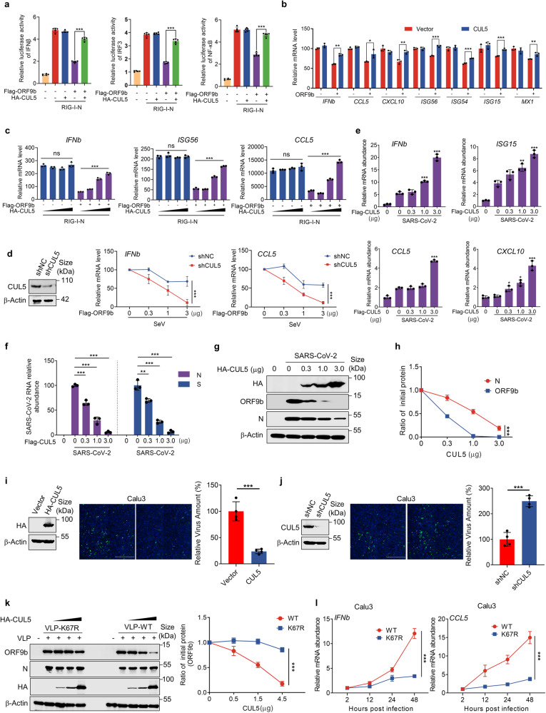
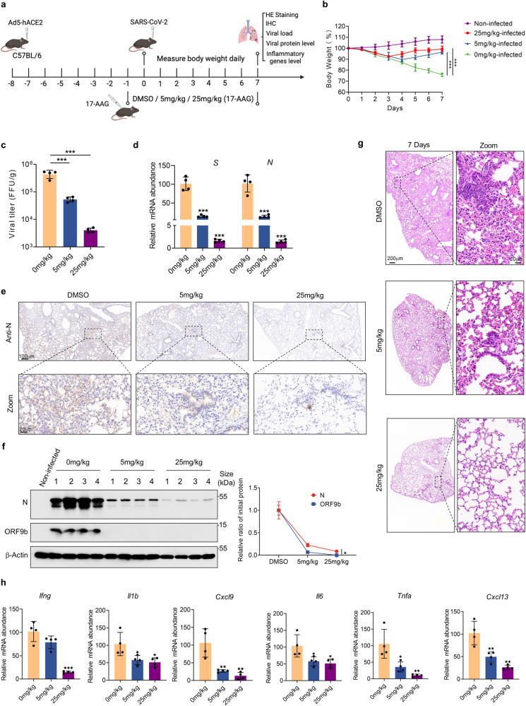
The ORF9b protein, derived from the nucleocapsid's open-reading frame in both SARS-CoV and SARS-CoV-2, serves as an accessory protein crucial for viral immune evasion by inhibiting the innate immune response. Despite its significance, the precise regulatory mechanisms underlying its function remain elusive. In the present study, we unveil that the ORF9b protein of SARS-CoV-2, including emerging mutant strains like Delta and Omicron, can undergo ubiquitination at the K67 site and subsequent degradation via the proteasome pathway, despite certain mutations present among these strains. Moreover, our investigation further uncovers the pivotal role of the translocase of the outer mitochondrial membrane 70 (TOM70) as a substrate receptor, bridging ORF9b with heat shock protein 90 alpha (HSP90α) and Cullin 5 (CUL5) to form a complex. Within this complex, CUL5 triggers the ubiquitination and degradation of ORF9b, acting as a host antiviral factor, while HSP90α functions to stabilize it. Notably, treatment with HSP90 inhibitors such as GA or 17-AAG accelerates the degradation of ORF9b, leading to a pronounced inhibition of SARS-CoV-2 replication. Single-cell sequencing data revealed an up-regulation of HSP90α in lung epithelial cells from COVID-19 patients, suggesting a potential mechanism by which SARS-CoV-2 may exploit HSP90α to evade the host immunity. Our study identifies the CUL5-TOM70-HSP90α complex as a critical regulator of ORF9b protein stability, shedding light on the intricate host-virus immune response dynamics and offering promising avenues for drug development against SARS-CoV-2 in clinical settings.


Conflict of interest statement

The authors declare no competing interests.

Figures

Fig. 1
Fig. 1
K48-linked polyubiquitin chain at the K67 site mediates the ubiquitinated degradation of SARS-CoV-2 ORF9b. a Half-life analyses of N and ORF9b proteins of SARS-CoV and SARS-CoV-2 in HEK293T cells. The cells were transfected with plasmids expressing the aforementioned viral proteins for 30 h and then split into 12-well plates. Following treatment with CHX, cells were collected as indicated time points for Western blot analysis. b The protein levels of ORF9b in HEK293T cells with treatment of DMSO, MG132, BTM, CQ, and NH4Cl. c Half-life analyses of SARS-CoV-2 ORF9b in the primary human airway epithelial (HAE) cells with treatment of CHX + MG132 or CHX + DMSO. d In vivo ORF9b ubiquitination assay in HEK293T cells. The HA-Ub WT, K48R, and K63R mutant plasmids were individually co-transfected with the Flag-ORF9b plasmid into HEK293T cells, followed by culturing and co-immunoprecipitation. Western blot was performed to detect ubiquitinated chains. e Half-life analyses of SARS-CoV-2 ORF9b-WT and SARS-CoV-2 ORF9b-3R mutant in HEK293T cells. f Flag-ORF9b was expressed in HEK293T and purified by anti-Flag beads, and then analyzed by mass spectrometry. One peptide containing lysine residues was identified. K67 was shown in red. g The protein levels of SARS-CoV-2 ORF9b-WT and indicated mutants expressed in HEK293T cells were detected after treatment with MG132 or DMSO. h In vivo ubiquitination assay of SARS-CoV-2 ORF9b-WT and SARS-CoV-2 ORF9b-K67R mutant in HEK293T cells. i and j Half-life analyses of indicated SARS-CoV-2 ORF9b mutants in HEK293T cells. Quantification was shown as mean±s.d. n = 3 independent experiments. Student’s t-test (unpaired, two-tailed) was used to compare two independent groups, and a two-way ANOVA test was performed for comparisons of multiple groups. **P < 0.01; ***P < 0.001; n.s. not significant
Fig. 2
Fig. 2
CUL5 induces the degradation of ORF9b. a The interacting proteins co-precipitated with different tagged ORF9b were identified by mass spectrometry. A protein–protein interaction network was constructed based on the identified E3 ligases. b Co-Immunoprecipitation was performed to test the interaction between CUL5 and ORF9b in HEK293T cells. The HEK293T cells were co-transfected with plasmids containing Flag-ORF9b and HA-CUL5. WCLs and precipitated proteins were analyzed by immunoblotting with indicated antibodies. c GST and GST-ORF9b were purified from E. coli and analyzed by Coomassie staining. d The HEK293T cells overexpressing the ORF9b protein were transfected with a gradually increasing amount of plasmids containing CUL5. Cells were collected for Western blot. e Half-life analyses of SARS-CoV-2 ORF9b were performed when CUL5 was overexpressed or not. f In vivo ORF9b ubiquitination assay when overexpressing CUL5 or not. g Half-life analyses of SARS-CoV-2 ORF9b when knocking down CUL5 or not. h In vivo ORF9b ubiquitination assay when CUL5 was knocked down or not in HEK293T cells. Quantification was shown as mean ± s.d. n = 3 independent experiments. *P < 0.05; **P < 0.01; ***P < 0.001. Student’s t-test (unpaired, two-tailed) was used to compare two independent groups, and a two-way ANOVA test was performed for comparisons of multiple groups. *P < 0.05; **P < 0.01; ***P < 0.001
Fig. 3
Fig. 3
CUL5 modulates the immunosuppressive effects of ORF9b and functions as a host antiviral factor. a Dual luciferase reporter gene assay was performed to test the relative activity of IFNβ, IRF3 and NF-κB when co-expressing indicated proteins in HEK293T cells. The indicated firefly luciferase reporter plasmid was transfected into HEK293T cells seeded in 12-well plates. Twenty-four hours after transfection, the innate immune pathway was activated by RIG-I-N transfection for 24 h. The Samples were collected to measure luciferase activity. n = 5 per group. b HEK293T cells were transfected with indicated plasmids and infected with SeV (100 HAU/ml) for 12 h before collection. Indicated genes were determined by qRT-PCR. c HEK293T cells carrying indicated plasmids were transfected with RIG-I-N plasmid for 12 h. IFNb, ISG56, and CCL5 were analyzed by qRT-PCR. d HEK293T cells stably expressing the shCUL5 or not were transfected with indicated plasmids. Cells were infected with SeV for 12 h. Expression of CUL5 was detected by Western blot (left). INFb and CCL5 were analyzed by qRT-PCR (right). eh Huh7 cells with the gradient overexpression of CUL5 were infected with SARS-CoV-2. Indicated antiviral genes (e), viral relative RNA abundance (f) and protein levels (g) were determined at 24 h after SARS-CoV-2 infection by qRT-PCR and Western blot. Quantification of ORF9b and N protein level normalized to β-actin (h). i Calu3 cells with overexpression of CUL5 or control were infected with SARS-CoV-2 for 24 h. The relative virus amount of SARS-CoV-2 in cells was detected by immunofluorescence assay with anti-N antibody. Scale bars, 2 mm. n = 4 per group. j The Calu3 cells stably expressing the shCUL5 or shNC were infected with SARS-CoV-2 for 24 h. The relative virus amount of SARS-CoV-2 in cells was detected by immunofluorescence assay with anti-N antibody. Scale bars, 2 mm. n = 4 per group. k Hela-hACE2 cells with the gradient overexpression of CUL5 were infected with SARS-CoV-2 K67R-VLP or WT-VLP for 24 h. Cells were collected and indicated protein levels were detected by Western blot. l Calu3 cells were infected with SARS-CoV-2 K67R-VLP or WT-VLP and collected as indicated time. The mRNA levels of INFb and CCL5 were determined by qRT-PCR. Quantification was shown as mean ± s.d. Student’s t-test (unpaired, two-tailed) was used to compare two independent groups and a two-way ANOVA test was performed for comparisons of multiple groups. *P < 0.05; **P < 0.01; ***P < 0.001; n.s. not significant
Fig. 4
Fig. 4
HSP90α maintains the stability of ORF9b. a Co-Immunoprecipitation was performed to test the interaction of SARS-CoV-2 ORF9b and ElonginB/C in HEK293T cells. b HEK293T cells overexpressing the ORF9b protein were transfected with ElonginB or ElonginC expressing plasmid as indicated. The ORF9b protein level was detected at 24 h after transfection. c HEK293T cells stably expressing the indicated siRNA were transfected with plasmids containing Flag-ORF9b, treated with CHX and collected at indicated time for Western blot. d In vivo ubiquitination assay of SARS-CoV-2 ORF9b when Elongin B or Elongin C was knocked down in HEK293T cells. e Co-Immunoprecipitation was performed to test the interaction of ORF9b and HSP90 in HEK293T cells. f and g HEK293T cells overexpressing Flag-ORF9b-WT (f) or Flag-ORF9b-K67R (g) were treated with increasing concentrations of GA or 17-AAG for 24 h and the ORF9b protein level was detected. h HEK293T cells expressing ORF9b were treated with DMSO, GA or 17-AAG for 12 h and then infected with SeV (100 HAU/ml). Total RNA was extracted to detect the relative levels of IFNb, CCL5, and CXCL10. i HEK293T cells overexpressing Flag-ORF9b were treated with 1.0 μM GA or 17-AAG, together treated with MG132 or not for 24 h. Cells were collected to detect the protein level of ORF9b. j In vivo ubiquitination assay of SARS-CoV-2 ORF9b in HEK293T cells when treated with HSP90 inhibitors (GA or 17-AAG) or not. k Half-life analyses of SARS-CoV-2 ORF9b when treated with GA or 17-AAG. l Co-Immunoprecipitation was performed to test the interaction of SARS-CoV-2 ORF9b and HSP90α/β in HEK293T cells. m In vivo ubiquitination assay of SARS-CoV-2 ORF9b when overexpressing HSP90α or HSP90β in HEK293T cells. n The ORF9b protein level was detected in HEK293T cells transfected with indicated plasmids. o The HEK293T, Calu3 and HCT116 cells expressing ORF9b were transfected with indicated siRNA for 36 h and collected to detect the HSP90α, HSP90β, and ORF9b protein levels. Quantification was shown as mean±s.d. n = 3 independent experiments. Student’s t-test (unpaired, two-tailed) was used to compare two independent groups, and a two-way ANOVA test was performed for comparisons of multiple groups. *P < 0.05; **P < 0.01; ***P < 0.001
Fig. 5
Fig. 5
TOM70 serves as a substrate receptor of CUL5-based E3 ligase for ORF9b. a HEK293T cells expressing siNC or siTOM70 were transfected with plasmids containing ORF9b and treated with DMSO, GA or 17-AAG for 24 h. The protein levels of ORF9b were detected by Western blot. b Half-life analyses of SARS-CoV-2 ORF9b when knocking down TOM70 or not in HEK293T cells. c In vivo ubiquitination assay of SARS-CoV-2 ORF9b when overexpressing TOM70 or not in HEK293T cells. d In vivo ubiquitination assay of SARS-CoV-2 ORF9b when knocking down TOM70 or not in HEK293T cells. eg Co-Immunoprecipitation was performed to test the interaction among TOM70, CUL5 and HSP90α in HEK293T cells. h The interactions between SARS-CoV-2 ORF9b and CUL5, or ORF9b and HSP90α were tested in HEK293T cells when siTOM70 was transfected or not. ik HEK293T cells expressing indicated siRNAs were transfected with plasmids expressing Flag-tagged proteins. The whole cell lysates and precipitated proteins were analyzed by immunoblotting with indicated antibodies. l A structural model of the ORF9b–TOM70–CUL5–HSP90α complex is presented in two orthogonal views. The complex includes two copies of HSP90α (light green and blue-white) and one copy each of ORF9b (red), CUL5 (magenta), and TOM70 (cyan). The model was generated using AlphaFold v2.3.2. Quantification was shown as mean ± s.d. n = 3 independent experiments. Student’s t-test (unpaired, two-tailed) was used to compare two independent groups, and a two-way ANOVA test was performed for comparisons of multiple groups. **P < 0.01; ***P < 0.001
Fig. 6
Fig. 6
HSP90 inhibitors restrict the replication of SARS-CoV-2 by promoting ORF9b degradation. a The plasmids containing Strep-tagged SARS-CoV-2 viral genes were transfected into HEK293T cells and treated with indicated concentrations of GA or 17-AAG for 24 h. The cells were lysed to detect the viral protein levels and quantification of different viral proteins was normalized to β-actin. b and c Calu3 cells treated with indicated increasing concentrations of GA (b-up), 17-AAG (b-down) or DMSO were inoculated with SARS-CoV-2 at MOI = 1 for 24 h. Quantification of relative virus amount was measured by immunofluorescence (c). dg Calu3 cells infected with SARS-CoV-2 at a MOI = 1 were treated with indicated concentrations of GA (d and e) or 17-AAG (f and g) for 24 h. Relative mRNA (left) and protein levels (right) were detected by qRT-PCR and Western blot as indicated. h A schematic diagram illustrates the regulation of the TOM70–CUL5–HSP90α complex on ORF9b. The interaction model of the complex was based on a predicted model generated using AlphaFold, as shown in Fig. 5l. Different components are represented by distinct colors: ORF9b is depicted in sky blue, TOM70 in yellow, CUL5 in yellow-green, HSP90α monomer 1 in cyan, HSP90α monomer 2 in aquamarine, and the nonfunctional HSP90α dimer in gray. This diagram demonstrates how, upon SARS-CoV-2 entry, host cells counteract viral immune evasion by targeting ORF9b for ubiquitination and subsequent degradation. In the absence of HSP90α, TOM70 and CUL5 mediate ORF9b degradation via the ubiquitin-proteasome pathway (left). Conversely, in the presence of HSP90α, ORF9b is shielded from degradation (middle). However, HSP90 inhibitors such as 17-AAG/GA deactivate HSP90αα, leading to the degradation of ORF9b (right). ik A dataset was obtained from GEO with accession number GSE171524 to analyze the difference in RNA levels of HSP90AA1 between lung epithelial cells of COVID-19 patients and healthy control. Group origins of cells (i) and RNA levels of HSP90AA1 in single cells (j) were shown with a UMAP plot. Violin plot (k) was performed to show the difference in HSP90AA1 RNA levels between lung epithelial cells of COVID-19 patients and healthy control. Quantification was shown as mean ± s.d. n = 3 independent experiments. Student’s t-test (unpaired, two-tailed) was used to compare two independent groups, and a two-way ANOVA test was performed for comparisons of multiple groups. *P < 0.05; **P < 0.01; ***P < 0.001
Fig. 7
Fig. 7
Inhibition of HSP90 attenuates the virulence of SARS-CoV-2 in the infection mouse model. a C57BL/6-hACE2 mice were intraperitoneally injected with 0, 5, or 25 mg/kg 17-AAG for 8 consecutive days. On the second day of drug treatment, the mice were nasally infected with SARS-CoV-2. b Daily recordings of mouse body weights commenced at the time of infection. c Lung tissues were collected seven days post-infection, lysed, and subjected to viral titer analysis using FFA. d Total RNA was extracted from mouse lung tissues, and relative levels of SARS-CoV-2 S and N genes were detected. e The levels of SARS-CoV-2 N protein in the lungs of infected mice treated with 17-AAG or DMSO were assessed by Immunohistochemistry with the anti-SARS-CoV-2 N antibody. f SARS-CoV-2 N proteins and ORF9b proteins in mouse lung tissues were detected by Western blot. g H&E staining of mouse lung tissues was performed to visualize the extent of inflammatory infiltration. h Relative RNA abundance of indicated cytokines or inflammatory factors in mouse lung tissues was measured by qRT-PCR. Quantification was shown as mean ± s.d. n = 4 independent experiments. Student’s t-test (unpaired, two-tailed) was used to compare two independent groups, and a two-way ANOVA test was performed for comparisons of multiple groups. *P < 0.05; **P < 0.01; ***P < 0.001

References

    1. Wu A, et al. Genome composition and divergence of the novel coronavirus (2019-nCoV) originating in China. Cell Host Microbe. 2020;27:325–328. doi: 10.1016/j.chom.2020.02.001. - DOI - PMC - PubMed
    1. Timani KA, et al. Cloning, sequencing, expression, and purification of SARS-associated coronavirus nucleocapsid protein for serodiagnosis of SARS. J. Clin. Virol. 2004;30:309–312. doi: 10.1016/j.jcv.2004.01.001. - DOI - PMC - PubMed
    1. Djukic T, et al. Expression, purification and immunological characterization of recombinant nucleocapsid protein fragment from SARS-CoV-2. Virology. 2021;557:15–22. doi: 10.1016/j.virol.2021.01.004. - DOI - PMC - PubMed
    1. Burbelo PD, et al. Sensitivity in detection of antibodies to nucleocapsid and spike proteins of severe acute respiratory syndrome coronavirus 2 in patients with coronavirus disease 2019. J. Infect. Dis. 2020;222:206–213. doi: 10.1093/infdis/jiaa273. - DOI - PMC - PubMed
    1. Cubuk J, et al. The SARS-CoV-2 nucleocapsid protein is dynamic, disordered, and phase separates with RNA. Nat. Commun. 2021;12:1936. doi: 10.1038/s41467-021-21953-3. - DOI - PMC - PubMed

Publication types

MeSH terms