. 2024 Jun 27;15(1):5239.
doi: 10.1038/s41467-024-48691-6.

Distributable, metabolic PET reporting of tuberculosis

Affiliations

Distributable, metabolic PET reporting of tuberculosis

R M Naseer Khan et al. Nat Commun. .

Abstract

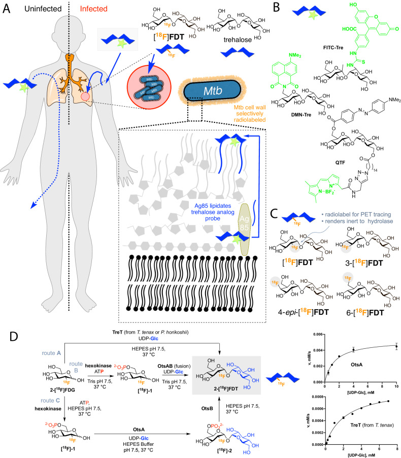
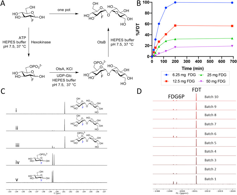
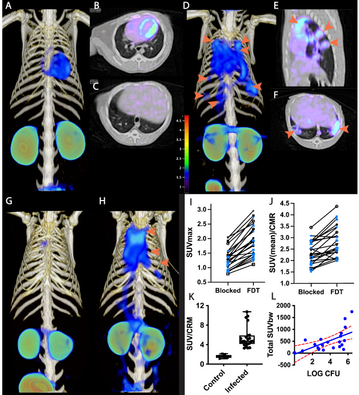
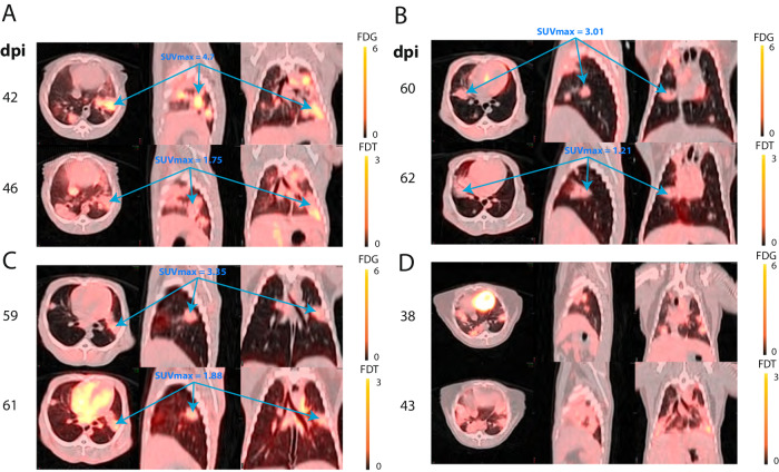
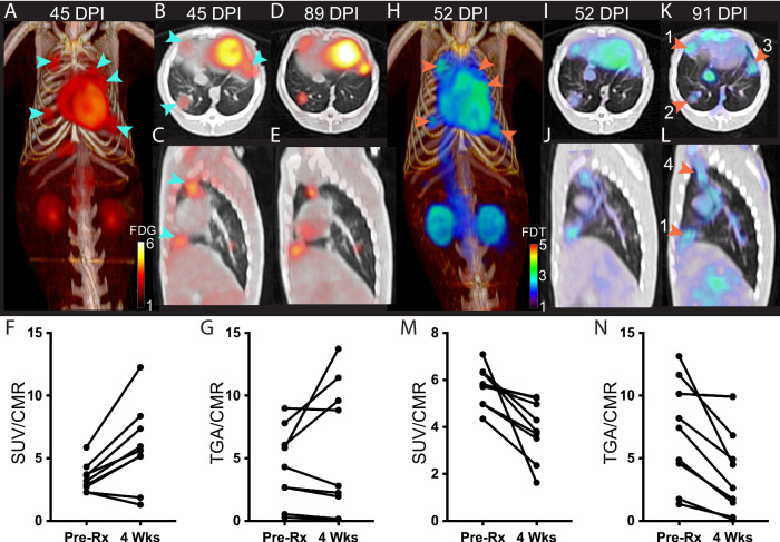
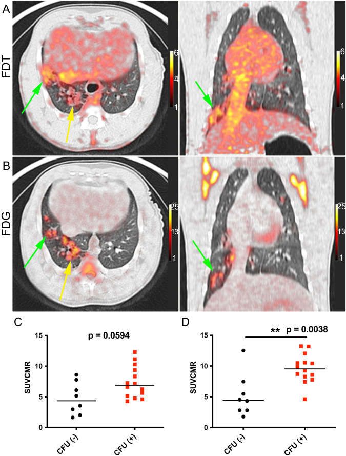
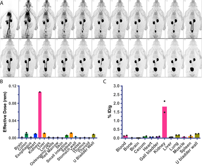
Tuberculosis remains a large global disease burden for which treatment regimens are protracted and monitoring of disease activity difficult. Existing detection methods rely almost exclusively on bacterial culture from sputum which limits sampling to organisms on the pulmonary surface. Advances in monitoring tuberculous lesions have utilized the common glucoside [18F]FDG, yet lack specificity to the causative pathogen Mycobacterium tuberculosis (Mtb) and so do not directly correlate with pathogen viability. Here we show that a close mimic that is also positron-emitting of the non-mammalian Mtb disaccharide trehalose - 2-[18F]fluoro-2-deoxytrehalose ([18F]FDT) - is a mechanism-based reporter of Mycobacteria-selective enzyme activity in vivo. Use of [18F]FDT in the imaging of Mtb in diverse models of disease, including non-human primates, successfully co-opts Mtb-mediated processing of trehalose to allow the specific imaging of TB-associated lesions and to monitor the effects of treatment. A pyrogen-free, direct enzyme-catalyzed process for its radiochemical synthesis allows the ready production of [18F]FDT from the most globally-abundant organic 18F-containing molecule, [18F]FDG. The full, pre-clinical validation of both production method and [18F]FDT now creates a new, bacterium-selective candidate for clinical evaluation. We anticipate that this distributable technology to generate clinical-grade [18F]FDT directly from the widely-available clinical reagent [18F]FDG, without need for either custom-made radioisotope generation or specialist chemical methods and/or facilities, could now usher in global, democratized access to a TB-specific PET tracer.


Conflict of interest statement

The authors declare no competing interests.

Figures

Fig. 1
Fig. 1. A Strategy for Non-Invasive Imaging Reporters using Trehalose-based, TB-specific Probes Derived Directly from [18F]FDG.
A Trehalose (blue) in Mtb is found in the outer portion of the mycobacterial cell envelope as its corresponding mycolate glycolipids. The biosynthesis of trehalose mycolates (lipidation) is catalyzed specifically in Mtb by abundant membrane-associated Antigen 85 (Ag85a, Ag85b and Ag85c) enzymes. The lack of naturally occurring trehalose in mammalian hosts as well as the uptake of exogenous trehalose by Mtb suggests that it could function as both a highly specific and sensitive probe, allowing here the development of an in vivo TB-specific, PET-radiotracer analog [18F]FDT that selectively labels lesions (red) in infected organisms (right). Uninfected organisms (left) do not process trehalose and so probe is not retained. B Prior work has established that Ag85s are sufficiently plastic in their substrate scope that they can process, for example, fluorescent analogs of trehalose, allowing them to be metabolically incorporated into the mycobacterial outer membrane in vitro for labeling. Fluorescence-based methods are not yet amenable to effective, non-invasive imaging in vivo. C Four 18F-labeled variants of trehalose were tested in which each of the available hydroxyls (OH−2, 3, 4, 6) were converted in turn to 18F. D Enzymatic synthesis of [18F]FDT. Three parallel routes (A, B and C) were evaluated for efficiency, rate and yield. Route A: TreT-mediated synthesis was evaluated using enzymes from two different sources (from T. tenax or P. horikoshii). Both proved functional but gave lower turnover under a range of conditions (see pseudo-single substrate plot for TreT (from T. tenax), lower right – See Fig. S1 and Source Data File for further details of kinetics). [Reagents and Conditions: 50 mM HEPES, 100 mM NaCl and 10 mM MgCl2, pH 7.5]. Route B: OtsAB-fusion enzyme-mediated synthesis was explored. An OtsAB fusion protein was constructed but proved to be more difficult to express and less stable under typical reaction conditions [Reagents and Conditions: 50 mM HEPES, 100 mM NaCl and 10 mM MgCl2, pH 7.5 C] Route C: Although a three-step, three-enzyme route, Route C proved to be more flexible and reliable. The selectivity of the biocatalysts allowed this to be performed in a convenient one-pot manner. The greater stability of enzyme components and the ability to vary catalyst amounts to control flux led to its choice over Route B. The higher turnovers and efficiencies led to its choice over Route A (see pseudo-single substrate plot for OtsA, upper right – See Fig. S1 and Source Data File for further details of kinetics). [Reagents and Conditions: 50 mM HEPES, 100 mM NaCl and 10 mM MgCl2, pH 7.5.]. The data points are average values from three replicates with error bars ± SD(n  =  3).
Fig. 2
Fig. 2. Reaction Optimization, Scale-up and Batch Synthesis in the Development of an Efficient Scaleable One-Pot Synthesis.
A Chosen Route C (from Fig. 1) was tested in a one-pot format [19F]FDT using pyrogen-free enzymes OtsASf and OtsBSf. B Example reaction optimization with fixed donor sugar Glc-UDP [30 mM] at different acceptor substrate concentrations (9.08, 18.17, 36.34 and 72.68 mM) of [19F]FDG; reactions were monitored in real time by calibrated 19F NMR. Please also see Source Data File. C Direct reaction monitoring of the steps of one-pot, 3-step [19F]FDT synthesis from [19F]FDG by 19F-NMR. NMR spectra: (i) 19F NMR spectrum of purified [19F]FDT; (ii) Crude 19F NMR spectrum of [19F]FDT with small amount of deoxy-fluoro-G6P ([19F]-1) still present; (iii) Crude 19F NMR spectrum of intermediates [19F]-1 and [19F]-2; (iv) Crude 19F NMR spectrum of [19F]-1 from [19F]FDG conversion; and (v) reference sample of starting material [19F]FDG. D Representative, 19F-NMR spectra of crude reaction mixtures containing [19F]FDT from repeated batches using fresh enzyme (including from newly expressed preparations) and reactants synthesized by 3-enzymes-3-steps, one-pot syntheses, prior to purification.
Fig. 3
Fig. 3. Pulmonary [18F]FDT PET uptake into Tuberculous Lung Lesions is Reproducible within 90 min with Correlated Signal in Lesions with Higher Bacterial Loads.
A–C [18F]FDT PET/CT scan of a naïve marmoset lung (administered 1 mCi) [18F]FDT and imaged 90 min post-injection (3D volume rendering, transverse and sagittal views). D–F [18F]FDT PET/CT scan of a representative, Mtb-infected marmoset lung with lesions indicated by orange arrows (3D, transverse,and sagittal views). The target dose was 2.2 mCi/kg ( ~ 1 mCi). G, H 3D volume renderings of [18F]FDT PET uptake scans for blocked (G) and unblocked (H) uptake in the same infected marmoset. Marmosets (n = 2) were randomized as to the order of blocked scan; two days later the same animals were imaged with the treatments reversed. [18F]FDT uptake was blocked by administering excess [19F]FDT, split into two administrations 1 h and 5 min prior to radiotracer administration. I, J Reduction of [18F]FDT SUVmax and SUVmean/CMR, respectively, in marmosets administered cold [19F]FDT blocker prior to being administered [18F]FDT (1.2 mCi) compared to when administered [18F]FDT alone. In both cases the animal receiving [19F]FDT blocking dose showed significantly lower accumulation of [18F]FDT into tubercular lesions (p < 0.0001, 2-tailed paired T for both measures; n = 24 lesions [12 individual lesions measured in each of the 2 animals scanned]). Different symbols have been used, colored black or blue, with one symbol-color combination for each animal. K The [18F]FDT PET signal from Mtb-infected marmoset lungs is significantly higher than the signal from uninfected (control) marmoset lungs. The mean Standard uptake values SUV/CRM are represented as boxplots (box bounds: lower and upper quartile range) where the central bars represent the median and whiskers show all points max to minimum. At least 4 lung regions of interest (ROI) were measured from 3 infected and 2 uninfected marmosets (p < 0.0001, two-tailed unpaired, T test [n = 22 ROIs in infected lungs; n = 9 in ROIs uninfected lungs]). L [18F]FDT uptake into tubercular lesion tends to increase with higher mycobacterial loads (p = 0.0019, Pearson’s ρ = 0.63). The total SUV of each lesion ROI was compared with mycobacterial colony forming units (log CFU) measured in the 22 lesions (dots) collected from two marmosets euthanized within 24 h of FDT scan collection. The mean and 95% confidence interval lines (red) are shown on a linear regression plot. Please also see Source Data File.
Fig. 4
Fig. 4. Differential Uptake and Labeling of Tubercular Lesions by [18F]FDT and [18F]FDG in Mtb-Infected Marmosets.
Serial [18F]FDG (0.8 mCi) and [18F]FDT (1.1 mCi) scans of four TB-infected marmosets were collected at 90 min post injection A–D. Animals were imaged as close in time as possible allowing for decay of FDG and for the animal to recover from anesthesia. Fused images are shown with maximum intensity for [18F]FDG set to 6 and for [18F]FDT set to 3. SUVmax for some selected lesions for each probe are shown; [18F]FDT labeling intensity was typically about half that of [18F]FDG. A and D, despite being only four and five days apart show evidence of disease progression with newly evolved lesion areas appearing to label more intensely with [18F]FDT.
Fig. 5
Fig. 5. [18F]FDT Scans of an Mtb-infected Marmoset Correlate with 4-week HRZE Treatment.
A–G 3D volume renderings and 2D axial and sagittal images and quantification of FDG uptake in Mtb-infected marmoset and the same marmoset given [18F]FDT scans with the number of days post infection (DPI) indicated above. AC) In the pretreatment scan with [18F]FDG PET (0.7 mCi) collected 60 min post-injection, cyan arrrows point to 5 lesions. D–E Marmoset imaged with [18F]FDG after 4 weeks of treatment showing the same 5 lesions. F [18F]FDG SUV before and after treatment of the lung lesions was variable and sometimes increased with treatment (p = 0.011, two-tailed paired T test, n = 9 lesions). G Total probe uptake as determined by total glycolytic activity (TGA) of the lesions showed a lesion-dependent pattern suggestive of avid [18F]FDG uptake, despite HRZE treatment (p = 0.239, two-tailed paired T test). H–N By contrast, similarly-timed (pretreatment and 4 weeks of treatment) [18F]FDT scans 90 min post injection (I 0.7 mCi; K 1 mCi) showed reduced SUV and total [18F]FDT uptake in the lesions after 4 weeks of treatment. Typical initial lesion bacterial load is 4 to 6 log CFU. Lower bacterial load was indeed observed in the lesions: lesion 1 = 2.3, lesion 2 = 2.6, lesion 3 = 3.1, lesion 4 = 0.9 log CFU and CFU is associated with FDT total uptake. M After HRZE combination drug therapy, the [18F]FDT SUV/CMR of the lung lesions was significantly decreased (p = 0.0069, two-tailed paired T test, n = 9 lesions) and N the total [18F]FDT uptake in the lesions, as determined by TGA, was also reduced (p = 0.0036, two-tailed paired T test). One representative image pair is shown of three similar treatment experiments. Please also see Source Data File.
Fig. 6
Fig. 6. [18F]FDT is also an Effective and Specific Radiotracer in an ‘Old World’ NHP TB Model.
A Axial and coronal views of the lung of a representative Mtb-infected cynomolgus macaque showing labeling of tubercular lesion clusters (green and yellow arrows) with [18F]FDT (5 mCi) scanned 60 min post-injection. Calibration scale in SUV. B Axial and coronal views of the same lung lesion clusters labeled with [18F]FDG (5 mCi) 60 min post-injection. Separate calibration scales in SUV. C The animal was necropsied and the individual lesions were plated. Ante mortum probe uptake of the lesions was compared to their culture status [colony-forming units negative CFU(-), black circles or positive CFU(+), red squares]. In images captured at 60 min there was a trend toward lesions with culturable bacteria having higher probe uptake. D By 120 min, the uptake of [18F]FDT was significantly higher among lesions with culturable mycobacteria than among sterile lesions. Quantitative data from two macaques (22 lesions total) imaged are shown with p values from two-tailed Mann-Whitney test. Imaging data from one of three representative animals are shown. Please also see Source Data File.
Fig. 7
Fig. 7. Dynamic Biodistribution of [18F]FDT in Naïve Rhesus Macaques and Infected Marmosets to Estimate Organ Exposure.
A Sequential coronal maximum intensity projections of [18F]FDT PET activity in a representative naïve rhesus macaque collected over approximately 115 min in scan frames of increasing duration in a dynamic PET scan (from top left) after administration of 151 MBq of [18F]FDT. B Organ radiation absorbed doses (mSv/MBq) calculated by extrapolating the rhesus macaque data (n = 1) to humans; organs receiving the highest doses were urinary bladder wall, kidneys, and adrenals. C Similar to the rhesus experiments, the quantification of the radioactivity in marmoset tissues (using gamma counting of [18F]FDT radioactivity in excised tissues of euthanised marmosets, n = 2) indicates the kidneys were the solid organs with the highest proportion of the injected dose (%ID/g) when marmosets were necropsied 120 min after being administered [18F]FDT. Please also see Source Data File.

Update of

  • Distributable, Metabolic PET Reporting of Tuberculosis.
    Naseer Khan RM, Ahn YM, Marriner GA, Via LE, D'Hooge F, Lee SS, Yang N, Basuli F, White AG, Tomko JA, Frye LJ, Scanga CA, Weiner DM, Sutphen ML, Schimel DM, Dayao E, Piazza MK, Gomez F, Dieckmann W, Herscovitch P, Mason NS, Swenson R, Kiesewetter DO, Backus KM, Geng Y, Raj R, Anthony DC, Flynn JL, Barry CE 3rd, Davis BG. Naseer Khan RM, et al. bioRxiv [Preprint]. 2023 Apr 3:2023.04.03.535218. doi: 10.1101/2023.04.03.535218. bioRxiv. 2023. Update in: Nat Commun. 2024 Jun 27;15(1):5239. doi: 10.1038/s41467-024-48691-6. PMID: 37333343 Free PMC article. Updated. Preprint.

References

    1. WorldHealthOrganization. Global Tuberculosis Report 2023. (Geneva (Switzerland), 2023).
    1. Maitra A, et al. Early diagnosis and effective treatment regimens are the keys to tackle antimicrobial resistance in tuberculosis (TB): A report from Euroscicon’s international TB Summit 2016. Virulence. 2017;8:1005–1024. doi: 10.1080/21505594.2016.1256536. - DOI - PMC - PubMed
    1. Bhoil A, et al. Challenges using PET-CT for international multicentre coordinated research projects in developing countries. Nucl. Med. Commun. 2019;40:93–95. doi: 10.1097/MNM.0000000000000958. - DOI - PubMed
    1. Gallach M, et al. Addressing global inequities in positron emission tomography-computed tomography (pet-ct) for cancer management: a statistical model to guide strategic planning. Med Sci. Monit. 2020;26:e926544–e926544. doi: 10.12659/MSM.926544. - DOI - PMC - PubMed
    1. TheInternationalAtomicEnergyAuthority. IAEA Medical imAGIng and Nuclear Medicine (IMAGINE), accessed 1/5/24, https://humanhealth.iaea.org/HHW/DBStatistics/IMAGINE.html