. 2024 Jun 27;7(1):144.
doi: 10.1038/s42004-024-01230-2.

Design of a covalent protein-protein interaction inhibitor of SRPKs to suppress angiogenesis and invasion of cancer cells

Affiliations

Design of a covalent protein-protein interaction inhibitor of SRPKs to suppress angiogenesis and invasion of cancer cells

Gongli Cai et al. Commun Chem. .

Abstract

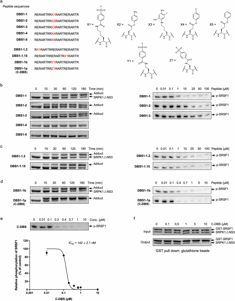
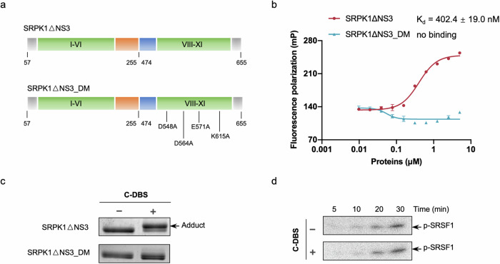
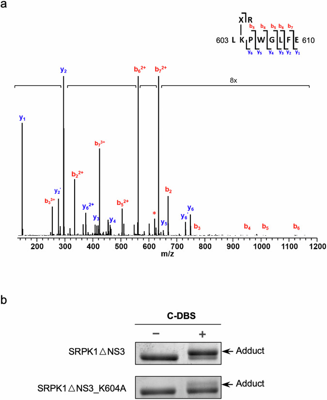
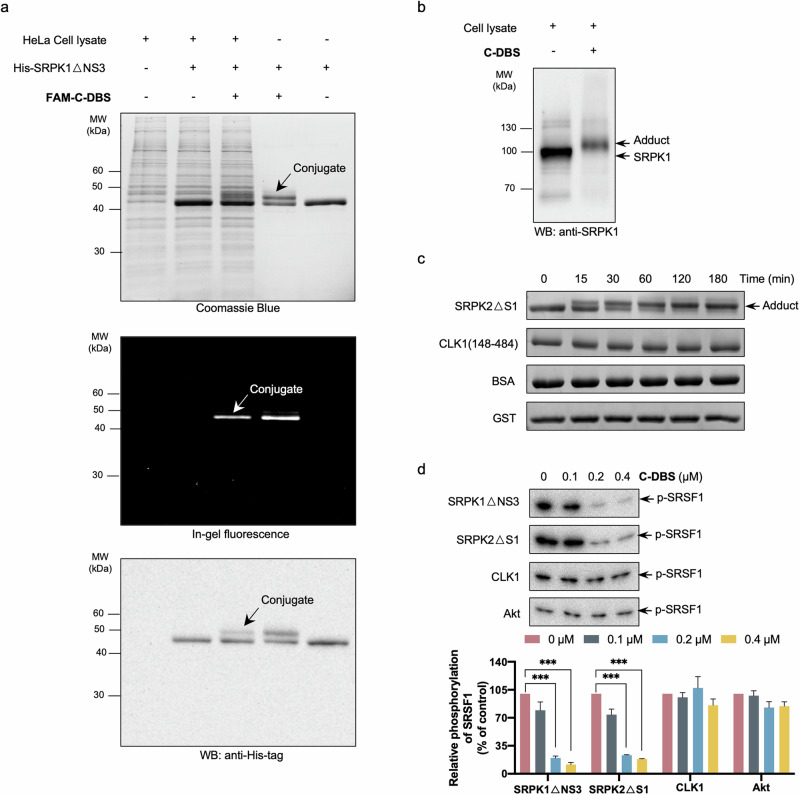
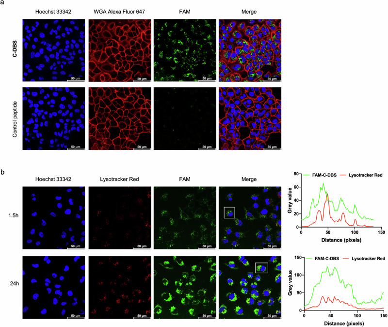
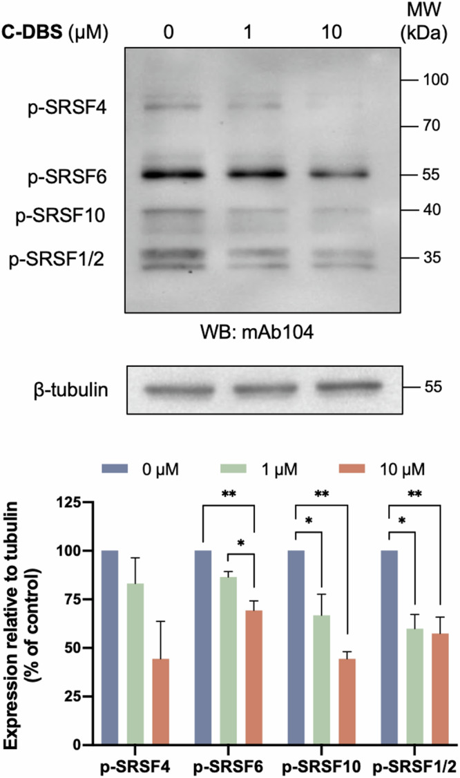
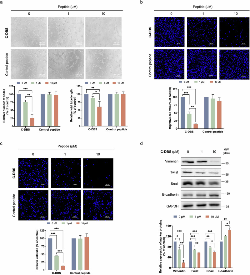
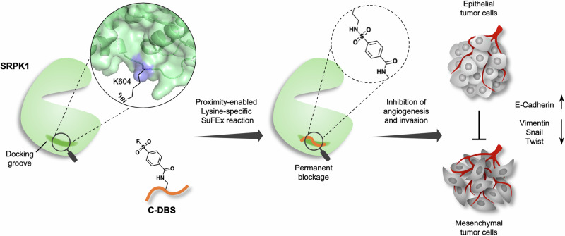
Serine-arginine (SR) proteins are splicing factors that play essential roles in both constitutive and alternative pre-mRNA splicing. Phosphorylation of their C-terminal RS domains by SR protein kinases (SRPKs) regulates their localization and diverse cellular activities. Dysregulation of phosphorylation has been implicated in many human diseases, including cancers. Here, we report the development of a covalent protein-protein interaction inhibitor, C-DBS, that targets a lysine residue within the SRPK-specific docking groove to block the interaction and phosphorylation of the prototypic SR protein SRSF1. C-DBS exhibits high specificity and conjugation efficiency both in vitro and in cellulo. This self-cell-penetrating inhibitor attenuates the phosphorylation of endogenous SR proteins and subsequently inhibits the angiogenesis, migration, and invasion of cancer cells. These findings provide a new foundation for the development of covalent SRPK inhibitors for combatting diseases such as cancer and viral infections and overcoming the resistance encountered by ATP-competitive inhibitors.


Conflict of interest statement

Gongli Cai, Yishu Bao, Qingyun Li, Jiang Xia, and Jacky Chi Ki Ngo are co-applicants for a provisional patent based on this work. All other authors declare no competing interests.

Figures

Fig. 1
Fig. 1. Rational design of an SRPK covalent inhibitor.
a List of peptides designed with lysine-reactive modifications. The peptides comprise 20 amino acids with reactive chemical warheads installed. The chosen reactive groups were aryl-sulfonyl fluoride (X1), 4-nitrophenyl (X2), 1-fluoro-2,4-dinitrobenzene (FDNB) (X3), 4-fluorophenyl (X4), and phenyl (X5) groups. b–d Adduct formation assays (left panel) and in vitro kinase activity assays (right panel) of the modified peptides. The adduction assays were performed in a concentration of 1:4 (protein: peptide) for the indicated time periods. The samples were resolved by SDS-PAGE and visualized by Coomassie Blue. Kinase activity assays were performed using [32P]ATP in the presence of the modified peptides for 2 min. The reaction samples were resolved by SDS-PAGE and visualized by autoradiography. DBS1-1p (hereafter named as C-DBS) showed the best conjugation and inhibition of SRPK1. e C-DBS inhibited SRPK1-mediated SRSF1 phosphorylation with an IC50 value of 142 nM. Data represent means ± SEM from three independent experiments. f C-DBS disrupted the interaction between SRPK1 and SRSF1 dose-dependently. SRPK1 was preincubated with C-DBS at the indicated concentrations before the addition of GST-SRSF1. Samples were resolved by SDS-PAGE and visualized by Coomassie Blue.
Fig. 2
Fig. 2. C-DBS targets the docking groove of SRPK1 and specifically conjugates to K604.
a Schematic representation of the SRPK1ΔNS3 and SRPK1ΔNS3_DM constructs. Four residues (D548, D564, E571, and K615) that are essential to substrate binding and phosphorylation were mutated to alanines in SRPK1ΔNS3_DM. b The binding affinity between SRPK1ΔNS3 and C-DBS was measured using fluorescence polarization. A constant concentration (100 nM) of FAM-C-DBS titrated with varying concentrations of SRPK1ΔNS3, yielding a Kd value of 402 nM. Data represent means ± SEM from three independent experiments. c Adduct formation assays of SRPK1ΔNS3 and SRPK1ΔNS3_DM were performed in the absence or presence of C-DBS. SRPK1ΔNS3_DM failed to conjugate with C-DBS. d The presence of C-DBS did not alter the phosphorylation of SRSF1 by SRPK1ΔNS3_DM, which is incapable of processive phosphorylation of SRSF1.
Fig. 3
Fig. 3. C-DBS specifically conjugates to K604.
a MS2 analysis of C-DBS labeled SRPK1ΔNS3 identified K604 at the docking groove was the residue covalently conjugated with C-DBS. The adduct band in SDS-PAGE was excised and digested by trypsin and Glu-C overnight for mass analysis. b Adduct formation assay of SRPK1ΔNS3 and SRPK1ΔNS3_K604A with C-DBS. The amount of the adduct formed with SRPK1ΔNS3_K604A was significantly reduced compared to that of SRPK1ΔNS3.
Fig. 4
Fig. 4. C-DBS is a selective pan-SRPK inhibitor.
a FAM-C-DBS conjugated with recombinant His-tagged SRPK1ΔNS3 in the absence or presence of HeLa cell lysate. The samples were resolved by SDS-PAGE and the adduct bands were visualized using Coomassie Blue (top panel), in-gel fluorescence (middle panel), and Western blotting (bottom panel). b C-DBS conjugated with endogenous SRPK1. MDA-MB-231 cell lysate samples before and after incubation with C-DBS were resolved by SDS-PAGE. SRPK1 and the adducts were detected using the anti-SRPK1 antibody. c C-DBS conjugated with SRPK2ΔS1. C-DBS was incubated with SRPK2ΔS1, CLK1 kinase domain, BSA, and GST for the indicated time periods. Only SRPK2 formed adducts. d C-DBS selectively inhibited SRPKs. Kinase activity assays were performed toward SRPK1ΔNS3, SRPK2ΔS1, CLK1, and Akt in the presence of the indicated concentrations of C-DBS. C-DBS inhibited both SRPK1 and SRPK2 but not CLK1 or Akt. The p-SRSF1 levels were quantified using ImageJ. Data represents means ± SEM from three independent experiments. Statistical analysis was performed using one-way ANOVA. ***p < 0.001.
Fig. 5
Fig. 5. C-DBS is self-cell permeable.
a Internalization of C-DBS and a control peptide in A549 cells. Both peptides were labeled with fluorescent FAM group (green). The cells were treated with 10 μM peptides for 24 h. Hoechst 33342 (blue) and Wheat Germ Agglutinin (WGA) Alexa Fluor 647 (red) were used to stain the nucleus and cell membrane, respectively. C-DBS, but not the control peptide, was readily up-taken by the cells. b Fluorescence analysis of FAM-C-DBS and Lysotracker. A549 cells were stained with Hoechst 33342 (blue) and Lysotracker Red (red) after C-DBS incubation for the indicated time. Increased diffused green signals were observed after prolonged incubation. The fluorescence intensity profiles of the region of interest (ROI, white box) were analyzed using ImageJ.
Fig. 6
Fig. 6. C-DBS inhibits SR protein phosphorylation.
A549 cells were treated with the indicated concentrations of C-DBS and the total cell lysate was subjected to Western blotting, where the phosphorylated SR proteins were probed with mAb104. The p-SR levels versus β-tubulin were quantified using ImageJ. Data represents means ± SEM from three independent experiments. Statistical analysis was performed using one-way ANOVA. *p < 0.05; **p < 0.01.
Fig. 7
Fig. 7. C-DBS suppresses angiogenesis, cell migration, and invasion.
a C-DBS inhibited endothelial cell tube formation. Conditioned medium collected from A549 cells treated with C-DBS (top panel) or the control peptide (bottom panel) was added to HUVECs seeded in Matrigel-coated wells. The images of capillary-like tubes were captured at three random views in each well. The inhibitory effect of C-DBS on angiogenesis was determined by counting the number of tube nodes and measuring the length of the capillary-like tubes that formed using ImageJ. Scale bar: 250 μm. C-DBS inhibited the migration b and invasiveness c of A549 cells. A549 cells were resuspended in an FBS-free medium containing different concentrations of C-DBS (top panel) or the control peptide (bottom panel). Full medium containing 10% FBS was used as attractants in the lower chamber. Migrated or invaded cells were fixed and stained with Hoechst 33342 and photographed at five random views using a fluorescent microscope. The number of migrated or invaded cells was counted using ImageJ. d C-DBS altered the expression of EMT markers. A549 cell lysates with or without C-DBS treatment were subjected to Western blotting. EMT markers were detected using the corresponding antibodies. Band intensity quantification was performed using ImageJ. All data represent means ± SEM from three independent experiments. Statistical analysis was performed using one-way ANOVA. *p < 0.05; **p < 0.01; ***p < 0.001.
Fig. 8
Fig. 8. Schematic illustration of C-DBS combating angiogenesis and invasion.
C-DBS permanently blocks the docking groove of SRPK1 via proximity-enabled SuFEx reaction with specific lysine, resulting in the anti-angiogenesis and anti-invasion effect by reversing the EMT of tumor cells.

References

    1. Long JC, Caceres JF. The SR protein family of splicing factors: master regulators of gene expression. Biochem. J. 2009;417:15–27. doi: 10.1042/BJ20081501. - DOI - PubMed
    1. Shepard PJ, Hertel KJ. The SR protein family. Genome Biol. 2009;10:242. doi: 10.1186/gb-2009-10-10-242. - DOI - PMC - PubMed
    1. Zhong XY, Wang P, Han J, Rosenfeld MG, Fu XD. SR proteins in vertical integration of gene expression from transcription to RNA processing to translation. Mol. Cell. 2009;35:1–10. doi: 10.1016/j.molcel.2009.06.016. - DOI - PMC - PubMed
    1. Karni R, et al. The gene encoding the splicing factor SF2/ASF is a proto-oncogene. Nat. Struct. Mol. Biol. 2007;14:185–193. doi: 10.1038/nsmb1209. - DOI - PMC - PubMed
    1. Das S, Krainer AR. Emerging Functions of SRSF1, Splicing Factor and Oncoprotein, in RNA Metabolism and Cancer. Mol. Cancer Res. 2014;12:1195–1204. doi: 10.1158/1541-7786.MCR-14-0131. - DOI - PMC - PubMed