. 2024 Aug;43(16):3450-3465.
doi: 10.1038/s44318-024-00150-0. Epub 2024 Jun 27.

Human mitochondrial carriers of the SLC25 family function as monomers exchanging substrates with a ping-pong kinetic mechanism

Affiliations

Human mitochondrial carriers of the SLC25 family function as monomers exchanging substrates with a ping-pong kinetic mechanism

Camila Cimadamore-Werthein et al. EMBO J. 2024 Aug.

Abstract

Members of the SLC25 mitochondrial carrier family link cytosolic and mitochondrial metabolism and support cellular maintenance and growth by transporting compounds across the mitochondrial inner membrane. Their monomeric or dimeric state and kinetic mechanism have been a matter of long-standing debate. It is believed by some that they exist as homodimers and transport substrates with a sequential kinetic mechanism, forming a ternary complex where both exchanged substrates are bound simultaneously. Some studies, in contrast, have provided evidence indicating that the mitochondrial ADP/ATP carrier (SLC25A4) functions as a monomer, has a single substrate binding site, and operates with a ping-pong kinetic mechanism, whereby ADP is imported before ATP is exported. Here we reanalyze the oligomeric state and kinetic properties of the human mitochondrial citrate carrier (SLC25A1), dicarboxylate carrier (SLC25A10), oxoglutarate carrier (SLC25A11), and aspartate/glutamate carrier (SLC25A13), all previously reported to be dimers with a sequential kinetic mechanism. We demonstrate that they are monomers, except for dimeric SLC25A13, and operate with a ping-pong kinetic mechanism in which the substrate import and export steps occur consecutively. These observations are consistent with a common transport mechanism, based on a functional monomer, in which a single central substrate-binding site is alternately accessible.

Keywords: Bioenergetics; Kinetic Analysis; Mitochondria; SLC25 Mitochondrial Carrier Family; Transport.


Conflict of interest statement

The authors declare no competing interests.

Figures
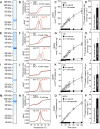
Figure 1
Figure 1. The human oxoglutarate, citrate, dicarboxylate, and aspartate/glutamate carriers are pure, folded, bind substrate, and are functional.
Instant-blue stained SDS-PAGE gel of purified protein (A) oxoglutarate carrier (OGC), (E) citrate carrier (CIC), (I) dicarboxylate carrier (DIC), (M) aspartate/glutamate carrier (AGC2). Typical unfolding curves of ~10 μg protein without compound (black trace), or with 10 mM compound (red trace) (B) OGC + 10 mM malate, (F) CIC + 10 mM citrate, (J) DIC + 10 mM malate, or (N) AGC2 + 10 mM aspartate, using nano differential scanning fluorimetry (Nanotemper Prometheus). The peak in the derivative of the unfolding curve (dR/dT) is at the apparent melting temperature (Tm). (C) [14C]-malate uptake curves of OGC reconstituted into proteoliposomes loaded with (white circles) or without (black circles) 1 mM malate. Transport was initiated by the external addition of 2.5 μM [14C]-malate. (G) [14C]-citrate uptake curves of CIC reconstituted into proteoliposomes loaded with (white circles) or without (black circles) 1 mM citrate. Transport was initiated by the external addition of 2.5 μM [14C]-citrate. (K) [14C]-malate uptake curves of DIC reconstituted into proteoliposomes loaded with (white circles) or without (black circles) 1 mM malate. Transport was initiated by the external addition of 2.5 μM [14C]-malate. (O) [14C]-aspartate uptake curves of AGC2 reconstituted into proteoliposomes loaded with (white circles) or without (black circles) 1 mM aspartate. Transport was initiated by the external addition of 2.5 μM [14C]-aspartate. (D, H, L, P) Initial rates were estimated by fitting the uptake curves to Eq. 1. The data represent the average and standard deviation of two biological repeats, each with three technical repeats except AGC2, which is the average of three technical repeats. Source data are available online for this figure.
Figure 2
Figure 2. Structures and oligomeric states of mitochondrial carriers.
(A) Structural models of the human dicarboxylate carrier (DIC), oxoglutarate carrier (OGC), citrate carrier (CIC), ADP/ATP carrier (AAC1), and aspartate/glutamate carrier (AGC2) were all determined by the Alphafold 3.0 server (Abramson et al, 2024), except for the experimentally determined structure of an uncoupling protein (UCP1) (pdb entry: 8G8W) (Jones et al, 2023). (B) Determination of the molecular weights in LMNG/TOCL by size exclusion chromatography. The normalized absorbance traces for monomeric AAC1 (blue), DIC (red), OGC (green), CIC (cyan), UCP1 (brown), and dimeric AGC2 (purple) are shown. The standards used for sizing were ferritin (440 kDa), aldolase (158 kDa), conalbumin (76 kDa), and ovalbumin (43 kDa). The dotted line indicates the elution volume of a hypothetical dimer peak for DIC, OGC, and CIC. Source data are available online for this figure.
Figure 3
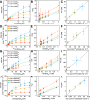
Figure 3. Two-substrate analysis of transport catalyzed by human mitochondrial carriers.
Initial rates were estimated by fitting uptake data to Eq. 1: OGC (Figs. 1C and  EV1), CIC (Figs. 1G and  EV2), DIC (Figs. 1K and  EV3), and AGC2 (Figs. 1O and  EV4). Michaelis–Menten plots of 0.10 mM (red traces), 0.25 mM (orange traces), 0.50 mM (green traces), and 1.00 mM (blue traces) internally-loaded (A) malate (OGC), (D) citrate (CIC), (G) malate (DIC), and (J) aspartate (AGC2). Transport was initiated by the addition of radiolabeled substrate externally ([14C]-malate (OGC and DIC), [14C]-citrate (CIC), and [14C]-aspartate (AGC2)) at the indicated concentration, and stopped by vacuuming and washing at defined time-intervals. Lineweaver–Burk analysis of homo-exchange for (B) OGC, (E) CIC, (H) DIC, and (K) AGC2, using the same color scheme as in (A). Km plotted against Vmax for the various chemical gradients for (C) OGC, (F) CIC, (I) DIC, and (L) AGC2, using the same color scheme as in (A). The two kinetic parameters were determined by fitting the Michaelis–Menten curves through iteration. The data were represented by the average and standard deviation of n = 6 (two independent experiments, each with three technical repeats, except for the 1 and 50 μM external [14C]-malate datasets for DIC, the 1.5 μM external [14C]-malate/1 mM internal malate datapoint for DIC, and the data for AGC2, which are the average of three technical repeats). Source data are available online for this figure.
Figure 4
Figure 4. Schematic representation of the ping-pong mechanism of mitochondrial carrier proteins.
Conformational changes between the cytoplasmic-open state (c-state), occluded state (o-state), and matrix-open state (m-state) with the carriers shown schematically, viewed laterally from the membrane. The imported substrate is shown in red and the exported substrate in blue. MM mitochondrial matrix, MIM mitochondrial inner membrane, IMS intermembrane space.
Figure EV1
Figure EV1. Uptake of transport catalyzed by reconstituted human oxoglutarate carrier.
Proteoliposomes containing human oxoglutarate carrier were loaded with either 0.10 mM (red traces), 0.25 mM (orange traces), 0.50 mM (green traces), or 1.00 mM (blue traces) malateint, and the transport was initiated by the externally added radiolabeled malate at either 0.5 μM (filled circles), 1.0 μM (filled squares), 1.5 μM (filled upward triangles), 2.5 μM (filled downward triangles), 5.0 μM (open circles), 10 μM (open squares), 15 μM (open upward triangles), or 20 μM (open downward triangles) [14C]-malate (malateext). Initial rates were estimated by fitting the uptake data to Eq. 1. The data represent the average and standard deviation of n = 6 (two independent experiments, each with three technical repeats). Source data are available online for this figure.
Figure EV2
Figure EV2. Uptake curves of the transport catalyzed by reconstituted human citrate carrier.
Proteoliposomes containing human citrate carrier were loaded with either 0.10 mM (red traces), 0.25 mM (orange traces), 0.50 mM (green traces), or 1.00 mM (blue traces) citrateint, and transport was initiated by the externally added radiolabeled sulfate at either 1.0 μM (filled circles), 1.5 μM (filled squares), 2.5 μM (filled upward triangles), 5 μM (filled downward triangles), 10 μM (open circles), 15 μM (open squares), 20 μM (open upward triangles), or 25 μM (open downward triangles) [14C]-citrate (citrateext). Initial rates were estimated by fitting the uptake data to Eq. 1. The data represent the average and standard deviation of n = 6 (two independent experiments, each with three technical repeats). Source data are available online for this figure.
Figure EV3
Figure EV3. Uptake curves of the transport catalyzed by reconstituted human dicarboxylate carrier.
Proteoliposomes containing human dicarboxylate carrier were loaded with either 0.10 mM (red traces), 0.25 mM (orange traces), 0.50 mM (green traces), or 1.00 mM (blue traces) malateint, and transport was initiated by the externally added radiolabeled malate at either 1.0 μM (filled circles), 1.5 μM (filled squares), 2.5 μM (filled upward triangles), 5 μM (filled downward triangles), 10 μM (open circles), 15 μM (open squares), 20 μM (open upward triangles), 25 μM (open downward triangles), or 50 μM (crosses) [14C]-malate (malateext). Initial rates were estimated by fitting the uptake data to Eq. 1. The data represent the average and standard deviation of n = 6 (two independent experiments, each with three technical repeats, except the 1 and 50 μM external [14C]-malate datasets, which are the average of three technical repeats). Source data are available online for this figure.
Figure EV4
Figure EV4. Uptake curves of the transport catalyzed by reconstituted human aspartate/glutamate carrier.
Proteoliposomes containing human aspartate/glutamate carrier were loaded with either 0.10 mM (red traces), 0.25 mM (orange traces), 0.50 mM (green traces), or 1.00 mM (blue traces) aspartateint, and the transport was initiated by the externally added radiolabeled aspartate at either 1.5 μM (filled squares), 2.5 μM (filled upward triangles), 5.0 μM (filled downward triangles), 10 μM (open circles), 15 μM (open squares), 20 μM (open upward triangles), or 25 μM (open downward triangles) [14C]-aspartate (aspartateext). Initial rates were estimated by fitting the uptake data to Eq. 1. The data represent the average and standard deviation of three technical repeats. Source data are available online for this figure.

References

    1. Abramson J, Adler J, Dunger J, Evans R, Green T, Pritzel A, Ronneberger O, Willmore L, Ballard AJ, Bambrick J et al (2024) Accurate structure prediction of biomolecular interactions with AlphaFold 3. Nature 630:493–500 - PMC - PubMed
    1. Alexander CG, Wanner R, Johnson CM, Breitsprecher D, Winter G, Duhr S, Baaske P, Ferguson N (2014) Novel microscale approaches for easy, rapid determination of protein stability in academic and commercial settings. Biochim Biophys Acta 1844:2241–2250 10.1016/j.bbapap.2014.09.016 - DOI - PMC - PubMed
    1. Azzi A, Chappell JB, Robinson BH (1967) Penetration of the mitochondrial membrane by glutamate and aspartate. Biochem Biophys Res Commun 29:148–152 10.1016/0006-291X(67)90556-6 - DOI - PubMed
    1. Bamber L, Harding M, Butler PJG, Kunji ERS (2006) Yeast mitochondrial ADP/ATP carriers are monomeric in detergents. Proc Natl Acad Sci USA 103:16224–16229 10.1073/pnas.0607640103 - DOI - PMC - PubMed
    1. Bamber L, Harding M, Monné M, Slotboom DJ, Kunji ERS (2007a) The yeast mitochondrial ADP/ATP carrier functions as a monomer in mitochondrial membranes. Proc Natl Acad Sci USA 104:10830–10834 10.1073/pnas.0703969104 - DOI - PMC - PubMed

MeSH terms

Substances

LinkOut - more resources