

Retrospective analysis of dogs and cats with a mixed form of pulmonary hypertension and suspected pulmonary capillary hemangiomatosis in comparison to animals with predomination of precapillary pulmonary hypertension
- PMID: 38938438
- PMCID: PMC11199750
- DOI: 10.5455/OVJ.2024.v14.i5.17
Retrospective analysis of dogs and cats with a mixed form of pulmonary hypertension and suspected pulmonary capillary hemangiomatosis in comparison to animals with predomination of precapillary pulmonary hypertension
Abstract
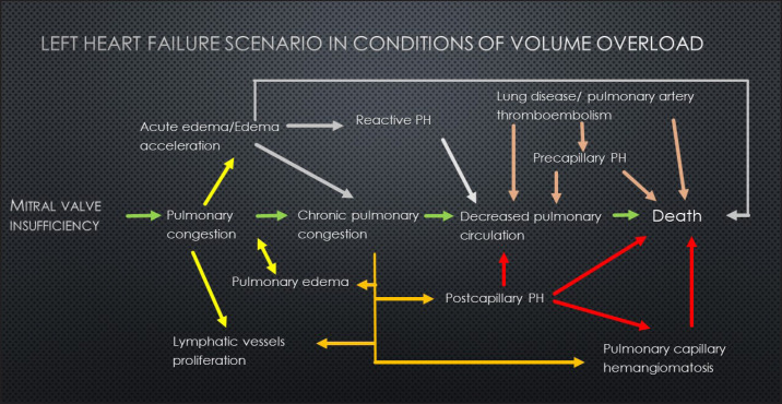
Background: Pulmonary capillary hemangiomatosis (PCH) is an idiopathic disease with the anomalous proliferation of a small capillary-like vessel in the pulmonary tissue, which can lead to a severe form of PH. There are only several cases of PCH described in veterinary literature: 27 cases in dogs and 2 cases in cats. In veterinary medicine, PH is mostly recognized as a consequence of left heart failure as a progression of the postcapillary PH to the precapillary form. PCH is mostly described as a primary disease, but resistant postcapillary PH with the high possibility of pulmonary edema raises speculation that PCH could be a secondary malformation to the left heart disease.
Aim: Discover the features associated with the shift between left- and right-sided heart disease in the context of PH development.
Methods: Retrospective analysis of materials from cats and dogs with histological markers of PCH (sPCH) versus those with right heart failure (RHF).
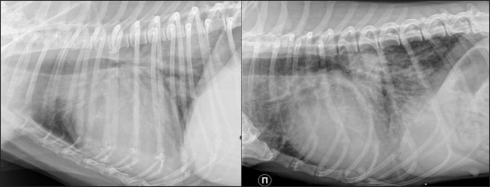
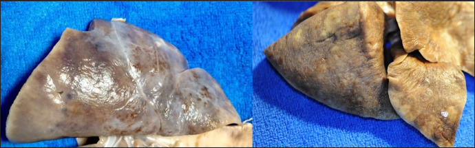
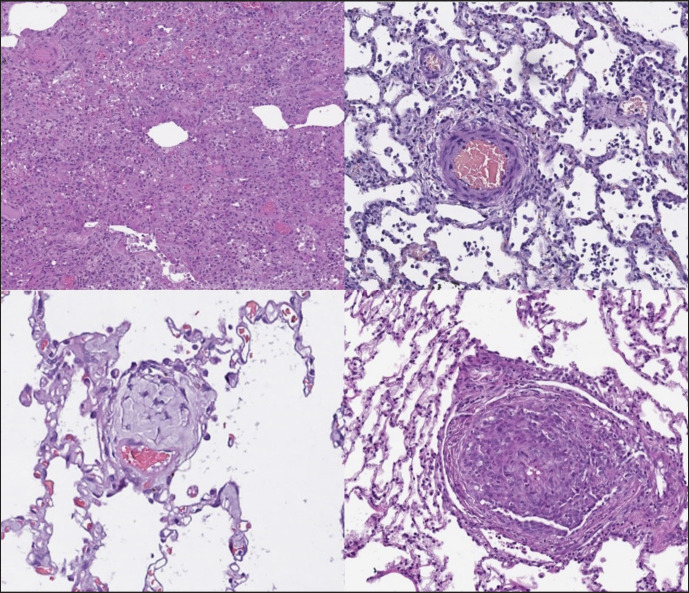
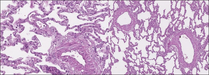
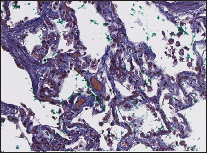
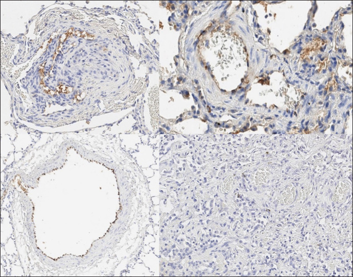
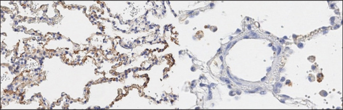
Results: Animals with histological and immunohistochemistry markers of PCH had a previous history of disease with left heart volume overload. There were no differences between the groups in radiography and gross pathology. Histologically, pulmonary fibrosis and arteriopathy could be found in RHF; in sPCH-a duplication of capillaries in alveolar septa and bizarre proliferation in surrounding structures.
Conclusion: PCH could be a secondary pattern of vascular remodeling due to volume overload.
Keywords: Pulmonary hypertension; canine; feline; pulmonary capillary hemangiomatosis.
Conflict of interest statement
The authors declare that there is no conflict of interest.
Figures
Similar articles
-
Pulmonary capillary hemangiomatosis and hypertrophic cardiomyopathy in a Persian cat.J Vet Diagn Invest. 2017 Nov;29(6):900-903. doi: 10.1177/1040638717723686. Epub 2017 Jul 28. J Vet Diagn Invest. 2017. PMID: 28754081
-
Pulmonary Veno-Occlusive Disease: A Newly Recognized Cause of Severe Pulmonary Hypertension in Dogs.Vet Pathol. 2016 Jul;53(4):813-22. doi: 10.1177/0300985815626572. Epub 2016 Feb 29. Vet Pathol. 2016. PMID: 26926086
-
Vasoproliferative process resembling pulmonary capillary hemangiomatosis in a cat.BMC Vet Res. 2017 Mar 20;13(1):72. doi: 10.1186/s12917-017-0984-9. BMC Vet Res. 2017. PMID: 28320395 Free PMC article.
-
An autopsy case of pulmonary capillary hemangiomatosis without evidence of pulmonary hypertension.Virchows Arch. 2001 Oct;439(4):586-92. doi: 10.1007/s004280100465. Virchows Arch. 2001. PMID: 11710647 Review.
-
Pulmonary capillary hemangiomatosis associated with connective tissue disease: a report of 4 cases and review of the literature.Ann Diagn Pathol. 2015 Jun;19(3):149-53. doi: 10.1016/j.anndiagpath.2015.03.006. Epub 2015 Mar 20. Ann Diagn Pathol. 2015. PMID: 25886868 Review.
References
-
- Adachi S., Hirashiki A., Kondo T., Nakaguro M., Ogawa A., Miyaji K., Matsubara H., Yokoi T., Murohara T. Imatinib is partially effective for the treatment of pulmonary capillary hemangiomatosis. Intern. Med. 2014;53(6):603–607. - PubMed
-
- Frazier A.A., Franks T.J., Mohammed T.L.H., Ozbudak I.H., Galvin J.R. From the archives of the AFIP: pulmonary veno-occlusive disease and pulmonary capillary hemangiomatosis. RadioGraphics. 2007;27:867–82. - PubMed
-
- Ginns L.C., Roberts D.H., Mark E.J., Brusch J.L., Marler J.J. Pulmonary capillary hemangiomatosis with atypical endotheliomatosis: successful antiangiogenic therapy with doxycycline. Chest. 2003;124(5):2017–2022. - PubMed
-
- Humbert M., Maitre S., Capron F., Rain B., Musset D., Simonneau G. Pulmonary edema complicating continuous intravenous prostacyclin in pulmonary capillary hemangiomatosis. Am. J. Respir. Crit. Care Med. 1998;157:1681–1685. - PubMed
MeSH terms
LinkOut - more resources
Full Text Sources
Medical
Miscellaneous