Randomized Controlled Trial
. 2024 Jun 24:12:e17567.
doi: 10.7717/peerj.17567. eCollection 2024.

Squatting biomechanics following physiotherapist-led care or hip arthroscopy for femoroacetabular impingement syndrome: a secondary analysis from a randomised controlled trial

Affiliations
Randomized Controlled Trial

Squatting biomechanics following physiotherapist-led care or hip arthroscopy for femoroacetabular impingement syndrome: a secondary analysis from a randomised controlled trial

Tamara M Grant et al. PeerJ. .

Abstract

Background: Femoroacetabular impingement syndrome (FAIS) can cause hip pain and chondrolabral damage that may be managed non-operatively or surgically. Squatting motions require large degrees of hip flexion and underpin many daily and sporting tasks but may cause hip impingement and provoke pain. Differential effects of physiotherapist-led care and arthroscopy on biomechanics during squatting have not been examined previously. This study explored differences in 12-month changes in kinematics and moments during squatting between patients with FAIS treated with a physiotherapist-led intervention (Personalised Hip Therapy, PHT) and arthroscopy.

Methods: A subsample (n = 36) of participants with FAIS enrolled in a multi-centre, pragmatic, two-arm superiority randomised controlled trial underwent three-dimensional motion analysis during squatting at baseline and 12-months following random allocation to PHT (n = 17) or arthroscopy (n = 19). Changes in time-series and peak trunk, pelvis, and hip biomechanics, and squat velocity and maximum depth were explored between treatment groups.

Results: No significant differences in 12-month changes were detected between PHT and arthroscopy groups. Compared to baseline, the arthroscopy group squatted slower at follow-up (descent: mean difference -0.04 m∙s-1 (95%CI [-0.09 to 0.01]); ascent: -0.05 m∙s-1 [-0.11 to 0.01]%). No differences in squat depth were detected between or within groups. After adjusting for speed, trunk flexion was greater in both treatment groups at follow-up compared to baseline (descent: PHT 7.50° [-14.02 to -0.98]%; ascent: PHT 7.29° [-14.69 to 0.12]%, arthroscopy 16.32° [-32.95 to 0.30]%). Compared to baseline, both treatment groups exhibited reduced anterior pelvic tilt (descent: PHT 8.30° [0.21-16.39]%, arthroscopy -10.95° [-5.54 to 16.34]%; ascent: PHT -7.98° [-0.38 to 16.35]%, arthroscopy -10.82° [3.82-17.81]%), hip flexion (descent: PHT -11.86° [1.67-22.05]%, arthroscopy -16.78° [8.55-22.01]%; ascent: PHT -12.86° [1.30-24.42]%, arthroscopy -16.53° [6.72-26.35]%), and knee flexion (descent: PHT -6.62° [0.56- 12.67]%; ascent: PHT -8.24° [2.38-14.10]%, arthroscopy -8.00° [-0.02 to 16.03]%). Compared to baseline, the PHT group exhibited more plantarflexion during squat ascent at follow-up (-3.58° [-0.12 to 7.29]%). Compared to baseline, both groups exhibited lower external hip flexion moments at follow-up (descent: PHT -0.55 N∙m/BW∙HT[%] [0.05-1.05]%, arthroscopy -0.84 N∙m/BW∙HT[%] [0.06-1.61]%; ascent: PHT -0.464 N∙m/BW∙HT[%] [-0.002 to 0.93]%, arthroscopy -0.90 N∙m/BW∙HT[%] [0.13-1.67]%).

Conclusion: Exploratory data suggest at 12-months follow-up, neither PHT or hip arthroscopy are superior at eliciting changes in trunk, pelvis, or lower-limb biomechanics. Both treatments may induce changes in kinematics and moments, however the implications of these changes are unknown.

Trial registration details: Australia New Zealand Clinical Trials Registry reference: ACTRN12615001177549. Trial registered 2/11/2015.

Keywords: Hip joint; Kinematics; Kinetics; Physical therapy; Squat.


Conflict of interest statement

David Lloyd has received research support from Arthrex and Orthopediatrics on an Australian Research Council Industrial Training and Transformation Centre grant, and from Orthocell on MTPConnect BioMedTech Horizons grant and Australian Research Council Industry Linkage grant. David J. Hunter has received consulting fees for scientific advisory roles from Pfizer, Lilly, Merck Serono, TLCBio, Kolon Tissuegene and Novartis. Nadine Foster is funded through an Australian National Health and Medical Research Council (NHMRC) Investigator Grant (ID: 2018182). No other potential Conflicts of Interest have been declared by any other authors.

Figures
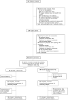
Figure 1
Figure 1. Study flow diagram.
FAIS, femoroacetabular impingement syndrome.
Figure 2
Figure 2. Overview of personalised hip therapy (PHT) core components.
FAIS, femoroacetabular impingement syndrome; FABER, flexion, abduction, external rotation; FADIR, flexion, adduction, internal rotation; ROM, range of motion.
Figure 3
Figure 3. Sagittal plane trunk (upper) and pelvis (lower) angles across squat descent and ascent.
Ensemble average (±1 standard deviation) for Personalised Hip Therapy (PHT, blue) and arthroscopy (red) groups at baseline (solid line) and follow-up (broken line) are presented. Statistical analyses were applied using statistical parametric or nonparametric mapping, as appropriate. Differences in 12-month changes between PHT and arthroscopy were examined using independent t-tests and a general linear model including speed as a covariate. Differences between baseline and follow-up for each treatment group were examined using paired t-tests and a general linear model including baseline and follow-up speed as a covariate. Darker and lighter coloured bars indicate significant differences (p < 0.05) detected using t-tests and general linear models, respectively, at follow-up, compared to baseline for PHT (blue and green) and arthroscopy (red and orange). No significant differences were detected between treatments.
Figure 4
Figure 4. Sagittal (top), frontal (middle) and transverse (bottom) plane hip angles and moments across squat descent and ascent.
Ensemble average (±1 standard deviation) for Personalised Hip Therapy (PHT, blue) and arthroscopy (red) groups at baseline (solid line) and follow-up (broken line) are presented. Statistical analyses were applied using statistical parametric or nonParametric mapping, as appropriate. Differences in 12-month changes between PHT and arthroscopy were examined using independent t-tests and a general linear model including speed as a covariate. Differences between baseline and follow-up for each treatment group were examined using paired t-tests and a general linear model including baseline and follow-up speed as a covariate. Coloured bars indicate significant differences (p < 0.05) detected using t-tests and general linear models, respectively, at follow-up, compared to baseline for PHT (blue and green) and arthroscopy (red and orange). No significant differences were detected between treatments, or within-groups after adjusting for speed.
Figure 5
Figure 5. Sagittal plane knee (upper) and ankle (lower) angles and moments across squat descent and ascent.
Ensemble average (±1 standard deviation) for Personalised Hip Therapy (PHT, blue) and arthroscopy (red) groups at baseline (solid line) and follow-up (broken line) are presented. Statistical analyses were applied using statistical parametric or nonParametric mapping, as appropriate. Differences in 12-month changes between PHT and arthroscopy were examined using independent t-tests and a general linear model, including speed as a covariate. Differences between baseline and follow-up for each treatment group were examined using paired t-tests and a general linear model including baseline and follow-up speed as a covariate. Coloured bars indicate significant differences (p < 0.05) detected using t-tests and general linear models, respectively, at follow-up, compared to baseline for PHT (blue and green) and arthroscopy (red and orange). No significant differences were detected between treatments, or within-groups after adjusting for speed.

References

    1. Adler RJ, Taylor JE. Random fields and geometry. New York: Springer; 2007.
    1. Bagwell JJ, Fukuda TY, Powers CM. Sagittal plane pelvis motion influences transverse plane motion of the femur: kinematic coupling at the hip joint. Gait & Posture. 2016a;43(9):120–124. doi: 10.1016/j.gaitpost.2015.09.010. - DOI - PubMed
    1. Bagwell JJ, Powers CM. The influence of squat kinematics and cam morphology on acetabular stress. Arthroscopy the Journal of Arthroscopic and Related Surgery. 2017;33(10):1797–1803. doi: 10.1016/j.arthro.2017.03.018. - DOI - PubMed
    1. Bagwell JJ, Powers CM. Persons with femoroacetabular impingement syndrome exhibit altered pelvifemoral coordination during weightbearing and non-weightbearing tasks. Clinical Biomechanics. 2019;65(6):51–56. doi: 10.1016/j.clinbiomech.2019.04.003. - DOI - PubMed
    1. Bagwell JJ, Snibbe J, Gerhardt M, Powers CM. Hip kinematics and kinetics in persons with and without cam femoroacetabular impingement during a deep squat task. Clinical Biomechanics. 2016b;31:87–92. doi: 10.1016/j.clinbiomech.2015.09.016. - DOI - PubMed

Publication types

LinkOut - more resources