. 2024 Jul 16:10:843.
doi: 10.12688/f1000research.55351.4. eCollection 2021.

Hypoxic Preconditioning Promotes Survival of Human Adipose Derived Mesenchymal Stem Cell

Affiliations

Hypoxic Preconditioning Promotes Survival of Human Adipose Derived Mesenchymal Stem Cell

I Gde Rurus Suryawan et al. F1000Res. .

Abstract

Background: Contributing factors for improved survival of human adipocytes mesenchymal stem cells (h-AMSCs) cultured through hypoxia preconditioning, in example apoptosis inhibition involving BCL2 and HSP27 expression, trigger signal expression (VEGF), SCF expression, OCT-4 expression, and CD44+ expression. The objective if this study was to explain the mechanism and role of hypoxic preconditioning and the optimal duration of hypoxic preconditioning exposure to improve survival of h-AMSCs. Methods: An experimental laboratory explorative study ( in vitro) with hypoxic preconditioning in h-AMSCs cultures. This research was conducted through four stages. First, isolation of h-AMSCs culture from adipose tissue of patients. Second, the characterization of h-AMSCs from adipose tissue by phenotype (flowcytometry) through CD44+, CD90+ and CD45-expression before being pre-conditioned for hypoxic treatment. Third, the hypoxic preconditioning in h-AMSCs culture ( in vitro) was performed with an oxygen concentration of 1% for 24, 48 and 72 hours. Fourth, observation of survival from h-AMSCs culture was tested on the role of CD44+, VEGF, SCF, OCT-4, BCL2, HSP27 with Flowcytometry and apoptotic inhibition by Tunnel Assay method. Results: The result of regression test showed that time difference had an effect on VEGF expression ( p<0.001; β=-0.482) and hypoxia condition also influenced VEGF expression ( p<0.001; β=0.774). The result of path analysis showed that SCF had effect on OCT-4 expression ( p<0.001; β=0.985). The regression test results showed that time effects on HSP27 expression ( p<0.001; β=0.398) and hypoxia precondition also affects HSP27 expression ( p<0.001; β=0.847). Pathway analysis showed that BCL2 expression inhibited apoptosis ( p=0.030; β=-0.442) and HSP27 expression also inhibited apoptosis ( p<0,001; β=-0.487). Conclusion: Hypoxic preconditioning of h-AMSC culture has proven to increase the expression of VEGF, SCF, OCT-4, and BCL2 and HSP27. This study demonstrated and explained the existence of a new mechanism of increased h-AMSC survival in cultures with hypoxic preconditioning (O2 1%) via VEGF, SCF, OCT-4, BCL2, and HSP 27.

Keywords: BCL-2; HSP27; SCF; VEGF expression; apoptosis; h-AMSCs.


Conflict of interest statement

No competing interests were disclosed.

Figures
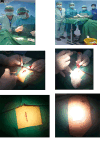
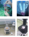
Figure 1.
Figure 1.
Isolation and culture of h-AMSCs from the patient's adipose tissue (human).
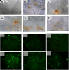
Figure 2.
Figure 2.
Observation of survival of h-AMSCs in the form of CD44+, VEGF, SCF, OCT-4, BCL2, HSP27 expression, and apoptotic inhibition:
  1. A.

    Phenotype expression of CD44+ was carried out by the flowcytometric method.

  2. B.

    Immuno-cytochemical expression of VEGF.

  3. C.

    Immunocytochemical expression of SCF from h-AMSCs culture.

  4. D.

    Phenotype of OCT-4 expression (Immunocytochemistry and Immunofluorescence).
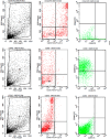
Figure 3.
Figure 3.
Flowcytometry results from human AMSCs based on cell culture for CD44+ CD90+ CD45- expression.
Figure 4.
Figure 4.
Immunohistochemical Characteristic of h-AMSCs based on VEGF expression at: A) normoxic condition for 24 hours; B) normoxic condition for 48 hours; C) normoxic condition for 72 hours; D) hypoxic condition for 24 hours; E) hypoxic condition for 48 hours; F) hypoxic condition for 72 hours.
Figure 5.
Figure 5.
Immunohistochemical Characteristic of h-AMSCs based on SCF expression at: A) normoxic condition for 24 hours; B) normoxic condition for 48 hours; C) normoxic condition for 72 hours; D) hypoxic condition for 24 hours; E) hypoxic condition for 48 hours; F) hypoxic condition for 72 hours.
Figure 6.
Figure 6.
Immunohistochemical Characteristic of h-AMSCs based on OCT-4 expression at: A) normoxic condition for 24 hours; B) normoxic condition for 48 hours; C) normoxic condition for 72 hours; D) hypoxic condition for 24 hours; E) hypoxic condition for 48 hours; F) hypoxic condition for 72 hours. Immunofluorescence assay of h-AMSCs based on OCT-4 expression at: G) normoxic condition for 24 hours; H) normoxic condition for 48 hours; I) normoxic condition for 72 hours; J) hypoxic condition for 24 hours; K) hypoxic condition for 48 hours; L) hypoxic condition for 72 hours.
Figure 7.
Figure 7.
Immunohistochemical Characteristic of h-AMSCs based on BCL2 expression at: A) normoxic condition for 24 hours; B) normoxic condition for 48 hours; C) normoxic condition for 72 hours; D) hypoxic condition for 24 hours; E) hypoxic condition for 48 hours; F) hypoxic condition for 72 hours.
Figure 8.
Figure 8.
Immunohistochemical Characteristic of h-AMSCs based on HSP27 expression at: A) normoxic condition for 24 hours; B) normoxic condition for 48 hours; C) normoxic condition for 72 hours; D) hypoxic condition for 24 hours; E) hypoxic condition for 48 hours; F) hypoxic condition for 72 hours.
Figure 9.
Figure 9.
Immunohistochemical Characteristic of h-AMSCs based on number of apoptotic cell amount at: A) normoxic condition for 24 hours; B) normoxic condition for 48 hours; C) normoxic condition for 72 hours; D) hypoxic condition for 24 hours; E) hypoxic condition for 48 hours; F) hypoxic condition for 72 hours.
Figure 10.
Figure 10.
Path analysis with MANOVA and multiple linear regression analysis for hypoxic preconditioning in h-AMSCs survive.

References

    1. Stępniewski J, Tomczyk M, Andrysiak K, et al. : Human Induced Pluripotent Stem Cell-Derived Cardiomyocytes, in Contrast to Adipose Tissue-Derived Stromal Cells, Efficiently Improve Heart Function in Murine Model of Myocardial Infarction. Biomedicines. 2020;8(12):1–21. 10.3390/biomedicines8120578 - DOI - PMC - PubMed
    1. Acquistapace A, Bru T, Lesault PF, et al. : Human mesenchymal stem cells reprogram adult cardiomyocytes toward a progenitor-like state through partial cell fusion and mitochondria transfer. Stem Cells. 2011;29(5):812–824. 10.1002/stem.632 - DOI - PMC - PubMed
    1. Lee J, Lee S, Lee CY, et al. : Adipose-derived stem cell-released osteoprotegerin protects cardiomyocytes from reactive oxygen species-induced cell death. Stem Cell Res Ther. 2017;8(1):4–9. 10.1186/s13287-017-0647-6 - DOI - PMC - PubMed
    1. Bagheri-Hosseinabadi Z, Mesbah-Namin SA, Salehinejad P, et al. : Fibrin scaffold could promote survival of the human adipose-derived stem cells during differentiation into cardiomyocyte-like cells. Cell Tissue Res. 2018;372(3):571–589. 10.1007/s00441-018-2799-9 - DOI - PubMed
    1. Lauvrud AT, Kelk P, Wiberg M, et al. : Characterization of human adipose tissue-derived stem cells with enhanced angiogenic and adipogenic properties. J Tissue Eng Regen Med. 2017;11(9):2490–2502. 10.1002/term.2147 - DOI - PubMed

MeSH terms

Substances