Case Reports
. 2024 May 6:6:ojae034.
doi: 10.1093/asjof/ojae034. eCollection 2024.

Minimally Invasive Beaded Electrosurgical Dissectors, Basic Science, and Pilot Studies

Case Reports

Minimally Invasive Beaded Electrosurgical Dissectors, Basic Science, and Pilot Studies

Taiyo C Weber et al. Aesthet Surg J Open Forum. .

Abstract

Background: Minimally invasive beaded electrosurgical dissectors ("BEED devices") provide simultaneous sharp dissection, blunt dissection, and electrosurgical coagulation while performing 100 cm2 porcine tissue plane dissections in 0.8 to 3 min with minimal bleeding and no perforations.

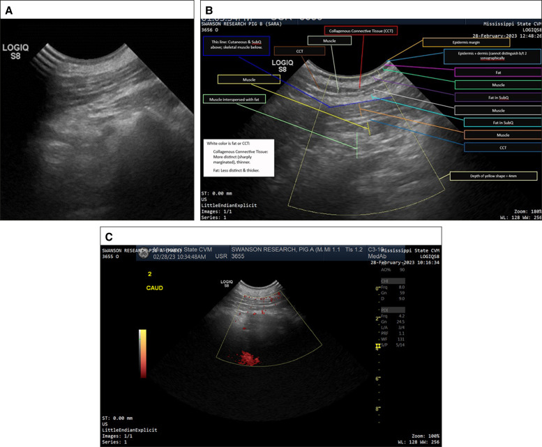
Objectives: The aim of the study was to report the basic science and potential clinical applications and to video document the speed and quality of planar dissections in in vivo and ex vivo porcine models with thermal damage quantified by thermal and histopathologic measurements. Additionally, in vivo porcine specimens were followed for 90 days to show whether adverse events occurred on a gross or macroscopic basis, as evidenced by photography, videography, physical examination, and dual ultrasonography.

Methods: Ex vivo porcine models were subjected to 20, 30, and 50 W in single-stroke passages with BEED dissectors (granted FDA 510(k) clearance (K233002)) with multichannel thermocouple, 3 s delay recordation combined with matching hematoxylin and eosin (H&E) histopathology. In vivo porcine models were subjected to eight 10 × 10 cm dissections in each of 2 subjects at 20, 30, and 50 W and evaluated periodically until 90 days, wherein histopathology for H&E, collagen, and elastin was taken plus standard and Doppler ultrasounds prior to euthanasia.

Results: Five to 8 mm width dissectors were passed at 1 to 2 cm/s in ex vivo models (1-10 cm/s in vivo models) with an average temperature rise of 5°C at 50 W. Clinically evidenced seromas occurred in the undressed, unprotected wounds, and resolved well prior to 90 days, as documented by ultrasounds and histopathology.

Conclusions: In vivo and ex vivo models demonstrated thermal values that were below levels known to damage subcutaneous adipose tissue or skin. Tissue histopathology confirmed healing parameters while Doppler ultrasound demonstrated normal blood flow in posttreatment tissues.


Figures

Figure 1.
Figure 1.
(A) Photographs of 2-BEED and 3-BEED models against 18 mm diameter US dime. (B) Ceramic tips atop 40 cm shafts with reversable and slidable snap handle. Top handle in unclamped position. (C, D) Computer-Aided Design (CAD) renderings of 2-BEED and 3-BEED devices show metallic conductive lysing segments in gold color detailing: distal lysing segments (d), proximal lysing segment (p) adjacent to bead (b) on ceramic tip (t). BEED, Beaded Enhanced Electrosurgical Dissectors.
Figure 2.
Figure 2.
(A) 3-BEED model in ballistic gelatin lysing row of pseudo-tendrils (white) on forward motion. (B) 3-BEED model lysing in rearward motion with remnant bubble paths. (C) 2-BEED model in forward motion with arcing visible. (D) 2-BEED model in rearward motion lysing pseudo-tendrils in proximal segment. BEED, Beaded Enhanced Electrosurgical Dissectors.
Figure 3.
Figure 3.
FLIR image of ex vivo abdominal wall with temperature minimum 18.7°C and maximum 32.1° C. FLIR, Forward Looking Infrared.
Figure 4.
Figure 4.
(A) 2-BEED intraoperative forward motion. (B) 2-BEED forward motion with operating room lights dimmed. Beaded Enhanced Electrosurgical Dissectors (BEED).
Figure 5.
Figure 5.
(A, B) Top views of porcine abdominal ventrums preoperative before tumescence; (C, D) preoperative before tumescence with superimposed neon-blue dotted parallelograms demonstrating total areas dissected as opposed to abdominal dimensions; (E, F) immediately postoperative; and (G, H) 90-days postoperative. Notice birthmark on left, mid-side of Pig B. The postoperative ecchymoses arose posttumescent infiltration and did not increase appreciably following BEED passage in the dense fibrotic tissue. Beaded Enhanced Electrosurgical Dissectors (BEED).
Figure 6.
Figure 6.
(A) Histologic evaluation of porcine 90 days postoperative sections demonstrated a linear band of fibrosis within the deep subcutaneous tissue (H&E, arrows). (B) Trichome stains collagen blue (arrows). (C) Elastin fibers were not present within the mature fibrosis (Verhoff Van Gieson, arrows); however, normal subcutaneous elastin fibers (black stain) were present within subcutaneous collagen bundles (*). Bar (bottom right) = 0.2 mm.
Figure 7.
Figure 7.
(A) Ninety days postoperative, diagnostic ultrasound image reveals no nodules, cysts, fluid collections, or tissue separations (as well as the remainder of the 16 dissected sites). (B) Labeled ultrasound layers display the normal, multilevel interpolation of numerous muscular bands throughout the subcutaneous fat. Note the labeling of the areas. (C) Ninety days postoperative in vivo Doppler ultrasound reveals slightly enhanced vascularity as per ultrasonographer and algorithm quantification.
Figure 8.
Figure 8.
A top intraoperative view of a 10 × 10 cm pig abdomen area (boundary highlighted by Sharpie marker) undergoing subcutaneous dissection by a 2.5 BEED device. The highest temperature displayed in the grid was 28°C. Beaded Enhanced Electrosurgical Dissectors (BEED).
Figure 9.
Figure 9.
In vivo porcine abdomen 10 × 10 cm dissection plane (which was created with 3-BEED device [top left] in under 90 s) is opened (within 30 s of formation) by 2 side cutting and flap reflected to display minimal bleeding as well as completeness of dissection. Beaded Enhanced Electrosurgical Dissectors (BEED).

References

    1. Meeuwsen F, Guédon A, Klein J, Elst MV, Dankelman J, Van Den Dobbelsteen J. Electrosurgery: short-circuit between education and practice. Minim Invasive Ther Allied Technol. 2019;28(4):247–253. doi: 10.1080/13645706.2018.1513945 - DOI - PubMed
    1. Pollinger HS, Mostafa G, Harold KL, Austin CE, Kercher KW, Matthews BD. Comparison of wound-healing characteristics with feedback circuit electrosurgical generators in a porcine model. Am Surg. 2003;69(12):1054–1060. doi: 10.1177/000313480306901207 - DOI - PubMed
    1. Chang EI, Carlson GA, Vose JG, Huang EJ, Yang GP. Comparative healing of rat fascia following incision with three surgical instruments. J Surg Res. 2011;167(1):e47–e54. doi: 10.1016/j.jss.2010.12.019 - DOI - PubMed
    1. Ruidiaz ME, Messmer D, Atmodjo DY, et al. . Comparative healing of human cutaneous surgical incisions created by the PEAK PlasmaBlade, conventional electrosurgery, and a standard scalpel. Plast Reconstr Surg. 2011;128(1):104–111. doi: 10.1097/PRS.0b013e31821741ed - DOI - PubMed
    1. Charoenkwan K, Iheozor-Ejiofor Z, Rerkasem K, Matovinovic E. Scalpel versus electrosurgery for major abdominal incisions. Cochrane Database Syst Rev. 2017;6(6):CD005987. doi: 10.1002/14651858.CD005987.pub3 - DOI - PMC - PubMed

Publication types

LinkOut - more resources