Review
. 2024 May 14;15(25):9408-9437.
doi: 10.1039/d3sc06549a. eCollection 2024 Jun 26.

Protein-based layer-by-layer films for biomedical applications

Affiliations
Review

Protein-based layer-by-layer films for biomedical applications

Muhammad Haseeb Iqbal et al. Chem Sci. .

Abstract

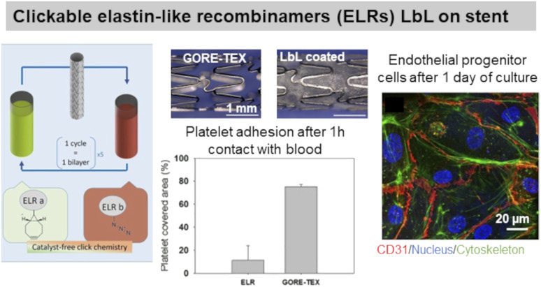
The surface engineering of biomaterials is crucial for their successful (bio)integration by the body, i.e. the colonization by the tissue-specific cell, and the prevention of fibrosis and/or bacterial colonization. Performed at room temperature in an aqueous medium, the layer-by-layer (LbL) coating method is based on the alternating deposition of macromolecules. Versatile and simple, this method allows the functionalization of surfaces with proteins, which play a crucial role in several biological mechanisms. Possessing intrinsic properties (cell adhesion, antibacterial, degradable, etc.), protein-based LbL films represent a powerful tool to control bacterial and mammalian cell fate. In this article, after a general introduction to the LbL technique, we will focus on protein-based LbL films addressing different biomedical issues/domains, such as bacterial infection, blood contacting surfaces, mammalian cell adhesion, drug and gene delivery, and bone and neural tissue engineering. We do not consider biosensing applications or electrochemical aspects using specific proteins such as enzymes.


Conflict of interest statement

There are no conflicts to declare.

Figures

Fig. 1
Fig. 1. Layer-by-layer (LbL) method and different processes of build-up. (a) Schematic representation of the alternated deposition of polyanions and polycations, each deposition is followed by a rinsing step. (b) The build-up of LbL films can be achieved by various processes like dipping, spin coating, spray coating, and brushing.
Scheme 1
Scheme 1. Schematic representation of protein-based LbL films and their various biomedical application addressed in this perspective.
Scheme 2
Scheme 2. Schematic representation of the three strategies of antibacterial LbL films.
Fig. 2
Fig. 2. Adhesion-resistant and contact-killing LbL films. (a) Schematic illustration of bacterial tests on native and LL-37 functionalized COL/HA LbL films. Antiadhesive properties of native LbL (0 μM LL-37) and antibacterial activity of LL-37 functionalized LbL against E. coli (green = live bacteria and red = dead bacteria). This figure has been adapted from ref. with permission from Elsevier, copyright 2016. (b) Scheme of the fabrication process of silk fibroin/nylon-6 nanofibrous mats by electrospinning and the build-up of COL/lysozyme (Ly) LbL films onto the nanofibrous mats, and the antibacterial activity as a function of the number of bilayers against S. aureus and E. coli. This figure has been adapted from ref. with permission from Elsevier, copyright 2020.
Fig. 3
Fig. 3. Release-killing LbL films. (a) Effect of the buffer on the build-up, topography, complexation, and antibacterial property of collagen/tannic acid (COL/TA) LbL films. This figure has been adapted from ref. with permission from American Chemical Society, copyright 2010. (b) Ca2+-binding mediated sustained release of minocycline (MN) from dextran sulfate/gelatin type A (DS/Gel) LbL shows antibacterial activity. This figure has been adapted from ref. with permission from PLOS ONE, copyright 2014. (c) The release of nitric oxide (NO) gas from gelatin/tannic acid (Gel/TA) LbL film shows antibacterial activity towards S. aureus. LbL and NO-LbL stand for Gel/TA and NO-generating moieties functionalized Gel/TA, respectively. This figure has been adapted from ref. with permission from Nature Springer, copyright 2019.
Scheme 3
Scheme 3. Schematic representation of the LbL strategies to promote cell adhesion.
Fig. 4
Fig. 4. Collagen-based LbL films. (a) Intrinsically crosslinked collagen-based LbL films with oxidized glycosaminoglycans (either hyaluronic acid or chondroitin sulfate) show preservation of COL fibrillar structures and enhanced adhesion, cell size, and polarization of embryonic fibroblasts, thanks to increased film stiffness due to the crosslinking. This figure has been adapted from ref. with permission from American Chemical Society, copyright 2014. (b) Scheme for build-up of oriented collagen/tannic acid (COL/TA) LbL film using a brushing LbL method, and use of the LbL for orientation and differentiation of human myoblasts into myotubes. This figure has been adapted from ref. with permission from American Chemical Society, copyright 2022.
Fig. 5
Fig. 5. Gelatin and fibronectin-based LbL films. Functionalization of polyethylene terephthalate (PET) ligaments with HA/Gel LbL film implanted in vivo in a porcine model showed regeneration of anterior cruciate ligament. This figure has been adapted from ref. with permission from PLoS One, copyright 2012.
Fig. 6
Fig. 6. Use of elastin in LbL films. Clickable elastin-like recombinamers (ELR), modified by either azide or cyclooctyne, LbL film on coronary stent reduces platelet adhesion and promotes endothelial layer formation, thanks to the presence of RGD-sequence on ELR-azide. This figure has been adapted from ref. with permission from Elsevier, copyright 2019.
Scheme 4
Scheme 4. Schematic illustration of LbL strategies to design hemocompatible surfaces.
Fig. 7
Fig. 7. LbL films inhibit platelet adhesion. (a) Bovine serum albumin/heparin (BSA/HEP) LbL on polyethersulfone foils (PES) reduces the adhesion of platelets and leukocytes upon incubation in slightly heparinized (1.5 IU heparin per mL) human whole blood in vitro. This figure has been adapted from ref. with permission from John Wiley and Sons, copyright 2006. (b) (Fn/PLL) LbL films deposited on different polyurethanes influence the behavior of monocytes. This figure has been adapted from ref. with permission from Elsevier, copyright 2018.
Fig. 8
Fig. 8. LbL films promote endothelial cell adhesion. (a) Human umbilical vein endothelial cells (HUVECs) formed a confluent layer on genipin cross-linked (ALG/COL) LbL after 5 days compared to glutaraldehyde ones. This figure has been adapted from ref. with permission from American Chemical Society, copyright 2012. (b) Collagen/heparin (COL/HEP) LbL ended by CD34 antibody coated on stents showed rapid endothelialisation and no neointimal hyperplasia in rabbit femoral arteries in vivo. This figure has been adapted from ref. with permission from Elsevier, copyright 2010.
Scheme 5
Scheme 5. Schematic illustration of drug/gene release from the LbL films under various stimuli i.e., diffusion or erosion, pH change, and enzymatic degradation.
Fig. 9
Fig. 9. Drug release by diffusion or erosion. (a) Efficient loading and sustained delivery of transforming growth factor beta 1, platelet-derived growth factor ββ, and insulin growth factor 1 from either growth factor/heparin LbL films. The films resulted in the proliferation, migration, and differentiation of fibroblast cells into myofibroblasts (α-SMA labelling). This figure has been adapted from ref. with permission from the Royal Society of Chemistry, copyright 2020. (b) Protamine sulfate/hepatocyte growth factor-pDNA (PRM/HGF-pDNA) LbL, on top of human umbilical vein endothelial cells (HUVEC) and human umbilical artery smooth muscle cells (SMC), induced the secretion of HGF by the cells and showed competitiveness of ECs over SMCs. This figure has been adapted from ref. with permission from Elsevier, copyright 2013. (c) Direct nano-encapsulation of furosemide microcrystals with Gel/PSS LbL to enable sustained drug release profiles. This figure has been adapted from ref. with permission from Elsevier, copyright 2003.
Fig. 10
Fig. 10. Drug release at physiological pH. (a) Scheme of gelatin A/epigallocatechin gallate (Gel/EGCG) LbL hollow capsule preparation, and fluorescence/AFM images. This figure has been adapted from ref. with permission from Elsevier, copyright 2009. (b) Hydrolysable polymer associated with ovalbumin and CpG, the release of ovalbumin, an optical image of LbL-coated PDMS substrate, and the scheme of its application onto tape-stripped ear skin and confocal images showing penetration of fluorophore-conjugated ova (red) and CpG (blue) released from LbL films into the skin. This figure has been adapted from ref. with permission from American Chemical Society, copyright 2009.
Fig. 11
Fig. 11. Drug release by enzymatic degradation of LbL films. Scheme of PRM/HA LbL coated bioactive micelles for targeting tumours, and biodistribution in vivo. This figure has been adapted from ref. with permission from Elsevier, copyright 2018.
Scheme 6
Scheme 6. Schematic illustration of LbL strategies to promote bone regeneration.
Fig. 12
Fig. 12. LbL films supporting osteoconduction. (a) Polyethylene imine/gelatin (Gel/PEI) LbL increased the proliferation of human osteoblast cells, thanks to the hydrophilic nature of the LbL film. This figure has been adapted from ref. with permission from Elsevier, copyright 2012. (b) Chitosan/gelatin/hydroxyapatite (CHI/Gel/HAP) composite LbL films on titanium alloy, a cross-section of the films by SEM, and mineralisation of osteoblasts after 7 and 14 days of culture. This figure has been adapted from ref. with permission from Elsevier, copyright 2014.
Fig. 13
Fig. 13. LbL films supporting osteoinduction. Gel/CHI/Gel/BMP2 LbL films were built on a titanium alloy rod for implantation in rabbit femur in vivo. Gel and BPM2 were released in a physiological medium. LbL-coated surfaces show better mesenchymal stem cell mineralization, and bone formation (MicroCT and histochemical analysis). This figure has been adapted from ref. with permission from Elsevier, copyright 2012.
Scheme 7
Scheme 7. Schematic illustration of neurogenesis, i.e., differentiation cascade of neural stem cells to form mature neurons, onto LbL films.
Fig. 14
Fig. 14. LbL films promote neural cell adhesion and differentiation. (a) Silicon microelectrode arrays (Si MEAs) coated with various LbL films, with compositions such as PEI/Gel, CHI/Gel, and PEI/laminin (Ln), show better chick cortical neuron differentiation only when using protein-based LbL, i.e., Gel or Ln, among which PEI/Ln was the best composition. This figure has been adapted from ref. with permission from Elsevier, copyright 2005. (b) Chitosan/laminin (CHI/Ln) LbL film on poly-l-lactide (PLLA) electrospun scaffold, with no preferential orientation of the nanofibers, increased the neurite outgrowth of neurons after 7 days. Increasing the number of bilayers from 1 to 5 enhanced the neurite outgrowth, thanks to a higher amount of Ln incorporated into the LbL film. This figure has been adapted from ref. with permission from John Wiley and Sons, copyright 2013.
None
Muhammad Haseeb Iqbal
None
Halima Kerdjoudj
None
Fouzia Boulmedais

References

    1. Decher G. Hong J. D. Schmitt J. Buildup of Ultrathin Multilayer Films by a Self-Assembly Process .3. Consecutively Alternating Adsorption of Anionic and Cationic Polyelectrolytes on Charged Surfaces. Thin Solid Films. 1992;210:831–835. doi: 10.1016/0040-6090(92)90417-A. - DOI
    1. vander Straeten A. Lefèvre D. Demoustier-Champagne S. Dupont-Gillain C. Protein-based polyelectrolyte multilayers. Adv. Colloid Interface Sci. 2020;280:102161. doi: 10.1016/j.cis.2020.102161. - DOI - PubMed
    1. Criado-Gonzalez M. Mijangos C. Hernández R. Polyelectrolyte Multilayer Films Based on Natural Polymers: From Fundamentals to Bio-Applications. Polymers. 2021;13:2254. doi: 10.3390/polym13142254. - DOI - PMC - PubMed
    1. Omidvar M. Zdarta J. Sigurdardóttir S. B. Pinelo M. Mimicking natural strategies to create multi-environment enzymatic reactors: From natural cell compartments to artificial polyelectrolyte reactors. Biotechnol. Adv. 2022;54:107798. doi: 10.1016/j.biotechadv.2021.107798. - DOI - PubMed
    1. Zhang J. Huang X. Zhang L. Si Y. Guo S. Su H. Liu J. Layer-by-layer assembly for immobilizing enzymes in enzymatic biofuel cells. Sustainable Energy Fuels. 2020;4:68–79. doi: 10.1039/C9SE00643E. - DOI