

Different MRI-based radiomics models for differentiating misdiagnosed or ambiguous pleomorphic adenoma and Warthin tumor of the parotid gland: a multicenter study
- PMID: 38939335
- PMCID: PMC11208325
- DOI: 10.3389/fonc.2024.1392343
Different MRI-based radiomics models for differentiating misdiagnosed or ambiguous pleomorphic adenoma and Warthin tumor of the parotid gland: a multicenter study
Abstract
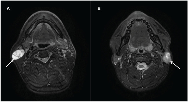
Purpose: To evaluate the effectiveness of MRI-based radiomics models in distinguishing between Warthin tumors (WT) and misdiagnosed or ambiguous pleomorphic adenoma (PA).
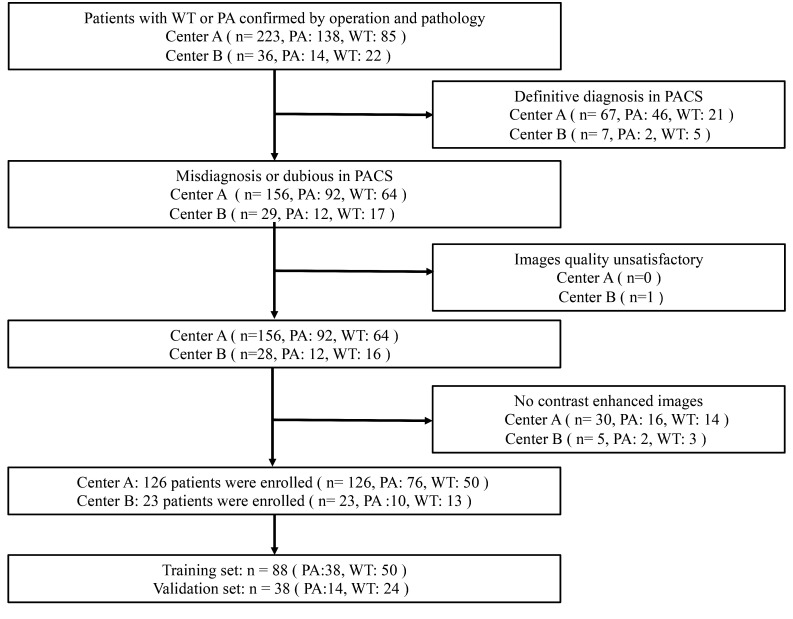
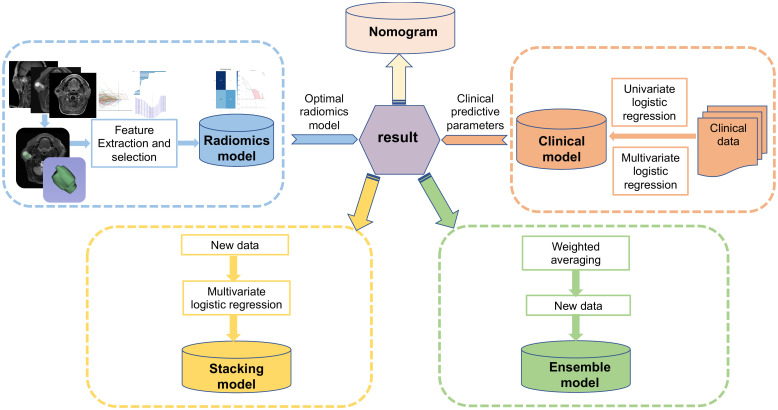
Methods: Data of patients with PA and WT from two centers were collected. MR images were used to extract radiomic features. The optimal radiomics model was found by running nine machine learning algorithms after feature reduction and selection. To create a clinical model, univariate logistic regression (LR) analysis and multivariate LR were used. The independent clinical predictors and radiomics were combined to create a nomogram. Two integrated models were constructed by the ensemble and stacking algorithms respectively based on the clinical model and the optimal radiomics model. The models' performance was evaluated using the area under the curve (AUC).
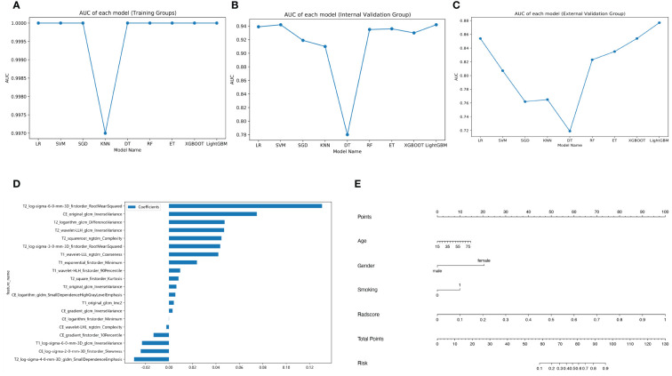
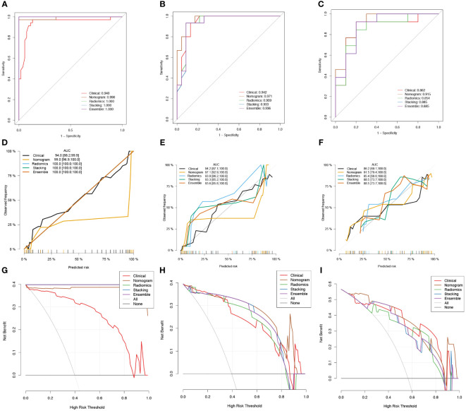
Results: There were 149 patients included in all. Gender, age, and smoking of patients were independent clinical predictors. With the greatest average AUC (0.896) and accuracy (0.839) in validation groups, the LR model was the optimal radiomics model. In the average validation group, the radiomics model based on LR did not have a higher AUC (0.795) than the clinical model (AUC = 0.909). The nomogram (AUC = 0.953) outperformed the radiomics model in terms of discrimination performance. The nomogram in the average validation group had a highest AUC than the stacking model (0.914) or ensemble model (0.798).
Conclusion: Misdiagnosed or ambiguous PA and WT can be non-invasively distinguished using MRI-based radiomics models. The nomogram exhibited excellent and stable diagnostic performance. In daily work, it is necessary to combine with clinical parameters for distinguishing between PA and WT.
Keywords: MRI; Warthin tumor; nomogram; parotid gland; pleomorphic adenoma; radiomics.
Copyright © 2024 Yang, Bi, Jin, Yang, Du, Zhang and Wu.
Conflict of interest statement
The authors declare the research was conducted in the absence of any commercial or financial relationships that could be construed as a potential conflict of interest.
Figures
References
LinkOut - more resources
Full Text Sources