. 2024 Jan 31;3(2):e92.
doi: 10.1002/jex2.92. eCollection 2024 Feb.

ISG15 mediates the function of extracellular vesicles in promoting ovarian cancer progression and metastasis

Affiliations

ISG15 mediates the function of extracellular vesicles in promoting ovarian cancer progression and metastasis

Kalpana Deepa Priya Dorayappan et al. J Extracell Biol. .

Abstract

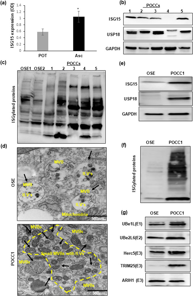
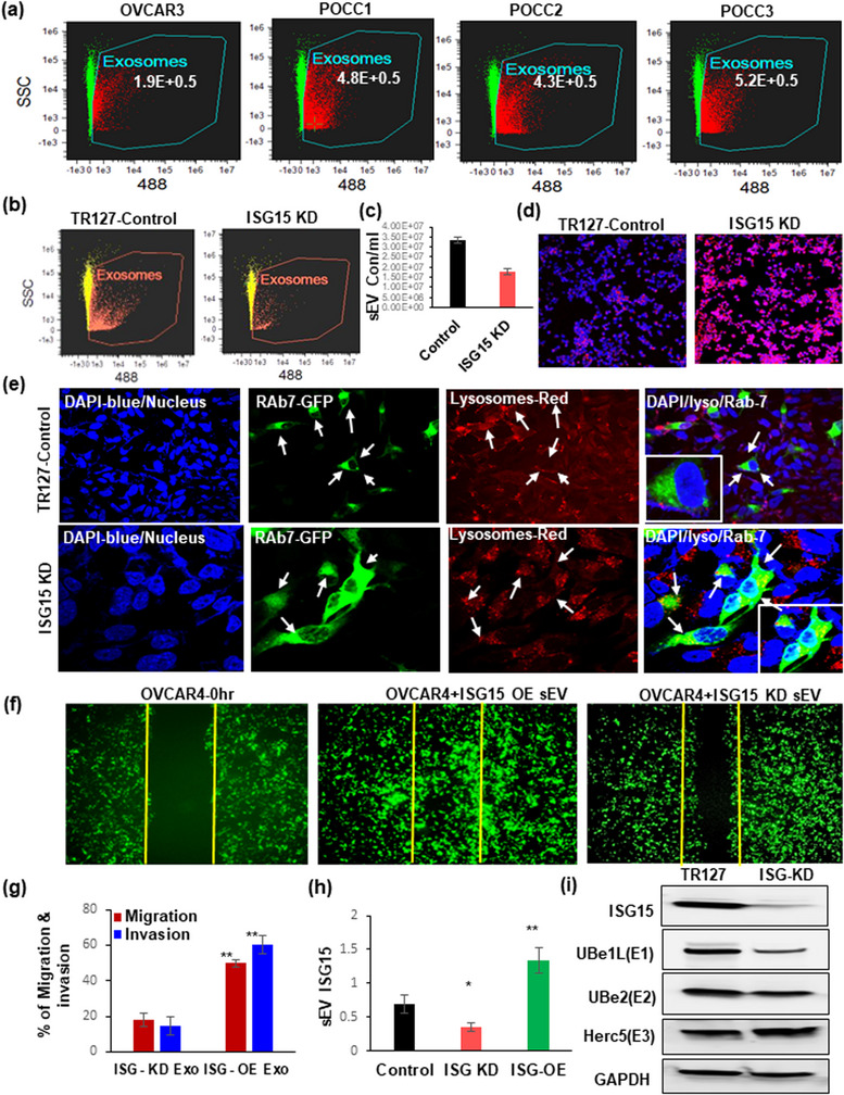
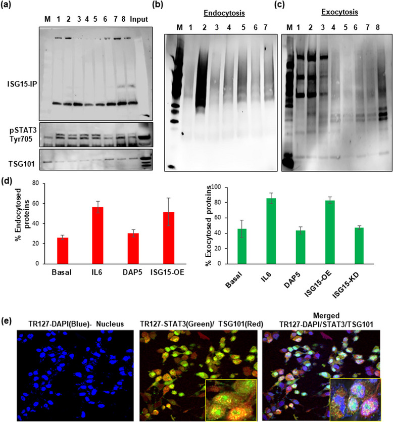
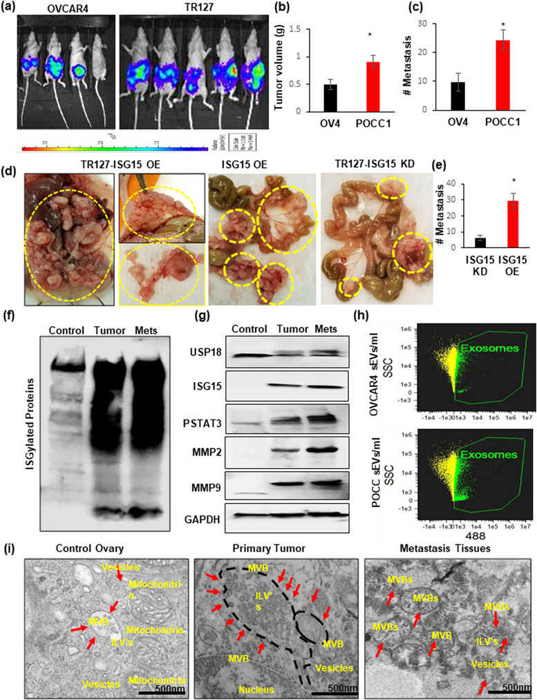
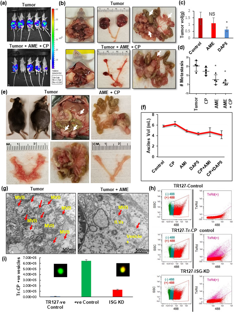
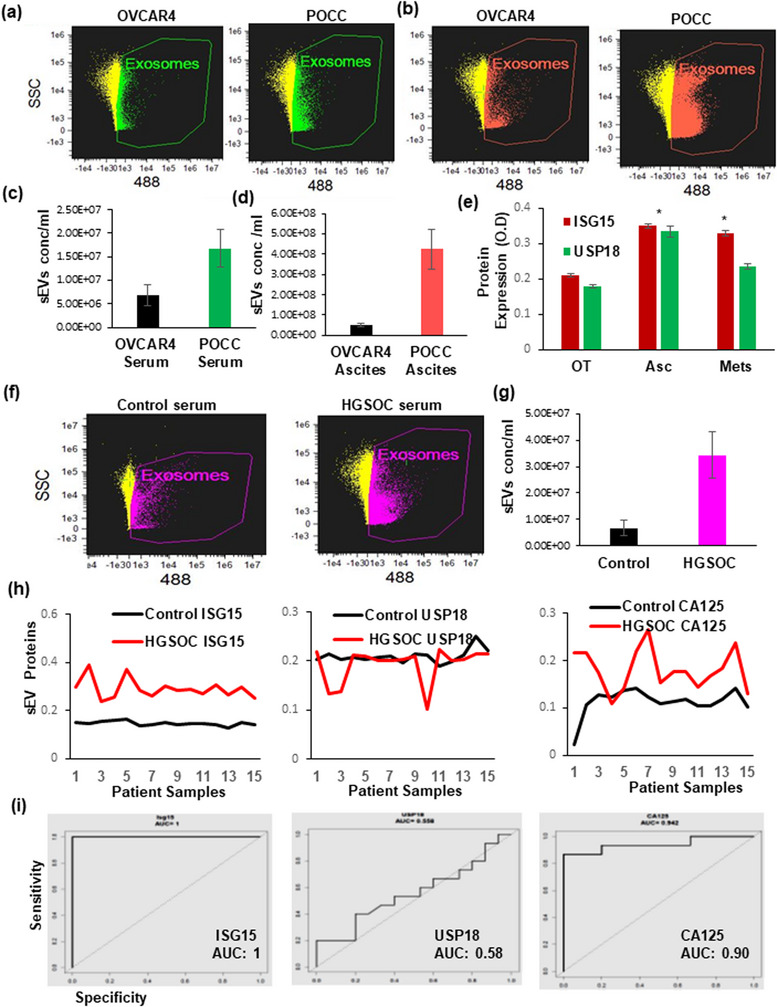
The interferon stimulated gene 15 (ISG15), a ubiquitin like protein and its conjugates have been implicated in various human malignancies. However, its role in ovarian cancer progression and metastasis is largely unknown. In high grade serous ovarian cancer (HGSOC), ascites is the major contributor to peritoneal metastasis. In this study, we identified significantly elevated ISG15 protein expression in HGSOC patient ascites, ascites derived primary ovarian cancer cells (POCCs), POCC small extracellular vesicles (sEVs) as well as metastatic tissue. Our results demonstrates that ISG15 increases exocytosis in ascites-derived POCCs by decreasing the endosome-lysosomal fusion, indicating a key role in sEV secretion. Further, knockdown (KD) of ISG15 resulted in a significant decrease in vesicles secretion from HGSOC cells and in vivo mouse models, leading to reduced HGSOC cell migration and invasion. Furthermore, our pre-clinical mouse model studies revealed the influence of vesicular ISG15 on disease progression and metastasis. In addition, knockdown of ISG15 or using the ISG15 inhibitor, DAP5, in combination therapy with carboplatin showed to improve the platinum sensitivity in-vitro and reduce tumour burden in-vivo. We also found that ISG15 expression within sEV represents a promising prognostic marker for HGSOC patients. Our findings suggest that ISG15 is a potential therapeutic target for inhibiting progression and metastasis in HGSOC and that vesicular ISG15 expression could be a promising biomarker in the clinical management of ovarian cancer. Significance: High-grade serous ovarian cancer (HGSOC) has high morbidity and mortality rates, but its progression and metastasis are still poorly understood, and there is an urgent need for early detection and targeted therapies. Our study presents novel findings that implicate ISG15-mediated vesicular proteins in the advancement and spread of HGSOC. These results offer pre-clinical evidence of potential new molecular targets, prognostic markers and therapeutic strategies for HGSOC that could ultimately enhance patient survival.

Keywords: ISG15; ISGylation; ascites; extracellular vesicles; metastasis and biomarker; ovarian cancer.


Conflict of interest statement

All authors declare that there are no conflicts of interest.

Figures

FIGURE 1
FIGURE 1
Expression of ISG15 and ISGylation enzymes in patient ascites and ascites derived cells: (a and b) Elevated ISG15 expression shown in HGSOC patient ascites and primary ovarian tumours (POT) by ELISA (n = 16) and in different primary ascites derived ovarian cancer cells (POCCs) by western blot (n = 5). (c) Comparison of ISGylated proteins in normal ovarian surface epithelial cells (OSE1&2) and patient derived ascites cells (POCC1‐5) (n = 5). (d) TEM images of extracellular vesicles in normal OSE cells and in ascites cells (POCC1) (n = 3). (e) Western blot comparison of ISG15 and USP18 expression in OSE cells and POCCs (n = 3). (f and g) Comparison of ISGylation profile and ISGylation enzymes’ expression in normal OSE cells and POCCs.
FIGURE 2
FIGURE 2
Extracellular vesicles’ concentration and their ISG15 expression influence ovarian cancer migration and invasion: (a) Comparison of small extracellular vesicle (sEV) quantification in immortalized HGSOC cell line –OVCAR3 and ascites derived POCCs by Image stream analysis (ISA) (n = 3 samples ± SD). (b and c) Knockdown of ISG15 in TR127‐ at 24 h decreases extracellular vesicle population as analysed by ISA (n = 3 samples ± SD). (d) Analysis of the lysosomal staining in ISG15‐KD TR127 cells at 24 h by confocal microscopy. (e) Co‐localization of Rab7‐GFP late endosomes (green) with lysosomes (red) observed in TR127‐ISG‐15‐KD cells after 24 h after transfection when compared with TR127 control cells at 40× magnification with 3× zoom images (inner square) in the merged channels. (f and g) Cell migration in vitro: Comparison of the wound healing effect in GFP labelled OVCAR4 cells after internalization of vesicles isolated from TR127 –ISG15 over expression (OE) or knockdown (KD) cells after 48 h. (h) Expression of vesicular ISG15 in ISG‐OE and ‐KD TR127 cells when compared to control TR127 cells at 24 h. (i) Influence of ISG15 knockdown in TR127 cells on the ISGylation enzymes by western blot analysis.
FIGURE 3
FIGURE 3
ISGylation of STAT3 and its interaction with ISG15 modulate the vesicle secretion. (a) Pull down of ISG15 by immunoprecipitation in different POCC cells cultured from different patient ascites (Lane 2‐8) along with Input and Marker lane (M) shows the ISGylation of STAT3 and TSG101,a MVB protein, when compared with immortalized HGSOC cancer cells (OVCAR4‐ Lane1) at 24 h after culture. (b) Cell surface biotinylation assay: Endocytosis in TR127 cells at 24 h shows the influence of STAT3 and ISG15 OE and inhibition in protein endocytosis in Basal (Lane 4), IL6 treated cells for STAT3 activation (Lane 5), ISG15 inhibition by DAP5 treatment (Lane 6), and ISG‐OE cells (Lane 7) as compared to their controls‐untreated (Lane 1), biotinylating control (Lane 2), strip control (Lane 3) and marker lane (M). (c) Exocytosis measured in TR127 cells at 24 h in different treatment conditions in Basal (Lane 4), IL6 treatment (Lane 5), ISG15 inhibition by DAP5 treatment (Lane 6), ISG‐OE cells (Lane 7) and ISG15 KD cells (Lane 8) as compared to their controls‐untreated (Lane 1), biotinylating control (Lane 2), strip control (Lane 3) and marker lane (M). (d) Graph showing the % of endo‐ and exocytosed proteins in different treatment conditions described above. (e) Confocal microscopy showing co‐localization of STAT3 and TSG101 in TR127 cells at 24 h cell culture by confocal microscopy at 40× magnification with 3× zoom images (inner square) in the merged channels.
FIGURE 4
FIGURE 4
ISG15 overexpression of HGSOC ascites cells increased ovarian tumour progression and metastasis: (a) Luciferase stable transfected OVCAR4 or TR127 (1 × 105 cells) were injected into the ovarian bursa in nude mice; (b and c) Tumour volume and metastasis analysed at week 5 in both groups (n = 5 mice ± SD). (d and e) HGSOC progression and metastasis observed in nude mice after intraperitoneal injections of ISG‐OE and ‐KD TR127 (2 × 106 cells) (n = 4 mice ± SD). (f and g) ISG15 with ISGylation products, USP18 and downstream target proteins STAT3, MMP2 and MMP9, expression analysed in control ovary, primary and metastatic ovarian tumour tissues in mice (n = 5 mice ± SD). (h) Extracellular vesicle quantification by image stream flow cytometry in ascites developed in OVCAR4 and TR127 injected mice (n = 3 samples ± SD). (i) Increased sEV formation observed by Transmission Electron Microscopy (TEM) in primary and metastatic mice ovarian tumour tissues when compared to control ovarian tissue.
FIGURE 5
FIGURE 5
Blocking sEV and inhibition of ISG15 reduces ovarian tumour progression and metastasis. (a–d) Significant reduction in tumour growth and metastasis was observed in DAP5 (100 ppm in animal feed) treatment (ISG15 inhibitor) and in amiloride (AME)+ carboplatin (CP‐ 2 mg/kg b.wt.) treated group in orthotopic nude mice injected with luciferase stable transfected TR127 cells when compared to treatment with sEV inhibitor (AME‐ 2 mg/kg/ twice a week) alone for 5 weeks (n = 4 mice ± SD). (e and f) Tumour progression in immunocompetent mouse models injected orthotopically with immortalized ID8 cells, (1 × 106 cells) treated with amiloride (2 mg/kg/twice a week) or DAP5‐100 ppm in combination with carboplatin (CP‐ 2 mg/kg b.wt., n = 3 mice ± SD). (g) sEV formation in amiloride treated mouse tissue compared to untreated tumour tissue as observed by TEM. (h and i) sEV quantification by image stream flow cytometry on POCC control and TR127‐ISG15‐Kd cells treated with Texas‐red labelled carboplatin (n = 3). The overlay of the Tr‐CP on the FITC+ve vesicle is shown in the inner square on the graph.
FIGURE 6
FIGURE 6
ISG15 expression in ascites and serum as prognostic biomarker in HGSOC mouse models and patients. (a–c) sEV quantification in OVCAR4 and TR127 transplanted mice serum and ascites (n = 3). (d) Relative expression of ISG15 and USP18 in primary and metastatic mouse ovarian cancer tissue compared to ascites by ELISA (n = 6). (e and f) sEV levels in control and HGSOC patient serum samples (n = 3). (g) ISG15, USP18 and CA125 expression in healthy control and HGSOC patient serum sEV (n = 15). (h) ROC curves based on ELISA results from small EVs isolated from controls and HGSOC serum samples analysed the ISG15, USP 18 and CA125 (n = 12).

Similar articles

Cited by

References

    1. Ahmed, N. , & Stenvers, K. L. (2013). Getting to know ovarian cancer ascites: Opportunities for targeted therapy‐based translational research. Frontiers in Oncology, 3(256), 1–12. 10.3389/fonc.2013.00256 - DOI - PMC - PubMed
    1. Alcalá, S. , Sancho, P. , Martinelli, P. , Navarro, D. , Pedrero, C. , Martín‐Hijano, L. , Valle, S. , Earl, J. , Rodríguez‐Serrano, M. , Ruiz‐Cañas, L. , Rojas, K. , Carrato, A. , García‐Bermejo, L. , Fernández‐Moreno, M. Á. , Hermann, P. C. , & Sainz, B. (2020). ISG15 and ISGylation is required for pancreatic cancer stem cell mitophagy and metabolic plasticity. Nature Communications, 11(2682), 1–17. 10.1038/s41467-020-16395-2 - DOI - PMC - PubMed
    1. Andersen, J. B. , Aaboe, M. , Borden, E. C. , Goloubeva, O. G. , Hassel, B. A. , & Ørntoft, T. F. (2006). Stage‐associated overexpression of the ubiquitin‐like protein, ISG15, in bladder cancer. British Journal of Cancer, 94, 1465–1471. 10.1038/sj.bjc.6603099 - DOI - PMC - PubMed
    1. Basters, A. , Geurink, P. P. , Röcker, A. , Witting, K. F. , Tadayon, R. , Hess, S. , Semrau, M. S. , Storici, P. , Ovaa, H. , Knobeloch, K. P. , & Fritz, G. (2017). Structural basis of the specificity of USP18 toward ISG15. Nature Structural & Molecular Biology, 24, 270–278. 10.1038/nsmb.3371 - DOI - PMC - PubMed
    1. Burks, J. , Reed, R. E. , & Desai, S. D. (2014). ISGylation governs the oncogenic function of Ki‐Ras in breast cancer. Oncogene, 33, 794–803. 10.1038/onc.2012.633 - DOI - PubMed