

Cancer metastasis through the lymphatic versus blood vessels
- PMID: 38940900
- PMCID: PMC11374872
- DOI: 10.1007/s10585-024-10288-0
Cancer metastasis through the lymphatic versus blood vessels
Abstract
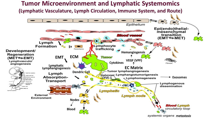
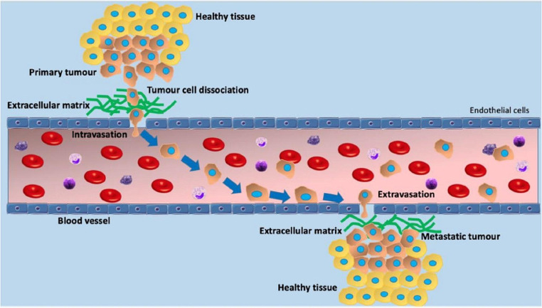
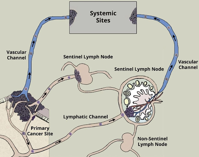
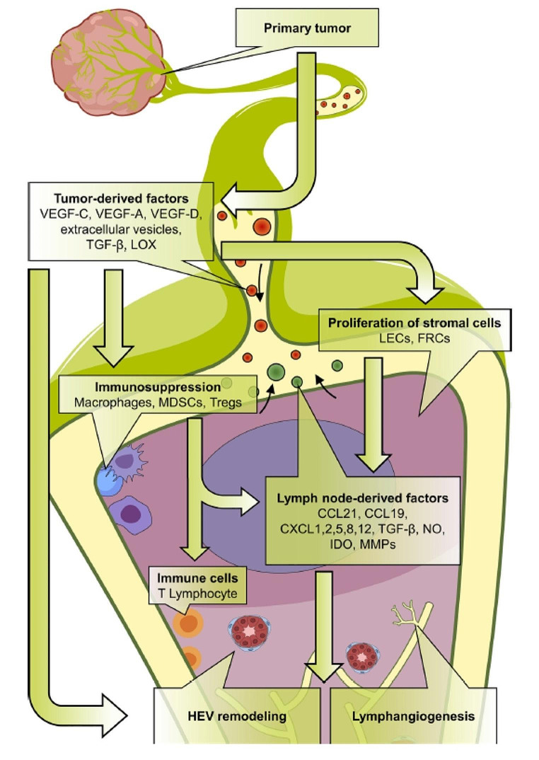
Whether cancer cells metastasize from the primary site to the distant sites via the lymphatic vessels or the blood vessels directly into the circulation is still under intense study. In this review article, we follow the journey of cancer cells metastasizing to the sentinel lymph nodes and beyond to the distant sites. We emphasize cancer heterogeneity and microenvironment as major determinants of cancer metastasis. Multiple molecules have been found to be associated with the complicated process of metastasis. Based on the large sentinel lymph node data, it is reasonable to conclude that cancer cells may metastasize through the blood vessels in some cases but in most cases, they use the sentinel lymph nodes as the major gateway to enter the circulation to distant sites.
Keywords: Cancer metastasis; Lymphatic and blood vessels.
© 2024. The Author(s).
Conflict of interest statement
The authors have no relevant financial or non-financial interests to disclose.
Figures
Similar articles
-
The lymphatic system and sentinel lymph nodes: conduit for cancer metastasis.Clin Exp Metastasis. 2022 Feb;39(1):139-157. doi: 10.1007/s10585-021-10123-w. Epub 2021 Oct 15. Clin Exp Metastasis. 2022. PMID: 34651243 Free PMC article. Review.
-
Molecular mechanisms of cancer metastasis via the lymphatic versus the blood vessels.Clin Exp Metastasis. 2022 Feb;39(1):159-179. doi: 10.1007/s10585-021-10120-z. Epub 2021 Nov 12. Clin Exp Metastasis. 2022. PMID: 34767139 Free PMC article. Review.
-
Evidence for disseminated tumor cells in lymphatic vessels afferent to sentinel lymph nodes in patients diagnosed with cervical cancer.Cancer Rep (Hoboken). 2021 Aug;4(4):e1366. doi: 10.1002/cnr2.1366. Epub 2021 Mar 14. Cancer Rep (Hoboken). 2021. PMID: 33719186 Free PMC article.
-
Reprogramming of sentinel lymph node microenvironment during tumor metastasis.J Biomed Sci. 2022 Oct 20;29(1):84. doi: 10.1186/s12929-022-00868-1. J Biomed Sci. 2022. PMID: 36266717 Free PMC article. Review.
-
From tumor lymphangiogenesis to lymphvascular niche.Cancer Sci. 2009 Jun;100(6):983-9. doi: 10.1111/j.1349-7006.2009.01142.x. Epub 2009 Feb 20. Cancer Sci. 2009. PMID: 19385973 Free PMC article. Review.
Cited by
-
Molecular Mechanisms of Lymphatic Metastasis in Breast Cancer: An Updated Review.Cancers (Basel). 2025 Jun 25;17(13):2134. doi: 10.3390/cancers17132134. Cancers (Basel). 2025. PMID: 40647432 Free PMC article. Review.
-
A rare case report on esophageal squamous cell carcinoma metastatic to the pancreas.Front Oncol. 2025 Jul 24;15:1602987. doi: 10.3389/fonc.2025.1602987. eCollection 2025. Front Oncol. 2025. PMID: 40777121 Free PMC article.
-
Immune responses and immunotherapeutic approaches in the treatment against cancer.Clin Exp Metastasis. 2024 Aug;41(4):473-493. doi: 10.1007/s10585-024-10300-7. Epub 2024 Aug 18. Clin Exp Metastasis. 2024. PMID: 39155358 Free PMC article. Review.
-
Incredible use of plant-derived bioactives as anticancer agents.RSC Adv. 2025 Jan 20;15(3):1721-1746. doi: 10.1039/d4ra05089d. eCollection 2025 Jan 16. RSC Adv. 2025. PMID: 39835210 Free PMC article. Review.
-
Circulating Tumor Cells in Cancer Diagnosis, Therapy, and Theranostics Applications: An Overview of Emerging Materials and Technologies.Curr Pharm Des. 2025;31(9):674-690. doi: 10.2174/0113816128328459241009191933. Curr Pharm Des. 2025. PMID: 39473210 Review.
References
-
- Simpson D, Ferguson R, Martinez CN, Kazlow E, Moran U, Heguy A, Hanniford D, Hernando E, Osman I, Kirchhoff T (2017) Mutation burden as a potential prognostic marker of melanoma progression and survival. American Society of Clinical Oncology
-
- Darwin C (1859) On the origin of species by means of natural selection. J. Murray, London
Publication types
MeSH terms
LinkOut - more resources
Full Text Sources
Medical
