. 2024 Aug 15;210(4):497-507.
doi: 10.1164/rccm.202402-0265OC.

The Renin-Angiotensin-Aldosterone System Regulates Sarcoidosis Granulomatous Inflammation

Affiliations

The Renin-Angiotensin-Aldosterone System Regulates Sarcoidosis Granulomatous Inflammation

Elliott D Crouser et al. Am J Respir Crit Care Med. .

Abstract

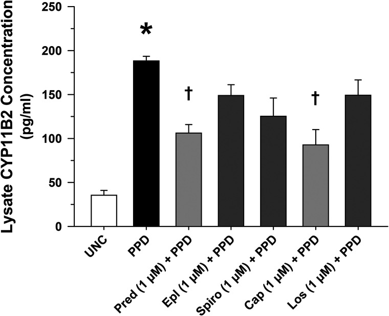
Rationale: Sarcoidosis is a granulomatous disorder of unclear cause notable for abnormal elevation of blood and tissue ACE1 (angiotensin converting enzyme 1) levels and activity. ACE1 regulates the renin-angiotensin-aldosterone system (RAAS), the terminal product of which is aldosterone, which selectively engages mineralocorticoid receptors to promote inflammation. Objectives: We sought to determine whether the RAAS promotes sarcoidosis granuloma formation and related inflammatory responses. Methods: Using an established ex vivo model, we first determined whether aldosterone was produced by sarcoidosis granulomas and verified the presence of CYP11B2, the enzyme required for its production. We then evaluated the effects of selective inhibitors of ACE1 (captopril), angiotensin type 1 receptor (losartan), and mineralocorticoid receptors (spironolactone, eplerenone) on granuloma formation, reflected by computer image analysis-generated granuloma area, and selected cytokines incriminated in sarcoidosis pathogenesis. Measurements and Main Results: Aldosterone was spontaneously produced by sarcoidosis peripheral blood mononuclear cells, and both intra- and extracellular levels steadily increased during granuloma formation. In parallel, peripheral blood mononuclear cells were shown to express more CYP11B2 during granuloma formation. Significant inhibition of sarcoidosis granulomas and related cytokines (TNFα, IL-1β, IFNγ, IL-10) was observed in response to pretreatments with captopril, losartan, spironolactone, or eplerenone, comparable to that of prednisone. Conclusions: The RAAS is intact in sarcoidosis granulomas and contributes significantly to early granuloma formation and to related inflammatory mediator responses, with important implications for clinical management.

Keywords: ACE1; CYP11B2; angiotensin receptor 1; granuloma; mineralocorticoid receptor.


Figures

Figure 1.
Figure 1.
Aldosterone production in sarcoidosis peripheral blood mononuclear cells during granuloma formation. (A) Extracellular supernatant aldosterone levels were higher in PPD-treated peripheral blood mononuclear cells (formed granulomas) than uncoated bead treatment (no granulomas) over time. (B) Aldosterone levels were much higher in granuloma lysates than in extracellular release, particularly during granuloma formation in response to PPD treatment beginning Day 3 and increasing through Day 7. n = 6 for each condition. *P < 0.05 versus uncoated bead treatment.
Figure 2.
Figure 2.
CYP11B2 protein expression in activated peripheral blood mononuclear cells is regulated by renin-angiotensin-aldosterone system components. CYP11B2 protein expression was significantly increased in sarcoidosis granuloma cell lysates 7 days after PPD-coated bead treatment compared with treatment with uncoated (UNC) beads, and CYP11B2 expression was significantly inhibited by captopril (Cap) and prednisone (Pred), but not in response to losartan (Los), spironolactone (Spiro), or eplerenone (Epl). n = 8 for each condition. *P < 0.01 (Cohen’s d > 5.0) compared with matching UNC bead treatment alone and P < 0.05 (Cohen’s d > 3.0) relative to corresponding PPD treatment alone.
Figure 3.
Figure 3.
Renin-angiotensin-aldosterone system and MR (mineralocorticoid receptor) inhibitors suppress ex vivo sarcoidosis granuloma formation. Representative photomicrographs of granuloma-like cell aggregates forming 7 days after sarcoidosis peripheral blood mononuclear cells were incubated with either uncoated beads (UNC) or PPD-coated beads (PPD) alone or after a 30-minute pretreatment with either prednisone (glucocorticoid receptor agonist), spironolactone or eplerenone (inhibitors of MR), captopril (inhibitor of ACE1 [angiotensin converting enzyme 1]), or losartan (inhibitor of ATR1 [angiotensin 1 receptor]). Images to the left are original, unfiltered images, whereas those in the center include MIPAR image analyses demonstrating color-coding of identified granulomas based on cell aggregate area, with larger cell aggregates represented in the red-yellow hues and smaller ones in the blue-green hues. The images to the right have all but the MIPAR-identified cell aggregates filtered out, showing the cumulative granuloma area as a fraction of the total area of the image (area fraction as determined by MIPAR analysis for each representative image). Scale bar, 100 μm (marked at the bottom in the top left image).
Figure 4.
Figure 4.
Suppression of ex vivo sarcoidosis granuloma formation by renin-angiotensin-aldosterone system inhibitors. Captopril (Cap; inhibitor of ACE1 [angiotensin converting enzyme 1]) or losartan (Los; inhibitor of ATR1 [angiotensin 1 receptor]) pretreatments for 30 minutes were equally effective in a dose-dependent manner to significantly suppress PPD bead–induced granuloma formation (as per MIPAR analysis of identified granuloma-like cell aggregates; area fraction = cumulative granuloma area as a fraction of the total area of the image) compared with prednisone (Pred). n = 14 for each condition. *P < 0.001 (Cohen’s d > 3.0) compared with UNC bead treatment, P < 0.001 (Cohen’s d > 2.5) relative to PPD treatment alone, and P < 0.01 (Cohen’s d > 1.5) compared with PPD treatment alone. UNC = uncoated.
Figure 5.
Figure 5.
Suppression of ex vivo sarcoidosis granuloma formation by MR (mineralocorticoid receptor) inhibitors. Area fraction of granuloma-like cell aggregates forming 7 days after peripheral blood mononuclear cell treatment with PPD-coated beads demonstrated significant dose-dependent inhibition after pretreatment with MR inhibitors spironolactone (Spiro), or eplerenone (Epl) compared with prednisone (Pred) (as per MIPAR analysis of identified granuloma-like cell aggregates; area fraction = cumulative granuloma area as a fraction of the total area of the image). n = 14 for each condition. *P < 0.001 (Cohen’s d > 3.0), compared with UNC bead treatment, P < 0.001 (Cohen’s d > 2.5) relative to PPD treatment alone, and P < 0.01 (Cohen’s d > 1.5) compared with PPD treatment alone. UNC = uncoated.
Figure 6.
Figure 6.
Delayed inhibition of renin-angiotensin-aldosterone system attenuates ex vivo sarcoidosis granuloma formation. Administration of MR (mineralocorticoid receptor) inhibitors, spironolactone (Spiro) or eplerenone (Epl), captopril (Cap, inhibitor of ACE1 [angiotensin converting enzyme 1]), or losartan (Los, inhibitor of ATR1 [angiotensin 1 receptor]) 4 days after PPD treatment significantly attenuated PPD-induced granuloma formation (as per MIPAR analysis of identified granuloma-like cell aggregates) similar to that of prednisone (Pred) at Day 7. n = 7 for each condition. *P < 0.001 (Cohen’s d > 2.0) relative to UNC bead treatment, P < 0.01 (Cohen’s d > 1.5) compared with PPD treatment alone, and P < 0.05 (Cohen’s d = 1.0) relative to PPD treatment alone. UNC = uncoated.
Figure 7.
Figure 7.
Renin-angiotensin-aldosterone system and MR (mineralocorticoid receptor) regulate cytokine release from activated sarcoidosis peripheral blood mononuclear cells. Extracellular cytokine release of (A) TNFα, (B) IFNγ, (C) IL-1β, and (D) IL-10 from PPD bead–treated sarcoidosis granulomas increased significantly relative to matching uncoated (UNC) bead–treated peripheral blood mononuclear cells 7 days after treatment. PPD-induced cytokine release was attenuated in varying degrees by pretreatment with inhibitor of ACE1 (angiotensin converting enzyme 1) captopril (Cap), inhibitor of ATR1 (angiotensin 1 receptor) losartan (Los), or by MR inhibitors spironolactone (Spiro) or eplerenone (Epl). Glucocorticoid receptor agonist prednisone (Pred), as expected, inhibited cytokine production under these conditions as well. n = 14 for each condition. *P < 0.01 (Cohen’s d > 2.0) relative to UNC bead treatment, P < 0.01 (Cohen’s d > 1.5) compared with PPD treatment alone, and P < 0.05 (Cohen’s d > 1.0) relative to PPD treatment alone.
Figure 8.
Figure 8.
Delayed suppression of renin-angiotensin-aldosterone system (RAAS) diminished sarcoidosis granuloma cytokine release. Administration of RAAS pathway (inhibitor of ACE1 [angiotensin converting enzyme 1] captopril [Cap] or inhibitor of ATR1 [angiotensin 1 receptor] losartan [Los]) or MR (mineralocorticoid receptor) inhibitors (spironolactone [Spiro] or eplerenone [Epl]) 4 days after PPD exposure reduced granuloma cytokine release (A) TNFα, (B) IFNγ, (C) IL-1β, and (D) IL-10 to a variable extent compared with the glucocorticoid receptor agonist prednisone (Pred) at Day 7. n = 7 for each condition. *P < 0.01 (Cohen’s d > 1.5) compared with UNC bead treatment, P < 0.01 (Cohen’s d > 1.5) relative to PPD treatment alone, and P < 0.05 (Cohen’s d > 0.8) compared with PPD treatment alone. UNC = uncoated.
Figure 9.
Figure 9.
Proposed mechanisms by which the renin-angiotensin-aldosterone system (RAAS) pathway promotes sarcoidosis granuloma formation. Renin enzymatically cleaves angiotensin (Ang) to form Ang I. ACE1 (angiotensin converting enzyme 1) then cleaves Ang I to form Ang II, which binds to ATR1 (angiotensin receptor 1) and is cleaved by ACE2 to form Ang 1–7. ATR1 induces aldosterone (Aldo) synthesis through nuclear transcription of CYP11B2 (the gene encoding the aldosterone-forming enzyme). The molecular and cellular mechanisms linking RAAS to granuloma formation remain to be determined, but mTOR (mammalian target of rapamycin) is a common signaling pathway regulated by both ATR1, via PI3K signaling (43), and MR (mineralocorticoid receptor), the cognate receptor for Aldo, which promotes sarcoidosis granuloma formation. Ang 1–7 suppresses mTOR and may thereby suppress granuloma formation (see text). MasR is a G-protein–coupled receptor that mediates Ang 1–7 suppression of other pathways.

Comment in

References

    1. Lieberman J. The specificity and nature of serum-angiotensin-converting enzyme (serum ACE) elevations in sarcoidosis. Ann N Y Acad Sci . 1976;278:488–497. - PubMed
    1. Lieberman J. Elevation of serum angiotensin-converting-enzyme (ACE) level in sarcoidosis. Am J Med . 1975;59:365–372. - PubMed
    1. Eklund A, Blaschke E, Danielsson B. Subcellular localization of angiotensin-converting enzyme in the human alveolar macrophage. Scand J Clin Lab Invest . 1987;47:47–54. - PubMed
    1. Silverstein E, Friedland J, Lyons HA, Gourin A. Elevation of angiotensin-converting enzyme in granulomatous lymph nodes and serum in sarcoidosis: clinical and possible pathogenic significance. Ann N Y Acad Sci . 1976;278:498–513. - PubMed
    1. Duan J, Xu Y, Zhu H, Zhang H, Sun S, Sun H, et al. Relationship between CT activity score with lung function and the serum angiotensin converting enzyme in pulmonary sarcoidosis on chest HRCT. Medicine (Baltimore) . 2018;97:e12205. - PMC - PubMed

MeSH terms