Clinical Trial
. 2024 Oct 3;144(14):1496-1507.
doi: 10.1182/blood.2024024144.

Inflammasome activation in patients with Kaposi sarcoma herpesvirus-associated diseases

Affiliations
Clinical Trial

Inflammasome activation in patients with Kaposi sarcoma herpesvirus-associated diseases

Silvia Lucena Lage et al. Blood. .

Abstract

Kaposi sarcoma herpesvirus (KSHV)-associated diseases include Kaposi sarcoma (KS), primary effusion lymphoma (PEL), KSHV-associated multicentric Castleman disease (MCD), and KS inflammatory cytokine syndrome (KICS). PEL, MCD, and KICS are associated with elevated circulating inflammatory cytokines. However, activation of the inflammasome, which generates interleukin-1β (IL-1β) and IL-18 via active caspase-1/4/5, has not been evaluated in patients with KSHV-associated diseases (KADs). Herein we report that patients with HIV and ≥1 KAD present with higher plasma levels of IL-18 and increased caspase-1/4/5 activity in circulating monocytes compared with HIV-negative healthy volunteers (HVs) or people with HIV (PWH) without KAD. Within KAD subtypes, KICS and MCD shared enhanced caspase-1/4/5 activity and IL-18 production compared with HVs and PWH, whereas patients with PEL showed remarkably high levels of inflammasome complex formation (known as apoptosis-associated speck-like protein containing a caspase recruitment domain). Moreover, caspase-1/4/5 activity and IL-18 plasma levels correlated with KSHV viral load, indicating KSHV-driven inflammasome activation in KAD. Accordingly, factors released by cells latently infected with KSHV triggered inflammasome activation and cytokine production in bystander monocytes in vitro. Finally, both supervised and unsupervised analyses with inflammasome measurements and other inflammatory biomarkers demonstrate a unique inflammatory profile in patients with PEL, MCD, and KICS as compared with KS. Our data indicate that detrimental inflammation in patients with KAD is at least partially driven by KSHV-induced inflammasome activation in monocytes, thus offering novel approaches to diagnose and treat these complex disorders. These trials were registered at www.ClinicalTrials.gov as #NCT01419561, NCT00092222, NCT00006518, and NCT02147405.


Conflict of interest statement

Conflict-of-interest disclosure: D.W. and A.R. are employed by Leidos Biomedical Research, Inc. R.R., K.L., and R.Y. report receiving research support from Celgene/Bristol Myers Squibb, CTI BioPharma (a Sobi A.B. company), PBS Biotech, and Janssen Pharmaceuticals through Cooperative Research and Development Agreements (CRADAs) with the National Cancer Institute (NCI). R.R., K.L., and R.Y. report receiving drugs for a clinical trial from Merck, EMD Serono, and Eli Lilly through CRADAs with the NCI. I.S., R.Y., and D.W. are coinventors on US patent 10,001,483 entitled “Methods for the treatment of Kaposi’s sarcoma or KSHV-induced lymphoma using immunomodulatory compounds and uses of biomarkers.” An immediate family member of R.Y. is a coinventor on patents or patent applications related to internalization of target receptors, epigenetic analysis, and ephrin tyrosine kinase inhibitors. All rights, title, and interest to these patents have been assigned to the US Department of Health and Human Services; the government conveys a portion of the royalties it receives to its employee inventors under the Federal Technology Transfer Act of 1986 (P.L. 99-502). The remaining authors declare no competing financial interests.

Figures

None
Graphical abstract
Figure 1.
Figure 1.
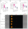
Exacerbated inflammasome activation is found in participants with KAD when compared with PWH and HVs. (A) Plasma levels of IL-18 were compared among participants with KAD (n = 42), PWH (n = 12), and HVs (n = 9). Lines represent median values and interquartile ranges. PBMCs from the same patients and distinct HVs (n = 10) were incubated with the FLICA, stained for monocyte identification and intracellular ASC, and acquired by imaging flow cytometry. (B) gMFI of FLICA within total circulating blood monocytes was assessed. Data are presented as median with interquartile range. (C) Representative images showing respectively CD14, ASC, and FLICA fluorescence followed by a composite image containing brightfield (BF), and the fluorescence of ASC and FLICA merged, were randomly selected from representative samples of the distinct KAD conditions. In these CD14+ cells, FLICA and ASC fluorescence are superimposed. (D) The number of monocytes showing spontaneous FLICA+ ASC-speck formation was quantified after application of Modulation_Morphology (M11,Ch11)_11-ASC feature, followed by Bright Detail Similarity R3_MC_11-ASC_2-FLICA, inside the monocyte gate, by using IDEAS software. Lines represent median values and interquartile ranges. Comparisons were made using the Kruskal-Wallis test followed by the Dunn multiple test. ∗∗P < .01; ∗∗∗P < .001; ∗∗∗∗P < .0001.
Figure 2.
Figure 2.
Correlation matrix of inflammasome activation flow cytometry markers, soluble biomarkers, and clinical laboratory tests within the KAD cohort. (A) Multiparameter Spearman correlation matrix of inflammasome activation markers, soluble biomarkers, and clinical laboratory tests from all participants with KAD. KSHV-VL represents copies per million PBMCs. (B-G) Individual correlation plots highlighting significant associations by Spearman r between caspase activity, KSHV-VL, and key KAD-related inflammatory cytokines. Hgb, hemoglobin; Plt, platelets; PTX3, pentraxin-3.
Figure 3.
Figure 3.
Inflammasome activation across different groups of patients presenting with KSHV-associated diseases stratified according to their clinical classification. (A) Plasma levels of IL-18 were compared among participants with KAD (n = 42, comprising patients with KS alone [10], MCD [11], MCD + PEL [5], KICS + KS [11], and PEL ± KS [5]; ∗means with or without KS), PWH (n = 12), and HV (n = 10). (B) PBMCs from the previously mentioned groups were incubated with the FLICA, and the gMFI of FLICA within total circulating blood monocytes was compared among groups. (C) Alternatively, PBMCs were also stained for monocyte identification and intracellular ASC and acquired by imaging flow cytometry. Data are presented as median with interquartile range. Data were analyzed using the Kruskal-Wallis test followed by Dunn multiple comparisons. ∗P < .05; ∗∗P < .01; ∗∗∗P < .001; ∗∗∗∗P < .0001.
Figure 4.
Figure 4.
Multidimensional analysis and heat map with unsupervised clustering of caspase-1/4/5 activity, speck formation, and inflammasome-associated biomarkers. (A) Principal component analysis incorporating inflammasome components (caspase-1/4/5 activity, FLICA+ ASC-speck formation) and 11 inflammatory biomarkers (ferritin, CRP, IFN-γ, IL-6, IL-10, IL-13, IL-18, IL18BP, CXCL9, CD163, and TNF-α) demonstrates significant overlap in clusters when defined by clinical syndrome. Individuals are represented by small colored circles for each group, whereas the overall group is represented by large colored circles. Participants with MCD and concomitant PEL are depicted as red circles with a blue outline. (B) Heat map with unsupervised clustering of inflammasome components, KSHV-VL (copies per million PBMCs), and monocyte-associated biomarkers identified that those with MCD primarily cluster together, including those with concomitant PEL (depicted with blue circle inside red box). Those with KS alone form a separate distinct cluster. Patients with KICS are distributed throughout. Individuals with MCD, PEL, and KICS may or may not have had concomitant KS. Analysis performed in R (version 4.1.2) using the FactoMineR, factoextra, and heatmap packages.
Figure 5.
Figure 5.
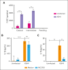
In vitro stimulation of monocytes with supernatants from KSHV-infected cells. HV-isolated monocytes were plated (1 × 106 cells per well) and incubated overnight with complete media containing 50% of supernatant from either uninfected or KSHV-infected HEK293T cells, in the presence or absence of the NLRP3 inflammasome inhibitor MCC950 (5 μM). After stimulation, culture supernatants were harvested for quantification of IL-1β and IL-18 by enzyme-linked immunosorbent assay (ELISA), and monocytes were stained for detection of inflammasome complex formation by imaging flow cytometry. (A) Numbers of monocytes showing FLICA+ ASC-speck formation per mL were quantified inside the monocyte gate. Lines represent mean values with standard error of the mean (SEM). Data were analyzed using the Welch t test to compare the uninfected with the KSHV-infected group within each monocyte subset. ∗∗P < .01; ∗∗∗P < .001. (B) The levels of (B) IL-1β and (C) IL-18 produced by stimulated monocytes were quantified by ELISA and compared across the different groups. Lines represent mean values with SEM. Data were analyzed using the Kruskal-Wallis test. ∗∗P < .01; ∗∗∗∗P < .0001. Data are presented as a pool of 2 different batches of HEK293T culture–derived supernatants.

Comment in

  • Is inflammation key in Kaposi sarcoma?
    Dittmer DP. Dittmer DP. Blood. 2024 Oct 3;144(14):1464-1465. doi: 10.1182/blood.2024025829. Blood. 2024. PMID: 39361305 Free PMC article. No abstract available.

References

    1. Hansen ME, Mangusan R, Lurain K, et al. Characteristics of patients admitted to the ICU with Kaposi sarcoma herpesvirus-associated diseases. AIDS. 2022;36(14):1969–1978. - PMC - PubMed
    1. Lurain K, Polizzotto MN, Aleman K, et al. Viral, immunologic, and clinical features of primary effusion lymphoma. Blood. 2019;133(16):1753–1761. - PMC - PubMed
    1. Yarchoan R, Uldrick TS. HIV-associated cancers and related diseases. N Engl J Med. 2018;378(22):2145. - PubMed
    1. Ramaswami R, Lurain K, Yarchoan R. Oncologic treatment of HIV-associated Kaposi sarcoma 40 years on. J Clin Oncol. 2022;40(3):294–306. - PMC - PubMed
    1. Goncalves PH, Ziegelbauer J, Uldrick TS, Yarchoan R. Kaposi sarcoma herpesvirus-associated cancers and related diseases. Curr Opin HIV AIDS. 2017;12(1):47–56. - PMC - PubMed

Publication types

MeSH terms

Supplementary concepts

Associated data