. 2024 Aug 7;44(32):e0140242024.
doi: 10.1523/JNEUROSCI.0140-24.2024.

d-Serine Inhibits Non-ionotropic NMDA Receptor Signaling

Affiliations

d-Serine Inhibits Non-ionotropic NMDA Receptor Signaling

Eden V Barragan et al. J Neurosci. .

Abstract

NMDA-type glutamate receptors (NMDARs) are widely recognized as master regulators of synaptic plasticity, most notably for driving long-term changes in synapse size and strength that support learning. NMDARs are unique among neurotransmitter receptors in that they require binding of both neurotransmitter (glutamate) and co-agonist (e.g., d-serine) to open the receptor channel, which leads to the influx of calcium ions that drive synaptic plasticity. Over the past decade, evidence has accumulated that NMDARs also support synaptic plasticity via ion flux-independent (non-ionotropic) signaling upon the binding of glutamate in the absence of co-agonist, although conflicting results have led to significant controversy. Here, we hypothesized that a major source of contradictory results might be attributed to variable occupancy of the co-agonist binding site under different experimental conditions. To test this hypothesis, we manipulated co-agonist availability in acute hippocampal slices from mice of both sexes. We found that enzymatic scavenging of endogenous co-agonists enhanced the magnitude of long-term depression (LTD) induced by non-ionotropic NMDAR signaling in the presence of the NMDAR pore blocker MK801. Conversely, a saturating concentration of d-serine completely inhibited LTD and spine shrinkage induced by glutamate binding in the presence of MK801 or Mg2+ Using a Förster resonance energy transfer (FRET)-based assay in cultured neurons, we further found that d-serine completely blocked NMDA-induced conformational movements of the GluN1 cytoplasmic domains in the presence of MK801. Our results support a model in which d-serine availability serves to modulate NMDAR signaling and synaptic plasticity even when the NMDAR is blocked by magnesium.

Keywords: NMDA receptors; co-agonist; d-serine; dendritic spine plasticity; long-term depression; silent synapses.


Conflict of interest statement

The authors declare no competing financial interests

Figures
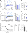
Figure 1.
Figure 1.
Co-agonist site antagonism increases the magnitude and reduces the variance of ion flux-independent NMDAR-mediated LTD. A, Left: averaged plasticity experiments using a 1 Hz, 900-pulse LTD induction protocol in the presence of 100 µM MK801 (gray circles; n = 19), 10 µM L689 (dark blue circles; n = 16), or 50 µM AP5 (light blue circles; n = 12). Right: compared with MK801, L689 resulted in an increased magnitude of LTD (unpaired t test). B, Coefficient of variation (CV) of the experiments in A demonstrating the reduction in variation with L689 compared with MK801. C, Left: averaged plasticity experiments using a 1 Hz, 900-pulse LTD induction protocol in the presence of 100 µM MK801 alone (gray circles; n = 16) or with the addition of either 10 µM L689 (dark blue circles; n = 9) or 50 µM AP5 (light blue circles; n = 9). Right: compared with MK801 alone, the addition of L689 resulted in an increased magnitude of LTD (unpaired t test). D, CV of the experiments in A demonstrating the reduction in variation with L689 compared with MK801 alone. E, Left: averaged NMDAR fEPSPs recorded in 0.2 mM Mg2+ normalized to baseline fEPSP amplitude prior to blocking AMPA receptors with 10 µM NBQX in slices preincubated with ACSF (black circles; n = 7) or 100 µM MK801 (gray circles; n = 6). Right: sample traces before (black) and after (gray) NBQX. F, summary data of E, MK801 preincubation completely blocks NMDAR fEPSPs (one sample t test compared with 0). Scale bars for all sample traces are 0.5 mV, 10 ms. All data represented as mean ± SEM. *p < 0.05, ***p < 0.001, nsp ≥ 0.05.
Figure 2.
Figure 2.
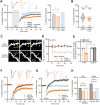
Enzymatic scavenging of endogenous co-agonists increases the magnitude and reduces the variance of ion flux-independent NMDAR-mediated LTD. A, Left: averaged plasticity experiments using a 1 Hz, 900-pulse LTD induction protocol in a recirculating bath with MK801 alone (gray circles; n = 11) or in the presence of scavenging enzymes (orange circles; n = 8) or 50 µM AP5 (light blue circles; n = 6). Right: compared with MK801 alone, the presence of scavenging enzymes resulted in an increased magnitude of non-ionotropic LTD (unpaired t test). B, CV of the experiments in A (left) and B (right) demonstrating the reduction in variation with either L689 or enzymes compared with MK801 alone. C, Images of dendrites of CA1 neurons from acute hippocampal slices from GFP-M mice (P16–P20) before and after sub-HFU (yellow crosses) at single spines across time (yellow arrowheads) in the presence of active scavenging enzymes (top) or heat-inactivated enzymes (bottom). D–E, Sub-HFU in the presence of active enzymes (orange-filled circles/bar; 5 spines/5 cells), but not inactive enzymes (black-filled circles/bar; 5 spines/5 cells), led to robust long-term spine shrinkage. Size of unstimulated neighbors (open circles/bars) did not change. F, Averaged plasticity experiments using a neutral 10 Hz, 900-pulse protocol in a recirculating bath with vehicle (black circles; n = 11), or in the presence of active scavenging enzymes (orange-filled circles; n = 9), or heat-inactivated enzymes (open orange circles; n = 7). G, Averaged plasticity experiments using a 50 Hz, 300-pulse LTP induction protocol in a recirculating bath with vehicle (black circles; n = 10) or in the presence of scavenging enzymes (orange circles; n = 10) or 50 µM AP5 (light blue circles; n = 10). H, Summary of data from C and D. One-way (electrophysiology data) and two-way (imaging data) ANOVA with Bonferroni’s post hoc multiple-comparisons test. Scale bars for all sample traces are 0.5 mV, 10 ms. All data represented as mean ± SEM. **p < 0.01, ***p < 0.001, ****p < 0.0001, nsp ≥ 0.05.
Figure 3.
Figure 3.
d-serine blocks ion flux-independent NMDAR-mediated LTD. A, Left: averaged plasticity experiments using a 1 Hz, 900-pulse LTD induction protocol in the presence of 100 µM MK801 alone (gray circles; n = 12) or with the addition of 10 µM d-serine (red circles; n = 11). Right: d-serine eliminated the non-ionotropic LTD observed with MK801 alone (unpaired t test). B, CV of the experiments in A demonstrating a reduction in variation with d-serine compared with MK801 alone. C, Averaged fEPSP recordings normalized to the baseline fEPSP slope and then 60 min of incubation with 10 µM d-serine (n = 7). Inset: d-serine did not change the fEPSP compared with baseline (one-sample t test). D, Whole-cell NMDAR-EPSC experiment showing the time course of MK801 block of synaptic receptors. A 10 µM d-serine was added at 20 min, and low-frequency (1 Hz) stimulation (LFS) was started at 25 min. Neither d-serine incubation alone nor in combination with LFS altered the extent of MK801 blockade (n = 10). Two-way ANOVA with Bonferroni’s post hoc multiple-comparisons test. Scale bars for all sample traces are 0.5 mV, 10 ms. All data represented as mean ± SEM. **p < 0.01, ***p < 0.001.
Figure 4.
Figure 4.
d-serine blocks LTD-associated spine shrinkage mediated by ion flux-independent NMDAR signaling. A, Images of dendrites of CA1 neurons from acute hippocampal slices from GFP-M mice (P16–P20) before and after LFU (yellow crosses) at single spines across time (yellow arrowheads) in the presence of MK-801 and vehicle (top) or d-serine (10 µM; bottom). B, C, LFU in the presence of 100 µM MK801 (black-filled circles/bar; 6 spines/6 cells) led to robust spine shrinkage, which was fully inhibited with the addition of d-serine (red-filled circles/bar; 7 spines/7 cells). Size of unstimulated neighbors (open circles/bars) did not change. D, Images of dendrites of CA1 neurons in acute hippocampal slices from GFP-M mice (P17–P21) before and after HFU (yellow crosses) at single spines in 1 mM Mg2+ and either vehicle (water; top) NBQX (50 µM; middle) or d-serine (10 µM; bottom). E, F, HFU-induced long-term spine shrinkage in 1 mM Mg2+ was blocked by d-serine (red-filled circles/bar; 9 spines/9 cells), but not by vehicle (black-filled circles/bar; 8 spines/8 cells) or by NBQX (beige-filled circles/bar; 12 spines/12 cells). Size of unstimulated neighbors (open circles/bars) did not change. G, Images of dendrites of CA1 neurons in acute hippocampal slices from WT/GFP-M (top) and SRKO/GFP-M (bottom) littermates (P17–P21) before and after HFU (yellow crosses) at single spines in 1 mM Mg2+. H, I, Both WT (black-filled circles/bar; 8 spines/8 cells) and SRKO (purple-filled circles/bar; 8 spines/8 cells) exhibited robust HFU-induced long-term spine shrinkage in 1 mM Mg2+. Size of unstimulated neighbors (open circles/bars) did not change. Two-way ANOVA with Bonferroni’s post hoc multiple-comparisons test. All data represented as mean ± SEM. *p < 0.01, **p < 0.001, nsp > 0.05.
Figure 5.
Figure 5.
d-serine inhibits NMDA-induced intracellular GluN1 conformational changes. A, Representative fluorescence lifetime images of neurons expressing GluN1-GFP and GluN1-mCherry (with GluN2B) before and during treatment with 25 µM NMDA. Neurons were incubated with MK801 alone (left) or with 10 µM d-serine (right). Pseudocolor scale indicates GFP lifetime at each pixel. Scale bar, 5 µm; dendritic segments masked for clarity. B, Average NMDA-induced spine GluN1-GFP lifetime change for indicated conditions, N (> 20 neurons, > 500 spines) for each condition, showing that 10 µM d-serine completely blocks the NMDA-induced lifetime change, unpaired t test. Data represented as mean ± SEM. **p < 0.01.

Update of

References

    1. Adesnik H, Li G, During MJ, Pleasure SJ, Nicoll RA (2008) NMDA receptors inhibit synapse unsilencing during brain development. Proc Natl Acad Sci U S A 105:5597–5602. 10.1073/pnas.0800946105 - DOI - PMC - PubMed
    1. Aman TK, Maki BA, Ruffino TJ, Kasperek EM, Popescu GK (2014) Separate intramolecular targets for protein kinase a control N-methyl- d-aspartate receptor gating and Ca2 + permeability. J Biol Chem 289:18805–18817.
    1. Aow J, Dore K, Malinow R (2015) Conformational signaling required for synaptic plasticity by the NMDA receptor complex. Proc Natl Acad Sci U S A 112:14711–14716. 10.1073/pnas.1520029112 - DOI - PMC - PubMed
    1. Babiec WE, Guglietta R, Jami SA, Morishita W, Malenka RC, O'Dell TJ (2014) Ionotropic NMDA receptor signaling is required for the induction of long-term depression in the mouse hippocampal CA1 region. J Neurosci 34:5285–5290. 10.1523/JNEUROSCI.5419-13.2014 - DOI - PMC - PubMed
    1. Basu AC, et al. (2009) Targeted disruption of serine racemase affects glutamatergic neurotransmission and behavior. Mol Psychiatry 14:719–727. 10.1038/mp.2008.130 - DOI - PMC - PubMed

MeSH terms