. 2024 Jun 28;14(1):14958.
doi: 10.1038/s41598-024-64924-6.

Hyaluronidase inhibitor delphinidin inhibits cancer metastasis

Affiliations

Hyaluronidase inhibitor delphinidin inhibits cancer metastasis

Jeremy McGuire et al. Sci Rep. .

Abstract

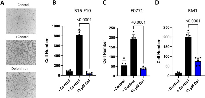
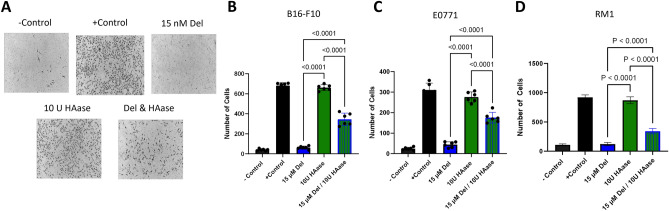
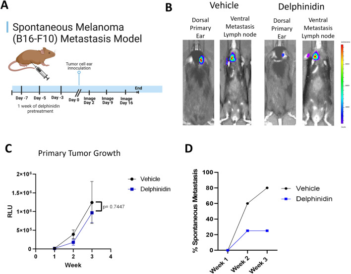
Cancer remains a formidable global health challenge, with metastasis being a key contributor to its lethality. Abundant high molecular mass hyaluronic acid, a major non-protein component of extracellular matrix, protects naked mole rats from cancer and reduces cancer incidence in mice. Hyaluronidase plays a critical role in degrading hyaluronic acid and is frequently overexpressed in metastatic cancer. Here we investigated the potential of targeting hyaluronidases to reduce metastasis. A high throughput screen identified delphinidin, a natural plant compound found in fruits and vegetables, as a potent hyaluronidase inhibitor. Delphinidin-mediated inhibition of hyaluronidase activity led to an increase in high molecular weight hyaluronic acid in cell culture and in mouse tissues, and reduced migration and invasion behavior of breast, prostate, and melanoma cancer cells. Moreover, delphinidin treatment suppressed melanoma metastasis in mice. Our study provides a proof of principle that inhibition of hyaluronidase activity suppresses cancer cell migration, invasion and metastasis. Furthermore, we identified a natural compound delphinidin as a potential anticancer therapeutic. Thus, we have identified a path for clinical translation of the cancer resistance mechanism identified in the naked mole rat.


Conflict of interest statement

VG is a co-founder of MatrixBio, leveraging the anti-cancer and pro-longevity effects of high molecular weight hyaluronic acid from naked mole rat to human. All other authors do not have any competing interest.

Figures

Figure 1
Figure 1
High throughput screening (HTS) for HAase inhibitor using fluorescence polarization & increases in HMW HA by delphinidin. (A) FP shows fluorescent HMW-HA at a higher FP value than LMW-HA (B) Representative data from HTS in a 384 well plate, red arrow indicates prodelphiniline. (C, D) Structures of Prodelphiniline and Delphinidin. (E) Equal numbers of human or mouse fibroblasts were incubated with increasing concentrations of Delphinidin for 24 h. HA was separated on agarose gel (F) Quantification of HA in mouse and human samples.
Figure 2
Figure 2
In vivo administration of delphinidin increased HA accumulation in mouse tissues: Mice were injected three times a week for three weeks with either vehicle, 50 mg/kg delphinidin or 100 mg/kg of delphinidin. Tissues were collected and embedded in paraffin before slide mounting and HABP staining. (A) Representative images (20X) showing HABP staining in mouse tissues. (B) Quantification of HABP staining in mouse tissue mean fluorescence intensity of 6 representative images (three biological replicates with two technical replicates from each tissue) using Imaris cell imaging software. Statistical analyses were generated from one-way ANOVA with multiple comparisons at 95% CI error bars are represented as mean with SEM.
Figure 3
Figure 3
Delphinidin inhibits proliferation of cancer cells. (A) Delphinidin had no significant effect on the growth of wildtype C57/Bl6 fibroblasts after 48 h. Proliferation was assayed using Cell Titer Glo 2.0 (BD) Delphinidin in a dose dependent manner significantly inhibited the growth of B16-F10, E0771 and RM1 after 48 h. (EG) When the cancer cells were treated with HAase in combination with delphinidin a reduction in growth inhibition was observed at 48 h. All data was collected as technical triplicates and statistical analyses were generated from one-way ANOVA with multiple comparisons at 95% CI error bars are represented as mean with SEM.
Figure 4
Figure 4
Delphinidin inhibits migration of cancer cells. (A) Representative images of membranes from Boyden chamber assay. Negative control was serum free media on both sides of the membrane and positive control was 1% FBS on the bottom well to promote chemotaxis delphinidin was added to top wells with cells and bottom wells contained 1% FBS (BD) All three cancer cell lines had significant inhibition of migration when incubated with 15 µM delphinidin. (B) B16-F10 (C) E0771 (D) RM1. All data were collected as technical triplicates and statistical analyses were generated from one-way ANOVA with multiple comparisons at 95% CI error bars are represented as mean with SEM.
Figure 5
Figure 5
Delphinidin inhibits invasion of cancer cell and HAase abrogates the effect of delphinidin. (A) Representative images of the membranes from the Boyden chamber invasion assay. The inside of the top membrane was coated with basement membrane to mimic metastatic invasion. Negative control was serum free media in top and bottom wells and positive control was 1% FBS on the bottom well to promote chemotaxis delphinidin and HAase was added to top wells with cells in serum free media and bottom wells contained 1% FBS. (BD) 15 µM Delphinidin inhibited the invasion of all three cancer cell lines compared with 10 U/ml of HAase when cells were incubated with delphinidin in combination with 100 U/ml HAase the amount of inhibition was reduced. (B) B16-F10 (C) E0771 (D) RM1. All data was collected as technical triplicates and statistical analyses were generated from one-way ANOVA with multiple comparisons at 95% CI error bars are represented as mean with SEM.
Figure 6
Figure 6
B16-F10 Spontaneous Metastasis model: (A) Model design adapted from “Mouse Experimental Timeline”, by BioRender.com (2023). 50 mg/kg of delphinidin administered by IP 3X a week starting one week before tumor cell (B16-F10) inoculation on the ear and continuing until tumor sizes reached endpoint (2 mm2). (B) Representative IVIS images of bioluminescence detection of primary tumor and metastases. (C) Primary tumor growth was not significant between delphinidin treated and vehicle mice. Statistical analysis was generated by unpaired t-test error bars are represented as mean with SEM. (D) At week three (Day 16) 80% (n = 4/5) of vehicle treated mice had detectable metastases while only 25% (n = 3/12) of delphinidin treated mice had detectable metastases.

References

    1. Guan X. Cancer metastases: Challenges and opportunities. Acta Pharm. Sin. B. 2015;5:402–418. doi: 10.1016/j.apsb.2015.07.005. - DOI - PMC - PubMed
    1. Chaffer CL, Weinberg RA. A perspective on cancer cell metastasis. Science. 2011;331:1559–1564. doi: 10.1126/science.1203543. - DOI - PubMed
    1. Kratzer TB, et al. Progress against cancer mortality 50 years after passage of the national cancer act. JAMA Oncol. 2022;8:156–159. doi: 10.1001/jamaoncol.2021.5668. - DOI - PMC - PubMed
    1. Welch, D. Defining a cancer metastasis. In AACR Education Book 2006 111–115 (2006).
    1. Welch DR, Hurst DR. Defining the hallmarks of metastasis. Cancer Res. 2019;79:3011–3027. doi: 10.1158/0008-5472.Can-19-0458. - DOI - PMC - PubMed