. 2024 Jun 28;29(1):92.
doi: 10.1186/s11658-024-00607-4.

A specific super-enhancer actuated by berberine regulates EGFR-mediated RAS-RAF1-MEK1/2-ERK1/2 pathway to induce nasopharyngeal carcinoma autophagy

Affiliations

A specific super-enhancer actuated by berberine regulates EGFR-mediated RAS-RAF1-MEK1/2-ERK1/2 pathway to induce nasopharyngeal carcinoma autophagy

Yao Wu et al. Cell Mol Biol Lett. .

Abstract

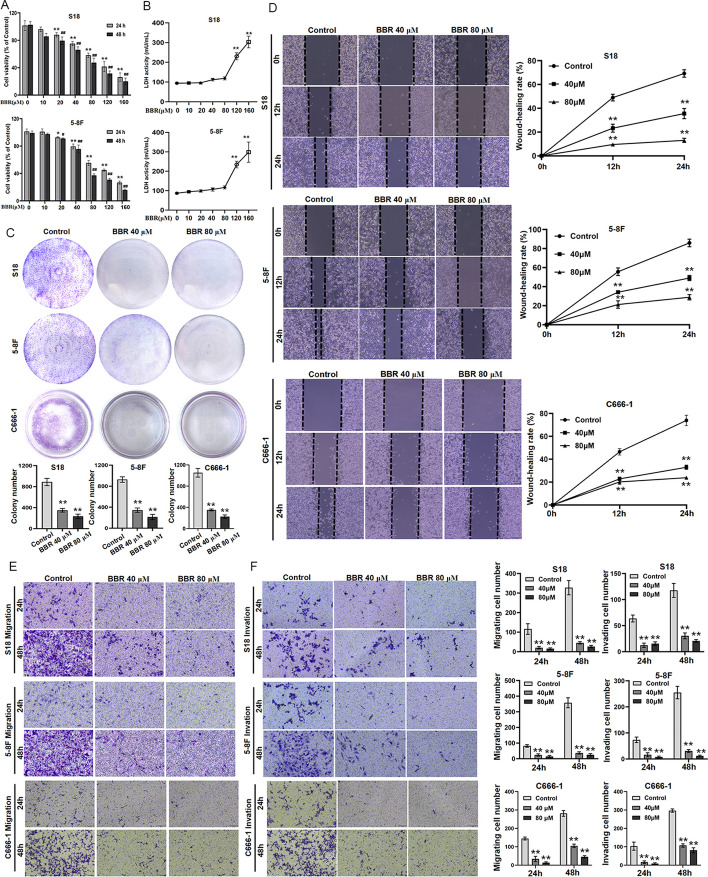
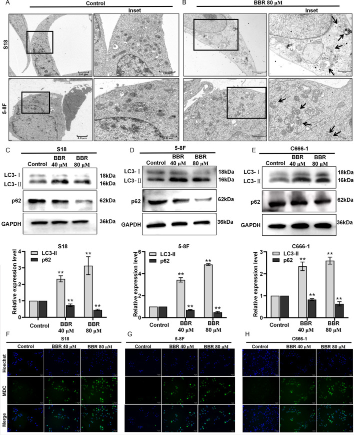
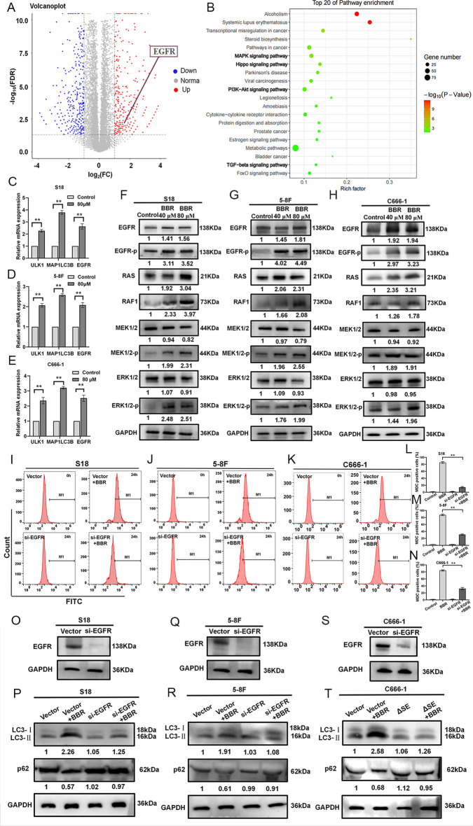
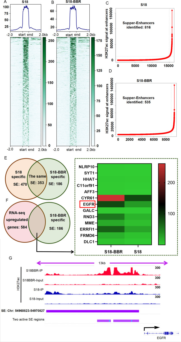
Nasopharyngeal carcinoma (NPC), primarily found in the southern region of China, is a malignant tumor known for its highly metastatic characteristics. The high mortality rates caused by the distant metastasis and disease recurrence remain unsolved clinical problems. In clinic, the berberine (BBR) compound has widely been in NPC therapy to decrease metastasis and disease recurrence, and BBR was documented as a main component with multiple anti-NPC effects. However, the mechanism by which BBR inhibits the growth and metastasis of nasopharyngeal carcinoma remains elusive. Herein, we show that BBR effectively inhibits the growth, metastasis, and invasion of NPC via inducing a specific super enhancer (SE). From a mechanistic perspective, the RNA sequencing (RNA-seq) results suggest that the RAS-RAF1-MEK1/2-ERK1/2 signaling pathway, activated by the epidermal growth factor receptor (EGFR), plays a significant role in BBR-induced autophagy in NPC. Blockading of autophagy markedly attenuated the effect of BBR-mediated NPC cell growth and metastasis inhibition. Notably, BBR increased the expression of EGFR by transcription, and knockout of EGFR significantly inhibited BBR-induced microtubule associated protein 1 light chain 3 (LC3)-II increase and p62 inhibition, proposing that EGFR plays a pivotal role in BBR-induced autophagy in NPC. Chromatin immunoprecipitation sequencing (ChIP-seq) results found that a specific SE existed only in NPC cells treated with BBR. This SE knockdown markedly repressed the expression of EGFR and phosphorylated EGFR (EGFR-p) and reversed the inhibition of BBR on NPC proliferation, metastasis, and invasion. Furthermore, BBR-specific SE may trigger autophagy by enhancing EGFR gene transcription, thereby upregulating the RAS-RAF1-MEK1/2-ERK1/2 signaling pathway. In addition, in vivo BBR effectively inhibited NPC cells growth and metastasis, following an increase LC3 and EGFR and a decrease p62. Collectively, this study identifies a novel BBR-special SE and established a new epigenetic paradigm, by which BBR regulates autophagy, inhibits proliferation, metastasis, and invasion. It provides a rationale for BBR application as the treatment regime in NPC therapy in future.

Keywords: Autophagy; BBR; EGFR; Nasopharyngeal carcinoma; Super-enhancer.


Conflict of interest statement

The authors declare no competing financial interests.

Figures

Fig. 1
Fig. 1
BBR inhibits NPC cell growth and migration. S18 and 5-8F cells were treated with BBR at 10, 20, 40, 80, 120, and 160 μM for 24 or 48 h and subjected to CCK-8 assay for cell viability (A). After BBR treatment for 24 h, LDH was detected with LDH assay (B) (**P < 0.01 versus BBR 80 μM). Cell proliferation was tested with cell colony growth (C). Quantitative analyses of colony numbers are shown (**P < 0.01 versus the control group). A wound-healing assay was used to detect cell migration (D). Representative pictures of S18, 5-8F, and C666-1 cells cultured with the different concentrations of BBR in 12 h or 24 h, and the relative migration values were presented as the means ± standard error of the mean (s.e.m.). The Transwell migration and invasion assays were used to detect the migratory and invasive capabilities of S18, 5-8F, and C666-1 cells treated with 40 and 80 μM, and the number of migrated and invaded cells were presented as the means ± s.e.m. (E, F). All experiments were done more than three times independently and statistically analyzed with one-way analysis of variance (**P < 0.01 versus the control group)
Fig. 2
Fig. 2
BBR induces autophagy in NPC cells. TEM was used to observe autophagy in S18 and 5-8F cells, and the scale bar represents 2 µM and 1 µM (A, B). The expressions of LC3 and p62 protein in S18, 5-8F, and C666-1 cells treated with BBR were examined by western blotting (CE). MDC-positive staining cells were observed by inverted fluorescence microscope (FH). All experiments were done more than three times independently and statistically analyzed with one-way analysis of variance (**P < 0.01 versus the control group)
Fig. 3
Fig. 3
The decreased-autophagy attenuates BBR-induced inhibition on the proliferation and metastasis of NPC cells. S18, 5-8F, and C666-1 cells were treated with BBR with or without CQ, 3-MA for 24 h, and the inhibition of growth was assayed (A, B) (** P < 0.01, versus the control group). A wound-healing assay was used to detect cell migration. Representative pictures of S18, 5-8F, and C666-1 cells were treated with BBR with or without CQ or 3-MA for 24 h are shown, and the relative migration values were presented as the means ± s.e.m. (CE). The transwell invasion assays were used to detect the invasive capabilities of NPC cells. The S18, 5-8F, and C666-1 cells were treated with BBR with or without 3-MA for 24 h, and the number of invaded cells were presented as the means ± s.e.m. (F). The MDC-positive staining cells were observed by inverted fluorescence microscope (GI). The expressions of LC3 and p62 protein in S18, 5-8F, and C666-1 cells treated with BBR with or without 3-MA for 24 h was examined by western blotting (JL). All experiments were done more than three times independently and statistically analyzed with one-way analysis of variance (**P < 0.01 versus the control group)
Fig. 4
Fig. 4
BBR induces autophagy by mediating EGFR and downstream signaling pathway activation. A the volcano plots of RNA-seq data (S18-BBR versus S18), FC represents fold change, and FDR represents false discovery rate. B Scatter plot of KEGG enrichment of differentially expressed genes in S18 and S18-BBR cells, rich factor represents the number of differential genes located in the KEGG/the total number of genes located in the KEGG. The expression of ULK1, microtubule-associated protein 1 light chain 3 beta (MAP1LC3B), and EGFR mRNA in S18, 5-8F, and C666-1 cells treated with BBR was examined by qRT–PCR. (n = 3–5 independent experiments) (CE). Data are expressed as mean ± s.e.m.; **P < 0.01. The expressions of EGFR, EGFR-p, RAS, RAF1, MEK1/2, MEK1/2-p, ERK1/2, and ERK1/2-p proteins in S18, 5-8F, and C666-1 cells treated with BBR were examined by western blotting (FH). The MDC-positive staining cells level in S18, 5-8F, and C666-1 cells treated with BBR were analyzed by a flow cytometer (IN). The expressions of LC3, p62 protein in S18, 5-8F, and C666-1 cells with knockout of EGFR or treated with BBR were examined by western blotting (OT)
Fig. 5
Fig. 5
A, B EGFR is driven by BBR-specific SE. H3K27ac signal in SE regions S18 cells with (A) or without BBR (B). C, D The numbers of identified SEs and the H3K27ac signal at enhancers in S18 (C) and S18-BBR cells (D). E the numbers of identical and distinct SEs in S18 and S18-BBR cells were analyzed. F Heatmap showed the normalized expression of significantly upregulated genes between S18 and S18-BBR cells. G the locations of the S18-BBR specific SE, and the active SE regions were visualized by IGV
Fig. 6
Fig. 6
Deletion of BBR-specific SE reverses BBR-induced NPC proliferation and metastasis in vitro. The SE upstream of EGFR promoter was deleted using CRISPR interference, the nested PCR was used to verify the knockout efficiency (A, B). EGFR mRNA and protein expression in ΔSE S18 cells and vector cells (C, D). ΔSE S18 cells treated with BBR binding SE deletion (n = 3–5 independent experiments). Data are expressed as mean ± s.e.m.; **P < 0.01. Cell viability in ΔSE S18 and 5-8F cells treated with BBR was detected by CCK-8 assay (E, F), (**P < 0.01 versus vector–BBR group). Transwell migration and invasion assay were used to detect the migratory and invasive capabilities of ΔSE S18 and 5-8F cells treated with BBR, and the number of migrated and invaded cells were presented as the means ± s.e.m. (GJ) (**P < 0.01 versus vector–BBR group)
Fig. 7
Fig. 7
BBR inhibits NPC growth and metastasis in vivo. A The S18 cells were injected into the tail vein of nude mice (n = 5). The mice were subjected to BBR (15 mg/kg/day) by intraperitoneal injection for 30 day (n = 5). The luminescence intensity of lung metastases was analyzed in vivo using an in vivo small animal imaging system (**P < 0.01 versus the control group). B Hematoxylin–eosin (HE) staining was used to detect the percent of tumor metastases per lung. The expression of LC3, p62, and EGFR in NPC lung metastatic tissues and NPC primary tissues were measured by IHC staining. C, D Gross image of subcutaneous tumors and tumor weight in control and BBR group (**P < 0.01 versus the control group). Tumor volume and body weight curve was measured every 5 days, (**P < 0.01 versus day 0) (E, F). G HE staining of the implanted tumors. The expressions of LC3, p62, and EGFR in NPC subcutaneous tumors were measured by IHC staining (**P < 0.01 versus the control group)
Fig. 8
Fig. 8
Schematic diagram of berberine-actuated SE regulating EGFR to induce NPC autophagy

Similar articles

Cited by

References

    1. Wong KCW, Hui EP, Lo KW, Lam WKJ, Johnson D, Li L, et al. Nasopharyngeal carcinoma: an evolving paradigm. Nat Rev Clin Oncol. 2021;18(11):679–695. doi: 10.1038/s41571-021-00524-x. - DOI - PubMed
    1. Guan S, Wei J, Huang L, Wu L. Chemotherapy and chemo-resistance in nasopharyngeal carcinoma. Eur J Med Chem. 2020;207:112758. doi: 10.1016/j.ejmech.2020.112758. - DOI - PubMed
    1. Gong L, Kwong DL, Dai W, Wu P, Wang Y, Lee AW, et al. The stromal and immune landscape of nasopharyngeal carcinoma and its implications for precision medicine targeting the tumor microenvironment. Front Oncol. 2021;11:744889. doi: 10.3389/fonc.2021.744889. - DOI - PMC - PubMed
    1. Jiang J, Ying H. Revealing the crosstalk between nasopharyngeal carcinoma and immune cells in the tumor microenvironment. J Exp Clin Cancer Res. 2022;41(1):244. doi: 10.1186/s13046-022-02457-4. - DOI - PMC - PubMed
    1. Tan R, Phua SKA, Soong YL, Oon LLE, Chan KS, Lucky SS, et al. Clinical utility of Epstein-Barr virus DNA and other liquid biopsy markers in nasopharyngeal carcinoma. Cancer Commun (Lond) 2020;40(11):564–585. doi: 10.1002/cac2.12100. - DOI - PMC - PubMed

MeSH terms