. 2024 Jun 28;24(1):80.
doi: 10.1186/s40644-024-00726-3.

Identification of T2W hypointense ring as a novel noninvasive indicator for glioma grade and IDH genotype

Affiliations

Identification of T2W hypointense ring as a novel noninvasive indicator for glioma grade and IDH genotype

Yawen Lu et al. Cancer Imaging. .

Abstract

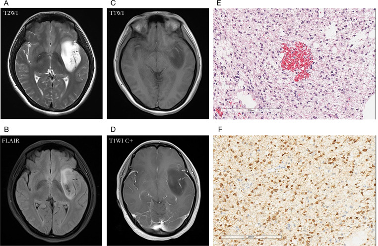
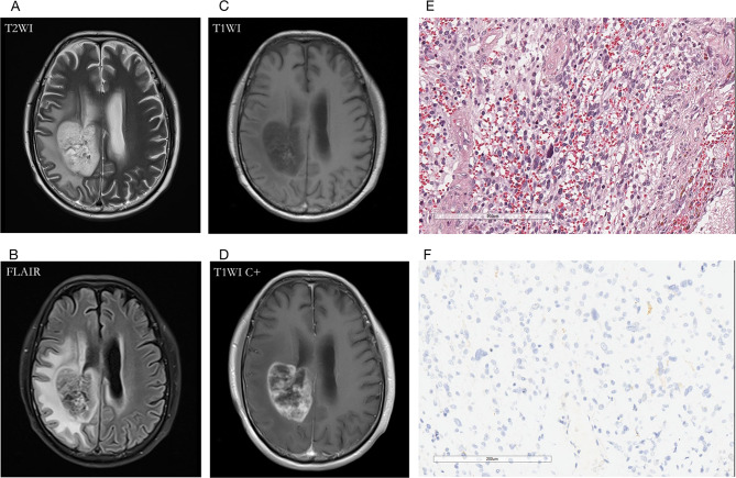
Background: This study aimed to evaluate the T2W hypointense ring and T2-FLAIR mismatch signs in gliomas and use these signs to construct prediction models for glioma grading and isocitrate dehydrogenase (IDH) mutation status.

Methods: Two independent radiologists retrospectively evaluated 207 glioma patients to assess the presence of T2W hypointense ring and T2-FLAIR mismatch signs. The inter-rater reliability was calculated using the Cohen's kappa statistic. Two logistic regression models were constructed to differentiate glioma grade and predict IDH genotype noninvasively, respectively. Receiver operating characteristic (ROC) analysis was used to evaluate the developed models.

Results: Of the 207 patients enrolled (119 males and 88 females, mean age 51.6 ± 14.8 years), 45 cases were low-grade gliomas (LGGs), 162 were high-grade gliomas (HGGs), 55 patients had IDH mutations, and 116 were IDH wild-type. The number of T2W hypointense ring signs was higher in HGGs compared to LGGs (p < 0.001) and higher in the IDH wild-type group than in the IDH mutant group (p < 0.001). There were also significant differences in T2-FLAIR mismatch signs between HGGs and LGGs, as well as between IDH mutant and wild-type groups (p < 0.001). Two predictive models incorporating T2W hypointense ring, absence of T2-FLAIR mismatch, and age were constructed. The area under the ROC curve (AUROC) was 0.940 for predicting HGGs (95% CI = 0.907-0.972) and 0.830 for differentiating IDH wild-type (95% CI = 0.757-0.904).

Conclusions: The combination of T2W hypointense ring, absence of T2-FLAIR mismatch, and age demonstrate good predictive capability for HGGs and IDH wild-type. These findings suggest that MRI can be used noninvasively to predict glioma grading and IDH mutation status, which may have important implications for patient management and treatment planning.

Keywords: Glioma; Isocitrate dehydrogenase; Magnetic resonance imaging; T2-FLAIR mismatch; T2W hypointense ring.


Conflict of interest statement

The authors declare that they have no competing interests.

Figures

Fig. 1
Fig. 1
Flow diagram of the patient selection process
Fig. 2
Fig. 2
T2W hypointense ring sign in three cases (white arrows). From left to right are T2WI, T1WI, and contrast-enhanced T1WI, respectively. Images (A-C) showed a patient (male, 51 years) with glioblastoma (IDHwt) in the left temporal lobe, no obvious edema around the tumor parenchyma on T2W, and no enhancement on the contrast-enhanced T1W image. (D-F) showed a patient (male, 59 years) with glioblastoma (IDHwt) in the right temporoparietal lobe. The T2W hypointense ring on T2WI was not completely consistent with the enhanced morphology, as indicated by the medial red arrow, where the T2W hypointense ring broke, which might suggest glioma invasion (red arrows). (G-I) A patient (male, 35 years) with an oligodendroglioma, IDHmut, showed a clear low signal arc on T2WI, and the tumor parenchyma was mildly enhanced
Fig. 3
Fig. 3
A 60-year-old male patient with oligodendroglioma (WHO Grade III, IDHmut) in the right temporal lobe. (A, B) axial T2WI showed T2W hyperintense lesions in tumor parenchyma, absence of T2W hypointense ring, and T2-FLAIR mismatch signs; (C, D) contrast-enhanced T1W image showed no obvious enhancement; (E) HE staining showed moderate to a severe increase in cell density, accompanied by the atypical nucleus, and perinuclear halo (×200); (F) IDH1 mutation positive expression
Fig. 4
Fig. 4
A 64-year-old female with glioblastoma (WHO Grade IV, IDHwt) in the left temporal lobe. (A) T2WI showed a hyperintense tumor, and the internal signal is inhomogeneous. The hypointense ring was located between the tumor parenchyma and edema as the red arrow indicated; (B) T2-FLAIR mismatch sign was absent; (C, D) Contrast-enhanced T1W image showed garland-like contrast enhancement; (E) HE staining showed dense cells, obvious atypia, and vascular endothelial hyperplasia (×200); (F) IDH1 mutation negative expression (×200)
Fig. 5
Fig. 5
A 30-year-old female with diffuse astrocytoma (WHO Grade II, IDHmut) in the left temporal lobe. (A) A hyperintense tumor showed in T2W MRI, without hypointense ring sign; (B) T2-FLAIR mismatch sign was positive; (C, D) Contrast-enhanced T1W image showed no obvious enhancement; (E) HE staining showed moderate increase in cell density with atypical nuclei (×200); (F) IDH1 mutation positive expression (×200)
Fig. 6
Fig. 6
A 62-year-old male with glioblastoma (WHO Grade IV, IDH-wt) in the right temporoparietal lobe. (A) The T2WI demonstrated a hypointense ring between the tumor parenchyma and edema; (B) T2-FLAIR mismatch sign was negative for the necrotic cavity; (C, D) Contrast-enhanced T1W image showed garland-like contrast enhancement; (E) HE staining with moderate to high cell proliferation (×200); (F) IDH1 mutation negative expression (×200)
Fig. 7
Fig. 7
The ROC curves using age、no-T2-FLAIR mismatch、hypointense ring, and union of these factors to predict HGGs (A) and IDHwt (B)

References

    1. Davis ME. Epidemiology and overview of Gliomas. Semin Oncol Nurs. 2018;34:420–9. doi: 10.1016/j.soncn.2018.10.001. - DOI - PubMed
    1. Bondy ML, Scheurer ME, Malmer B, Barnholtz-Sloan JS, Davis FG, Il’yasova D, et al. Brain tumor epidemiology: consensus from the Brain Tumor Epidemiology Consortium. Cancer. 2008;113:1953–68. doi: 10.1002/cncr.23741. - DOI - PMC - PubMed
    1. Ostrom QT, Cioffi G, Waite K, Kruchko C, Barnholtz-Sloan JS. CBTRUS Statistical Report: primary brain and other Central Nervous System tumors diagnosed in the United States in 2014–2018. Neuro Oncol. 2021;23:iii1–105. doi: 10.1093/neuonc/noab200. - DOI - PMC - PubMed
    1. Louis DN, Perry A, Reifenberger G, von Deimling A, Figarella-Branger D, Cavenee WK, et al. The 2016 World Health Organization Classification of Tumors of the Central Nervous System: a summary. Acta Neuropathol. 2016;131:803–20. doi: 10.1007/s00401-016-1545-1. - DOI - PubMed
    1. Vigneswaran K, Neill S, Hadjipanayis CG. Beyond the World Health Organization grading of infiltrating gliomas: advances in the molecular genetics of glioma classification. Ann Transl Med. 2015;3:95. - PMC - PubMed

Substances