. 2024 Jun 28;12(1):109.
doi: 10.1186/s40478-024-01810-2.

Retinal peri-arteriolar versus peri-venular amyloidosis, hippocampal atrophy, and cognitive impairment: exploratory trial

Affiliations

Retinal peri-arteriolar versus peri-venular amyloidosis, hippocampal atrophy, and cognitive impairment: exploratory trial

Oana M Dumitrascu et al. Acta Neuropathol Commun. .

Abstract

The relationship between amyloidosis and vasculature in cognitive impairment and Alzheimer's disease (AD) pathogenesis is increasingly acknowledged. We conducted a quantitative and topographic assessment of retinal perivascular amyloid plaque (AP) distribution in individuals with both normal and impaired cognition. Using a retrospective dataset of scanning laser ophthalmoscopy fluorescence images from twenty-eight subjects with varying cognitive states, we developed a novel image processing method to examine retinal peri-arteriolar and peri-venular curcumin-positive AP burden. We further correlated retinal perivascular amyloidosis with neuroimaging measures and neurocognitive scores. Our study unveiled that peri-arteriolar AP counts surpassed peri-venular counts throughout the entire cohort (P < 0.0001), irrespective of the primary, secondary, or tertiary vascular branch location, with a notable increase among cognitively impaired individuals. Moreover, secondary branch peri-venular AP count was elevated in the cognitively impaired (P < 0.01). Significantly, peri-venular AP count, particularly in secondary and tertiary venules, exhibited a strong correlation with clinical dementia rating, Montreal cognitive assessment score, hippocampal volume, and white matter hyperintensity count. In conclusion, our exploratory analysis detected greater peri-arteriolar versus peri-venular amyloidosis and a marked elevation of amyloid deposition in secondary branch peri-venular regions among cognitively impaired subjects. These findings underscore the potential feasibility of retinal perivascular amyloid imaging in predicting cognitive decline and AD progression. Larger longitudinal studies encompassing diverse populations and AD-biomarker confirmation are warranted to delineate the temporal-spatial dynamics of retinal perivascular amyloid deposition in cognitive impairment and the AD continuum.

Keywords: Amyloid imaging; Cognition; Hippocampal volume; Neurodegeneration; Peri-arteriolar; Peri-venular; Perivascular; Retina.


Conflict of interest statement

Koronyo-Hamaoui, Koronyo, Verdooner, and Black are co-founding members of NeuroVision Imaging Inc., 1395 Garden Highway, Suite 250, Sacramento, CA 95833, USA. Johnson and Verdooner are employed by NeuroVision Imaging Inc. NeuroVision’s sole involvement was to provide the processed retinal images. NeuroVision did not fund this study and was not involved in the development of the novel perivascular AP methodology or analysis. All authors declare that the research study was conducted in the absence of any commercial or financial relationships that could be construed as a potential conflict of interest.

Figures
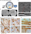
Fig. 1
Fig. 1
Retinal vascular amyloid imaging. A Pipeline of fluorescent imaging using Retia® SLO, Afina™ cloud storage, and a fully automated image processing and analyses. B Representative retinal fluorescent image of before and after image processing. C Representative retinal image of an AD patient demonstrating putative retinal amyloid deposits (white) along the blood vessels. D Representative histopathological images from a confirmed AD patient, that did not undergo retinal imaging pre-mortem. Postmortem retinal flatmount immunolabeled with anti-c monoclonal antibody (12F4; brown) and peroxidase-base 3,3′ diaminobenzidine (DAB) immunostaining. Typical Aβ plaque structures and Aβ deposition along and inside blood vessels (red arrows) and ‘plaque-free’ regions of retinal blood vessels (blue arrows). E High magnification images of perivascular and vascular Aβ deposits in retinal flatmounts of an AD patient that did not undergo retinal imaging pre-mortem
Fig. 2
Fig. 2
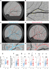
Retinal peri-venular and peri-arteriolar amyloid plaque distribution. AB Representative retinal fluorescent fundus image illustrating retinal curcumin-positive amyloid hyperfluorescent plaques in the left eye supero-temporal quadrant (A, magnification B). Illustration of the primary, secondary, and tertiary retinal venular (C) and arteriolar (D) branches. Magnifications of the peri-venular (C’) and peri-arteriolar (D’) area used for the amyloid plaque (AP) quantification; boundary zone delineated by dotted lines producing a perivascular area of one equivalent vessel diameter on either side. E Quantitative analysis of retinal perivascular area stratified by venules (V) and arterioles (A) showing no significant difference between the total area for each vessel type in the analyzed supero-temporal region. FJ Quantitative analyses of retinal perivascular AP count stratified by V versus A in the total branches (F), in females (G), males (H), individuals with normal cognition (I) and impaired cognition (J). Individual data points are shown. * P < 0.05, ** P < 0.01, *** P < 0.001, **** P < 0.0001 by paired two-tailed Student’s t test. NC, Normal cognition; IC, Impaired cognition; 1°V, primary venular branch; 2°V, secondary venular branch; 3°V tertiary venular branch; 1°A, primary arteriolar branch, 2°A, secondary arteriolar branch; 3°A, tertiary arteriolar branches. Color code vessel type: blue—peri-venular; red—peri-arteriolar
Fig. 3
Fig. 3
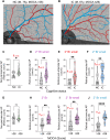
Retinal peri-arteriolar and peri-venular amyloid plaque count stratified by cognitive status and MOCA scores. AB Representative fundus images of retinal perivascular amyloid plaque (AP) showing their density and distribution along arterioles (red tracing) and venules (blue tracing), in individuals with normal cognition (NC; A) or impaired cognition (IC; B). C Quantitative analyses of retinal non-perivascular AP count stratified by cognitive status, NC versus IC. DF Quantitative analyses of retinal AP count stratified by cognitive status, in perivascular (D), peri-arteriolar (E), and peri-venular (F), for the secondary (2°) small branches. G Quantitative analyses of retinal non-perivascular AP count stratified by MOCA scores of 26 or lower compared with greater than 26. HJ Quantitative analyses of AP count stratified by MOCA, in perivascular 2° branches (H), 2° small branches (I), and peri-venular 2° small branches (J). Violin plots are showing individual data points, median and interquartile range. Statistics: *P < 0.05, ** P < 0.01, **** P < 0.0001, by unpaired two-tailed Student’s t test or Mann–Whitney. M, male; MOCA, Montreal Cognitive Assessment; y, years. Color code for vessel type: purple—perivascular; blue—peri-venular; red—peri-arteriolar
Fig. 4
Fig. 4
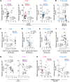
Correlations between retinal perivascular amyloid plaque distribution with cognitive and neuroimaging measures. Pearson’s r correlation analyses between retinal perivascular AP count and CDR (A), MOCA (B), RCFT-copy registration (C), Trail A-paper and pencil (D), ACS-TOFF (E), CVLT-II Long delay (F), hippocampal volume (G) and white matter hyperintensities lesions (H). AP, Amyloid plaques; CDR, Clinical dementia rating; MOCA, Montreal cognitive assessment; RCFT, Rey Complex Figure Test and Recognition Trial; ACS-TOPF, test of Premorbid Functioning; CVLT, California Verbal Learning Test; HV, hippocampal volume, WMH, White matter hypertensity; 2° Br, secondary branches; 3° Br, tertiary branches. Color code for vessel type: purple—perivascular; blue—peri-venular; red—peri-arteriolar

Update of

References

    1. Nation DA, et al. Blood-brain barrier breakdown is an early biomarker of human cognitive dysfunction. Nat Med. 2019;25(2):270–276. doi: 10.1038/s41591-018-0297-y. - DOI - PMC - PubMed
    1. Li M, et al. Increase of blood-brain barrier leakage is related to cognitive decline in vascular mild cognitive impairment. BMC Neurol. 2021;21(1):159. doi: 10.1186/s12883-021-02189-6. - DOI - PMC - PubMed
    1. Nikolakopoulou AM, et al. Pericyte loss leads to circulatory failure and pleiotrophin depletion causing neuron loss. Nat Neurosci. 2019;22(7):1089–1098. doi: 10.1038/s41593-019-0434-z. - DOI - PMC - PubMed
    1. Riphagen JM, et al. Linking APOE-epsilon4, blood-brain barrier dysfunction, and inflammation to Alzheimer's pathology. Neurobiol Aging. 2020;85:96–103. doi: 10.1016/j.neurobiolaging.2019.09.020. - DOI - PubMed
    1. Montagne A, et al. APOE4 leads to blood-brain barrier dysfunction predicting cognitive decline. Nature. 2020;581(7806):71–76. doi: 10.1038/s41586-020-2247-3. - DOI - PMC - PubMed

Publication types

MeSH terms