
Abstract

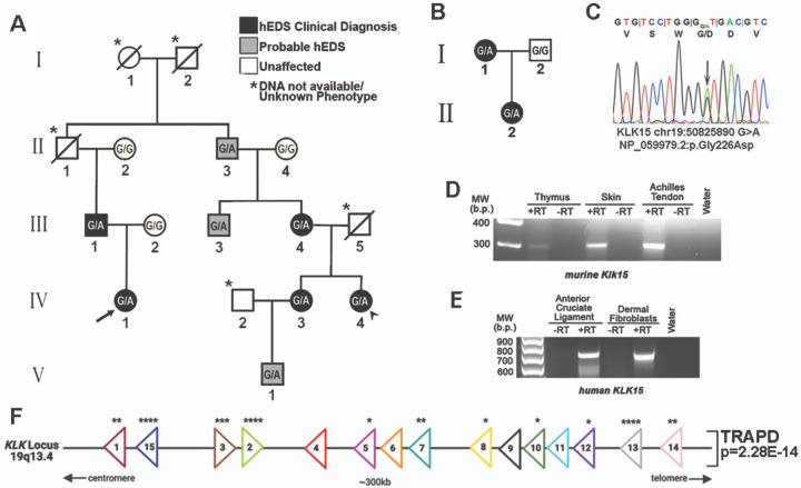
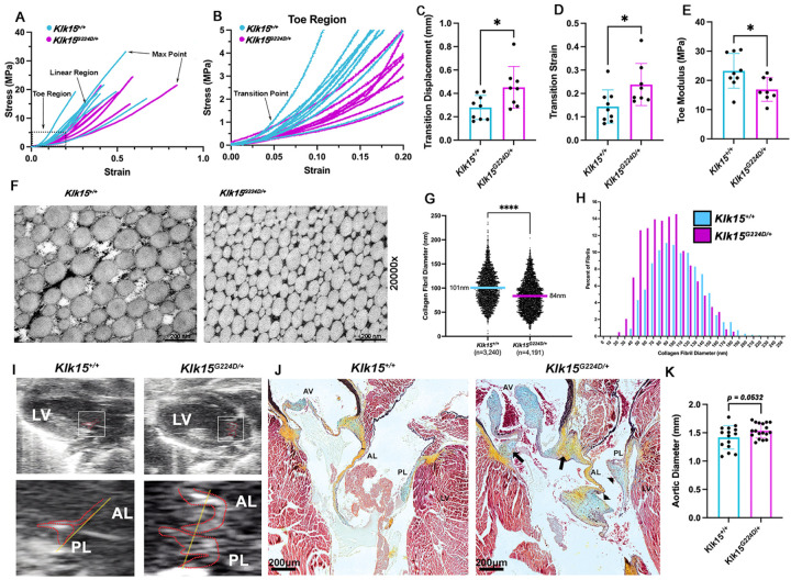
Hypermobile Ehlers-Danlos syndrome (hEDS) is a common heritable connective tissue disorder that lacks a known genetic etiology. To identify genetic contributions to hEDS, whole exome sequencing was performed on families and a cohort of sporadic hEDS patients. A missense variant in Kallikrein-15 (KLK15 p. Gly226Asp), segregated with disease in two families and genetic burden analyses of 197 sporadic hEDS patients revealed enrichment of variants within the Kallikrein gene family. To validate pathogenicity, the variant identified in familial studies was used to generate knock-in mice. Consistent with our clinical cohort, Klk15 G224D/+ mice displayed structural and functional connective tissue defects within multiple organ systems. These findings support Kallikrein gene variants in the pathogenesis of hEDS and represent an important step towards earlier diagnosis and better clinical outcomes.

Keywords: Connective tissues; Hypermobile Ehlers Danlos Syndrome; hEDS.


Figures

Figure 1
Figure 1. Kallikrein gene variant identification and enrichment in hEDS
(A) Pedigree of a multigenerational family with autosomal dominant hEDS. Black circles and squares represent those with a clinical hEDS diagnosis. Grey indicates those who are probable-hEDS. Unaffected individuals are black and unknown phenotypes and those whose DNA was not available are marked with an asterisk. Circles and squares represent females and males, respectively. WES was performed on the proband (IV-1) and IV-4 (arrow and arrowhead). (B) Pedigree of Family 2 showing genotype of KLK15G226D/+ allele (G/A) in affected I-1 and II-1. (C) Chromatogram showing missense KLK15 variant (G/A). (D, E) RT-PCR for Klk15 mRNA in a subset of relevant murine and human connective tissues. RT(+/−) is presence or absence (negative control) of reverse transcriptase enzyme (F) KLK locus at 19q13.33 shows the proximity and directionality of individual KLK genes. Asterisks for each gene represent statistical enrichment in hEDS patients for KLK variants (*<0.05, **<0.01, *** <0.001 ****<0.0001), and the p-value of the entire locus (P=2.28E-14).
Figure 2
Figure 2. Pathogenicity of the familial KLK15 variant.
(A) Stress-Strain curves from tensile testing of Klk15+/+ (n=9) and Klk15G224D/+ (n=8) tendons with a focus on the toe region (B). (C-E) Statistical analysis of transition point displacement, transition strain and toe modulus from tensile testing curves. *<0.05. (F) Representative TEMs of Achilles tendon from Klk15+/+ and Klk15G224D/+ mice showing smaller collagen fibrils in the mutant tendons. (G) Quantification of fibril diameters from wildtype (n=3,240) and mutant (n=4,191) tendons showing a significant decrease in average fibril diameter in Klk15G224D/+ (84nm vs. 101nm). (H) Distribution of fibril diameters in 10 nm increments showing a leftward shift in diameter. ****p<0.0001. (I) Echocardiography of 4-month-old wildtype (Klk15+/+;N=5) and mutant mice (Klk15G224D/+; N=6) showing mitral valve prolapse in 5/6 Klk15G224D/+ mice above the level of the annulus (yellow line) (J) Movats pentachrome stains revealed myxomatous mitral (arrow heads) and aortic leaflets (AV). Areas of chondrodysplasia and proteoglycan accumulation are evident in the hinge region of the aortic valve (arrows). Red= myocytes, blue= proteoglycans, black=elastin. (K) Diameter measurements of Klk15+/+ (N=15) and Klk15G224D/+ (N=18) aorta’s show a trend toward sign cant enlargement in mutant mice (p=0.0532)

References

    1. Blackburn P.R. et al. Bi-allelic Alterations in AEBP1 Lead to Defective Collagen Assembly and Connective Tissue Structure Resulting in a Variant of Ehlers-Danlos Syndrome. Am J Hum Genet 102, 696–705 (2018). - PMC - PubMed
    1. Gensemer C. et al. Hypermobile Ehlers-Danlos syndromes: Complex phenotypes, challenging diagnoses, and poorly understood causes. Dev Dyn (2020). - PMC - PubMed
    1. Petrucci T, B.J., Gensemer C, Morningstar J, Daylor V, Byerly K, Bistran E, Griggs M, Elliott JM, Kalechi T, Phillips S, Nichols M, Shapiro S, Patel S, Bouatia-Naji N, Norris RA Phenotypic Clusters and Multimorbidity in Hypermobile Ehlers-Danlos Syndrome. Mayo Clinic Proceedings: Innovations, Quality & Outcomes 8, 253–262 (2024). - PMC - PubMed
    1. Demmler J.C. et al. Diagnosed prevalence of Ehlers-Danlos syndrome and hypermobility spectrum disorder in Wales, UK: a national electronic cohort study and case-control comparison. BMJ Open 9, e031365 (2019). - PMC - PubMed
    1. Guo M.H., Plummer L., Chan Y.M., Hirschhorn J.N. & Lippincott M.F. Burden Testing of Rare Variants Identified through Exome Sequencing via Publicly Available Control Data. Am J Hum Genet 103, 522–534 (2018). - PMC - PubMed

Publication types

LinkOut - more resources