[Preprint]. 2024 Jun 16:2024.06.15.24308845.
doi: 10.1101/2024.06.15.24308845.

NLRP3 inflammasome activation and altered mitophagy are key pathways in inclusion body myositis

Affiliations

NLRP3 inflammasome activation and altered mitophagy are key pathways in inclusion body myositis

Elie Naddaf et al. medRxiv. .

Update in

Abstract

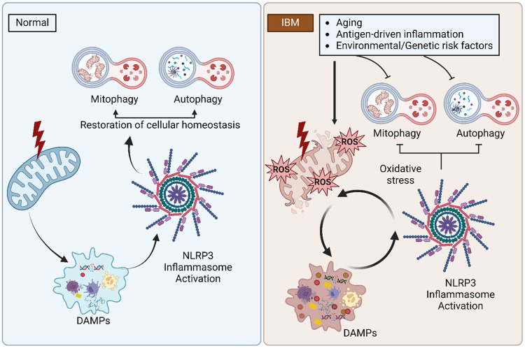
Background: Inclusion body myositis (IBM) is the most prevalent muscle disease in adults for which no current treatment exists. The pathogenesis of IBM remains poorly defined. Inflammation and mitochondrial dysfunction are the most common histopathological findings. In this study, we aimed to explore the interplay between inflammation and mitochondrial dysfunction in IBM patients, highlighting sex differences.

Methods: We included 38 IBM patients and 22 age- and sex-matched controls without myopathy. Bulk RNA sequencing, Meso Scale Discovery ELISA, western blotting, histochemistry and immunohistochemistry were performed on frozen muscle samples from the study participants.

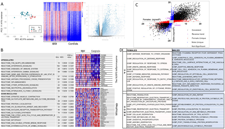
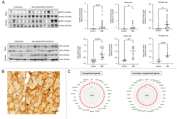
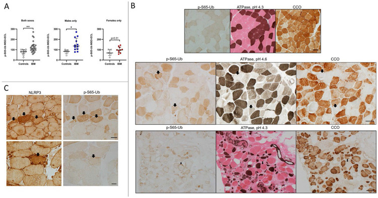
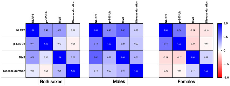
Results: We demonstrated activation of the NLRP3 inflammasome in IBM muscle samples, with the NLRP3 inflammasome pathway being the most upregulated. On muscle histopathology, there is increased NRLP3 immunoreactivity in both inflammatory cells and muscle fibers. Mitophagy is critical for removing damaged mitochondria and preventing the formation of a vicious cycle of mitochondrial dysfunction-NLRP3 activation. In the IBM muscle samples, we showed altered mitophagy, most significantly in males, with elevated levels of p-S65-Ubiquitin, a mitophagy marker. Furthermore, p-S65-Ubiquitin aggregates accumulated in muscle fibers that were mostly type 2 and devoid of cytochrome-c-oxidase reactivity. Type 2 muscle fibers are known to be more prone to mitochondrial dysfunction. NLRP3 RNA levels correlated with p-S65-Ubiquitin levels in both sexes but with loss of in muscle strength only in males. Finally, we identified sex-specific molecular pathways in IBM, with females having activation of pathways that could offset some of the pathomechanisms of IBM.

Conclusions: NLRP3 inflammasome is activated in IBM, along with altered mitophagy particularly in males, which is of potential therapeutic significance. These findings suggest sex-specific mechanisms in IBM that warrant further investigation.


Conflict of interest statement

Competing interests: Mayo Clinic, F.C.F. and W.S. have filed a patent related to PRKN mitophagy activators. All other authors report no conflicts of interest.

Figures

Figure 1:
Figure 1:. IBM transcriptome
Transcriptomic analysis of muscle tissue from 38 patients with inclusion body myositis and 22 controls. A) PCA (to the left) and heat map (middle) showing clear separation between IBM patients and controls. Volcano plot (to the right) representing differentially expressed genes (blue). B) Top 10 upregulated and top 10 downregulated pathways in IBM. The NLRP3 inflammasome and mitophagy pathways are detailed in the heatmaps with downregulated genes shown in blue and upregulated genes shown in red. Transcriptomic sex differences are shown in C&D. C) Volcano plot showing the signed −log10(p value) for each gene in females on the x axis and in males on y axis. Differentially expressed genes that are consistent in both sexes are shown in black, female-unique genes are shown in red, male-unique genes are shown in blue, and genes with reverse trends are shown in purple. Genes that did not reach fold change or p-value cutoffs in either sex are shown in gray (not significant). D) The top 10 upregulated and downregulated pathways for sex-specific genes.
Figure 2:
Figure 2:. NLRP3 inflammasome activation in inclusion body myositis.
A) Western blot of muscle lysates from IBM patients and controls showing increased NLRP3 and ASC protein expression in both males and females. Western blot quantification results comparing IBM group to controls were as follows (Mann-Whitney U, two-tailed p-value): NLRP3 for both sexes (20, p = 0.0009), NLRP3 for males (6, p = 0.045), NLRP3 for females (3, p = 0.011), ASC for both sexes (4, p < 0.0001), ASC for males (3, p = 0.0047), and ASC for females (0, p = 0.0007). B) NLRP3 immunohistochemistry showing increased NLRP3 immunoreactivity in scattered muscle fibers (examples shown as white arrows) as well as in inflammatory cells (examples shown as black arrowheads). C) Radar maps displaying the top coexpressed and inversely coexpressed genes with NLRP3 using Spearman rank correlation analysis. The correlation coefficient corresponds to the distance from the center. All represented genes had a p-value less than 0.0001.
Figure 3:
Figure 3:. Altered mitophagy in inclusion body myositis patients
A) Measurement of p-S65-Ub levels via sandwich ELISA in muscle samples from IBM patients and controls. Results comparing IBM samples to controls were as follows (Mann-Whitney U, two-tailed p-value): both sexes (43, p = 0.0054), males only (7, p = 0.0136), and females only (16, p = 0.31). MSD-ECL: Meso Scale Discovery- Electrochemiluminescence. B&C) Frozen muscle sections from IBM samples and controls reacted to p-S65-Ub, NLRP3, ATPase at pH 4.3 (type 1 fibers: dark brown, type 2 fibers: pink) and at pH 4.6 (type 1 fibers: dark brown; type 2a fibers: light brown; type 2b fibers: in between shade), and cytochrome c oxidase. MSD: Mesoscale discovery; B) Top row: muscle samples from controls, p-S65-Ub shows clear differentiation of fiber types, reflecting the difference in baseline mitophagy and mitochondrial content between fiber types. Type 1 fibers, dark brown on ATPase 4.3, had increased p-S65-Ub and CCO reactivity. Middle and bottom rows: muscle samples from a male and a female IBM patients respectively, demonstrating p-S65-Ub positive aggregates in scattered muscle fibers (examples shown as arrows). These fibers were almost exclusively type 2 fibers (ATPase stains) that were also devoid of CCO activity (CCO negative fibers). Also noted, all CCO negative fibers, with or without observable abnormal p-S65-Ub reactivity, were type 2 fibers. Scale bar for panel B: top row: 50 μm; middle row: 50 μm; bottom row: 200 μm. C) Muscle samples from IBM patients, reacted for NLRP3 and p-S65-Ub, demonstrating muscle fibers with increased NLRP3 and p-S65-Ub reactivity (arrows). Scale bar: top row 50 μm, bottom row 20 μm
Figure 4:
Figure 4:. Correlation of NLRP3 inflammasome activation with altered mitophagy and muscle weakness.
Pearson correlation plot demonstrating correlation coefficient values for the NLRP3 RNA level, p-S65-Ub level, manual muscle testing (MMT) score and disease duration, by sex. Both sexes: NLRP3 and p-S65-Ub, p = 0.059; NLRP3 and MMT score, p = 0.072; NLRP3 and disease duration, p = 0.543; p-S65-Ub and MMT score, p = 0.58; p-S65-Ub and disease duration, p = 0.699. Males: NLRP3 and p-S65-Ub, p = 0.116; NLRP3 and MMT score, p = 0.032; NLRP3 and disease duration, p = 0.699; p-S65-Ub and MMT score, p = 0.359; p-S65-Ub and disease duration, p = 0.49. Females: NLRP3 and p-S65-Ub, p = 0.106; NLRP3 and MMT score, p = 0.706; NLRP3 and disease duration, p-=0.781); p-S65-Ub and MMT score, p = 0.642; p-S65-Ub and disease duration, p = 0.898.
Figure 5:
Figure 5:. The vicious cycle of inflammasome activation-mitochondrial dysfunction/altered mitophagy in inclusion body myositis.
The release of mitochondrial damage-associated molecular patterns (DAMPs) results in the activation of the NLRP3 inflammasome. Under normal conditions, damaged mitochondria and the NRLP3 inflammasome are both subsequently removed by mitophagy and autophagy, reestablishing cellular homeostasis. In IBM, mitophagy and autophagy are altered, establishing a feedforward loop in which the inflammatory milieu results in additional oxidative stress and mitochondrial dysfunction with further release of mitochondrial DAMPs and subsequent aberrant NLRP3 inflammasome activation.

References

    1. Weihl CC. Sporadic Inclusion Body Myositis and Other Rimmed Vacuolar Myopathies. Continuum (Minneapolis, Minn) 2019;25:1586–1598. - PubMed
    1. Naddaf E. Inclusion body myositis: Update on the diagnostic and therapeutic landscape. Front Neurol 2022;13:1020113. - PMC - PubMed
    1. Shelly S, Mielke MM, Mandrekar J, et al. Epidemiology and Natural History of Inclusion Body Myositis: A 40-Year Population-Based Study. Neurology 2021. - PMC - PubMed
    1. Naddaf E, Shelly S, Mandrekar J, et al. Survival and associated comorbidities in inclusion body myositis. Rheumatology (Oxford, England) 2022;61:2016–2024. - PMC - PubMed
    1. Engel AG, Arahata K. Monoclonal antibody analysis of mononuclear cells in myopathies. II: Phenotypes of autoinvasive cells in polymyositis and inclusion body myositis. Ann Neurol 1984;16:209–215. - PubMed

Publication types

LinkOut - more resources