Case Reports
. 2024 Jun 28;2(7):luae107.
doi: 10.1210/jcemcr/luae107. eCollection 2024 Jul.

Inferior Laryngeal Nerve Paraganglioma With Norepinephrine Hypersecretion Diagnosed Shortly After Pregnancy

Affiliations
Case Reports

Inferior Laryngeal Nerve Paraganglioma With Norepinephrine Hypersecretion Diagnosed Shortly After Pregnancy

David Kishlyansky et al. JCEM Case Rep. .

Abstract

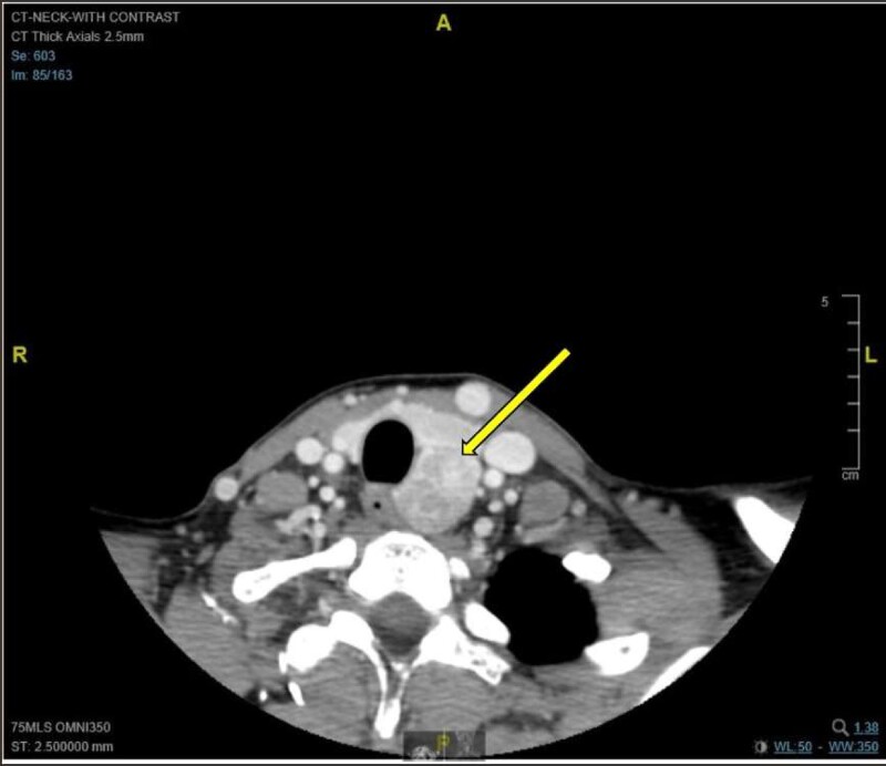
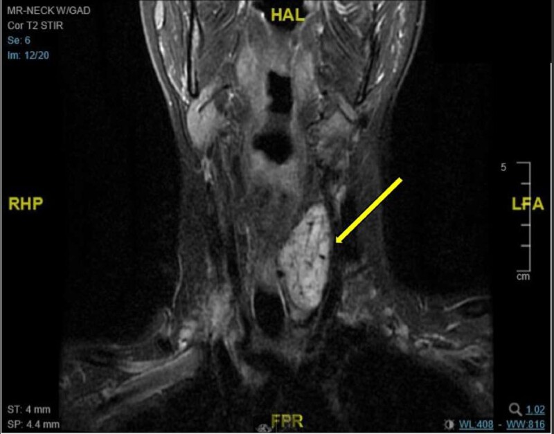
The diagnosis of pheochromocytoma or paraganglioma (PGL) during pregnancy is extremely rare, with 2 large case series suggesting that the prevalence is between 0.0002% and 0.007%. Here, we present a case of a 38-year-old woman who presented during pregnancy with clinical features suggestive of preeclampsia and was found to have a norepinephrine-secreting inferior laryngeal nerve PGL, which was diagnosed after pregnancy. She underwent uncomplicated surgical resection and genetic testing revealed a succinate dehydrogenase subunit B (SDHB) pathogenic variant. In conclusion, PGLs diagnosed during pregnancy and hypersecreting head and neck PGLs are both rare clinical entities. Hyperfunctioning PGLs may mimic pregnancy-induced hypertension or preeclampsia. Metanephrine testing should be considered in patients with atypical features and can be reliably assessed using nonpregnant reference ranges. Overall, maternal and fetal mortality has improved considerably with early diagnosis and treatment.

Keywords: laryngeal nerve; norepinephrine; paraganglioma; pregnancy.


Figures

Figure 1.
Figure 1.
Axial CT image with contrast demonstrating a moderately avid heterogeneously enhancing exophytic mass at the posterior aspect of the left thyroid lobe, which measures 5.2 cm in the craniocaudal dimension, 2.3 cm in the mediolateral dimension, and 2.6 cm in the anteroposterior dimension. The mass extends to the left posterior paraesophageal location with mild deviation of the trachea and the esophagus to the right. There is no invasion of the surrounding structures. The remainder of the thyroid gland is normal.
Figure 2.
Figure 2.
MRI scan of the neck with gadolinium (coronal MRI) demonstrating a mass in the left infrahyoid neck that is 5.6 cm in the maximal craniocaudal dimension, 2.8 cm in the anteroposterior dimension, and 2.4 cm in the mediolateral dimension. The mass is isointense to muscle on the T1-weighted sequence and demonstrates moderate T2 hyperintensity with avid postcontrast enhancement in keeping with a hypervascular mass with arterial supply that arose from the thyroid inferior mesenteric artery and left inferior thyroid artery.

References

    1. Neumann HPH, Young WF, Jr, Eng C. Pheochromocytoma and paraganglioma. N Engl J Med. 2019;381(6):552‐565. - PubMed
    1. Lenders JWM, Eisenhofer G. Update on modern management of pheochromocytoma and paraganglioma. Endocrinol Metab (Seoul). 2017;32(2):152‐161. - PMC - PubMed
    1. Pellitteri PK, Rinaldo A, Myssiorek D, et al. Paragangliomas of the head and neck. Oral Oncol. 2004;40(6):563‐575. - PubMed
    1. Patel D, Phay JE, Yen TWF, et al. Update on pheochromocytoma and paraganglioma from the SSO endocrine/head and neck disease-site work group. Part 1 of 2: advances in pathogenesis and diagnosis of pheochromocytoma and paraganglioma. Ann Surg Oncol. 2020;27(5):1329‐1337. - PMC - PubMed
    1. Valero C, Ganly I, Shah JP. Head and neck paragangliomas: 30-year experience. Head Neck. 2020;42(9):2486‐2495. - PMC - PubMed

Publication types

LinkOut - more resources