. 2024 May 21;27(6):110061.
doi: 10.1016/j.isci.2024.110061. eCollection 2024 Jun 21.

A non-symmetrical p97 conformation initiates a multistep recruitment of Ufd1/Npl4

Affiliations

A non-symmetrical p97 conformation initiates a multistep recruitment of Ufd1/Npl4

Michal Arie et al. iScience. .

Abstract

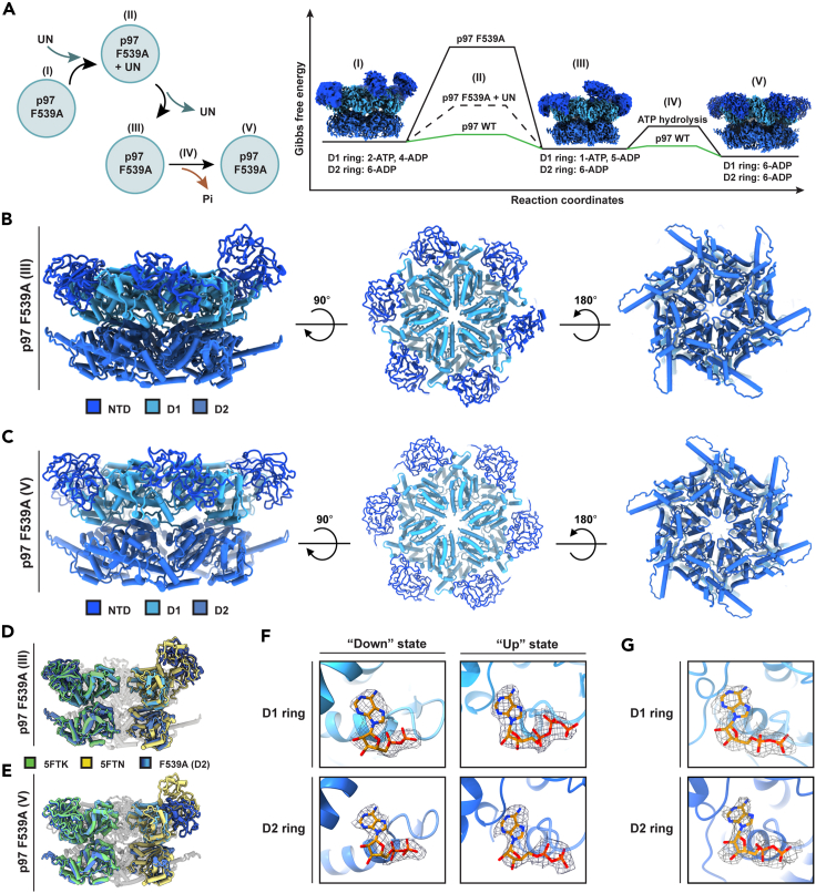
In vitro experiments and cryo-EM structures of p97 and its cofactor, Ufd1/Npl4 (UN), elucidated substrate processing. Yet, the structural transitions and the related ATPase cycle upon UN binding remain unresolved. We captured two discrete conformations: One in which D1 protomers are ATP bound, while the D2 subunits are in the ADP state, presumably required for substrate engagement with the D2 pore; and a heterologous nucleotide state within the D1 ring in which only two NTDs are in the "up" ATP state that favors UN binding. Further analysis suggests that initially, UN binds p97's non-symmetrical conformation, this association promotes a structural transition upon which five NTDs shift to an "up" state and are poised to bind ATP. The UBXL domain of Npl4 was captured bound to an NTD in the ADP state, demonstrating a conformation that may provide directionality to incoming substrate and introduce the flexibility needed for substrate processing.

Keywords: Molecular Structure; Proteomics; Structural Biology.


Conflict of interest statement

The authors declare no competing interests.

Figures

None
Graphical abstract
Figure 1
Figure 1
Characterization of p97 phenylalanine mutants (A) The four-phenylalanine signature conservation map overlaid on a human p97 D2 ring protomer, generated using ConSurf. See also Figure S2. (B) Schematic illustration of the phenylalanine residues studied. (C) p97 phenylalanine mutants are stable and retain the ability to bind ATP. p97 and its mutants were incubated at room temperature in the absence and presence of ATPγS (2mM) and subjected to thermal shift assay (35°C–95°C). The data are represented as mean ± SD. (D) The phenylalanine mutants display lower ATP hydrolysis rate. The indicated p97 species were incubated (0.3 μg each) at 37°C in reaction buffer containing 2 mM ATP; equal samples were collected at the indicated time pointes and the ATPase activity was measured and normalized to wild type p97. The data are represented as mean ± SD. (E) p97 F266A, p97 F539A, and p97 F266A F539A are functional in pQC substrate processing. Left: schematic illustration of the VCAM-1 substrate used before and upon glycosylation and signal sequence removal. Right: western blot analysis of HEK293T cells lysate expressing VCAM-1 and the indicated p97 variants, treated as indicated. Actin served as loading control. (F) Growth rate of yeast harboring Cdc48 D1, or the D1 & D2 double mutant resembles that of wild type, while the D2 mutant gains wild type-like growth rate at elevated temperatures. Growth rate of each strain was normalized to 100%, and the data were fitted using a logistic model. The data are represented as mean ± SD. See also Figure S3.
Figure 2
Figure 2
Wild type and phenylalanine p97 mutants affinity and cooperative binding of UN (A) In contrast to wild type p97 the phenylalanine mutants bind UN without the addition of exogenous ATPγS. Top: binding response curve of p97 deduced from UN ELISA binding assay. The fraction of each p97 variant bound to UN was normalized to 100% and the data were fitted using the Hill equation. Except for the wild type p97 that was assayed in the presence of ATPγS (dashed line), all p97 mutants were assayed in the absence of exogenous nucleotide. Bottom: calculated dissociation and Hill coefficients constants. The data are represented as mean ± SD. (B) p97 D2 mutant (F539A) binds UN with the highest affinity which is not further enhanced in the presence of ATPγS. Top: Binding curves for p97 F539A in the presence and absence of ATPγS. Bottom: calculated dissociation and Hill coefficients constants. The data are represented as mean ± SD. (C) The phenylalanine at positions 266 and 539 are allosterically associated in UN binding. Double mutant cycle analysis calculated from the disassociation constants (Kd), obtained in A. (D) The binding mechanism of UN is a two-state model. p97 F539A in the absence of exogenous nucleotide binds UN with higher affinity compared with wild type in the presence of ATP. Wild type p97 (top) and p97 F539A (bottom) binding response curves measured by SPR. Wild type p97 or p97 F539A were immobilized to the chip surface, whereas UN was used as analyte. The calculated rates of association and dissociation constants are presented below the response curves. See also Figure S4. (E) Upon UN binding D2 ring is activated while D1 is inhibited. p97 variants ATPase assay was performed in the presence or absence of UN. The data are represented as mean ± SD.
Figure 3
Figure 3
Cryo-EM structures of p97 F266A and F266A-F539A mutants Coordinates model shown from side (left), top (middle) and bottom (right) views of (A) p97 F266A at 2.7 Å resolution and (B) F266A F539A double mutant at 3.4 Å resolution. See also Figures S5‒S8. (C) and (D) Comparison of the NTDs of p97 F266A and p97 F266A F539A, respectively, to the NTDs of wild type p97 3D structure in the presence of ATPγS (PDB ID:5FTN) or ADP (PDB ID:5FTK). As shown in C and D middle and right panels, the D2 rings aligned with the ADP conformation. (E) The D2 ring pore loop 1 (P545-N558) of p97 F266A and p97 F266A F539A double mutant also resemble the ADP state, as evident from the pore dimension and the interactions between neighboring subunits constituting the pore.
Figure 4
Figure 4
The structure of p97 F539A reveals an asymmetrical conformation within the D1 ring (A) Coordinates model of p97 F539A shown from side (left), top (middle) and bottom (right) views at 3.6 Å resolution. See also Figures S9 and S11. (B) Two of the D1 ring protomers are in the ATP state and four are ADP bound, while the D2 protomers are all in the ADP bound conformation. As shown in the left panel two of the D1 ring NTDs align with the ATPγS bound conformation (PDB ID:5FTN) and four of the NTDs align with the ADP structure (PDB ID:5FTK). In the middle and right panels, the D2 ring architecture aligns with the ADP conformation in agreement with the nucleotides assigned to the D2 protomers. (C and D) p97 F539A coordinates model focusing on the nucleotide pockets in D1 and D2 rings reveals that two of the D1 ring protomers align with the ATP state (D) and the additional four with the ADP bound state (C). All D2 protomers are bound to ADP (C and D).
Figure 5
Figure 5
UN binding by p97 F539A favors transition of five NTDs to the "up" state even in the absence of exogenous nucleotide Upon binding of p97 F539A to the cofactor, five of its NTDs transition to the “up” state. (A) cryo-EM map of p97 F539A-adaptor complex at different views (3.0 Å resolution). See also Figures S12 and S13. p97’s six protomers (a-f) are presented in rainbow colors and Npl4 is shown in pink. The NTDs (unsharpened map) are shown at σ = 0.0029, D1 and D2 domains (sharpened map) are presented at σ = 0.0289 and Npl4 (unsharpened map) is presented at σ = 0.0289. Dashed lines (gray) indicate the “up” or “down” state. (B) Side and (C) Top views of (left) Cdc48-Ufd1/Npl4-substrate map (EMD-0665; additional map 2) and (middle) p97 F539A-adaptor map (from panel A), shown following Gaussian filter. The merged maps are presented on the right. The UBXL domains are shown in pink, while the NTDs marked in yellow are in the “up” state and in green are in the “down” state.
Figure 6
Figure 6
p97 F539A completes the nucleotide cycle upon UN binding and release (A) Left: an illustration of p97 F539A-UN binding and dissociation cycle. State I: Noncomplexed p97 F539A (D1: 2-ATP, 4-ADP). State II: Upon incubation of p97 F539A with UN @37°C. State III: p97 F539A following UN release (D1:1-ATP, 5-ADP). State IV: hydrolysis of the remaining ATP bound molecule. State V: p97 F539A followed ATP hydrolysis (D1: 6-ADP). Presumably, in the absence of ATP (in vitro), the nucleotide cycle of p97 F539A terminates at an ADP state (V), while in the presence of ATP an additional subunit would exchange for ATP to facilitate association with incoming UN-substrate complex. Right: Gibbs free energy diagram demonstrating the stabilizing effects of p97 F539A prior to UN binding (D1:2-ATP, 4-ADP) and following UN release (D1:1-ATP, 5-ADP) (black line) compared with WT p97 (green line). (B) Coordinates model of non-complexed p97 F539A (state III) obtained upon dissociation from UN, shown from side, top and bottom views at 3.3 Å. See also Figures S14 and S16. (C) Coordinates model of the ADP bound state p97 F539A (state V) obtained following UN incubation and ATP hydrolysis, shown from side, top and bottom views at 3.4 Å. See also Figures S15 and S16. (D) One of the D1 ring protomers are in the ATP state (PDB ID:5FTN, yellow) and five are ADP bound (PDB ID:5FTK, green), while the D2 protomers are all in the ADP bound conformation. (E) All six p97 protomers align with the ADP bound state (PDB ID:5FTK, green) assumed upon UN release and ATP hydrolysis. (F) Nucleotide pockets of p97 F539A state III: densities that accommodate ADP are detected in all D1 and D2 “down” protomers nucleotide binding pockets, in contrast to the “up” protomer where density that may accommodate ATP molecule was observed. D2 ring of this protomer is occupied by ADP. (G) Nucleotide pockets of p97 F539A state V: A representative “down” monomer encompassing both the D1 and D2 rings is shown.
Figure 7
Figure 7
An updated model for UN-substrate recruitment and processing by p97/Cdc48 UN is recruited to p97 in which two of the D1 ring NTDs are in the ATP bound “up” conformation. Initially, upon association with UN the D2 ring protomers are ADP bound and the D2 ring pore is dilated, allowing the substrate to transverse and engage with the D2 ring. As UN binds p97, p97’s five NTDs shift to the “up” (ATP) state, while the “down” protomer associates with UBXL domain of Npl4, presumably directing substrate binding. Once ubiquitinated substrate is committed, D2 ring ATP binding and hydrolysis is activated, while the ATPase activity of D1 domain is suppressed, to avoid UN premature release. Multiple ATP binding and hydrolysis cycles promote substrate unfolding and translocation. Once substrate processing is completed, the D1 ring undergoes sequential ATP hydrolysis promoting substrate and UN release. p97 assumes its “ground” state in which two of the D1 ring protomers are in the ATP state, while the remaining four are ADP bound. In this state all six D2 ring protomers are ADP bound.

References

    1. Xia D., Tang W.K., Ye Y. Structure and function of the AAA+ ATPase p97/Cdc48p. Gene. 2016;583:64–77. doi: 10.1016/j.gene.2016.02.042. - DOI - PMC - PubMed
    1. Ye Y., Meyer H.H., Rapoport T.A. The AAA ATPase Cdc48/p97 and its partners transport proteins from the ER into the cytosol. Nature. 2001;414:652–656. doi: 10.1038/414652a. - DOI - PubMed
    1. Dai R.M., Li C.C. Valosin-containing protein is a multi-ubiquitin chain-targeting factor required in ubiquitin-proteasome degradation. Nat. Cell Biol. 2001;3:740–744. doi: 10.1038/35087056. - DOI - PubMed
    1. Bodnar N., Rapoport T. Toward an understanding of the Cdc48/p97 ATPase. F1000Res. 2017;6:1318. doi: 10.12688/f1000research.11683.1. - DOI - PMC - PubMed
    1. Davies J.M., Brunger A.T., Weis W.I. Improved structures of full-length p97, an AAA ATPase: implications for mechanisms of nucleotide-dependent conformational change. Structure. 2008;16:715–726. doi: 10.1016/j.str.2008.02.010. - DOI - PubMed

LinkOut - more resources