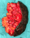
A Rare Case of a Pedunculated Lipoma in the Perianal Region: A 20-Year Journey
- PMID: 38947595
- PMCID: PMC11212838
- DOI: 10.7759/cureus.61304
A Rare Case of a Pedunculated Lipoma in the Perianal Region: A 20-Year Journey
Abstract
Lipomas are common benign soft tissue tumors, typically presenting as painless, slow-growing masses of mature adipose tissue. However, their occurrence as pedunculated lesions in the perianal region is rare. We present a case of a 70-year-old male with a 20-year history of a painless, cosmetically concerning mass in the perianal region. Clinical examination and ultrasonographic findings were consistent with a pedunculated lipoma. Surgical excision was performed successfully, and histopathological examination confirmed the diagnosis of lipofibroma. This case highlights the importance of considering unusual presentations of lipomas in the differential diagnosis of perianal masses. It emphasizes the role of surgical excision for symptomatic or cosmetically concerning lesions. Long-term follow-up is essential to monitor for recurrence and ensure optimal patient outcomes.
Keywords: histopathological examination; lipofibroma; lipoma; pedunculated; perianal; surgical excision.
Copyright © 2024, Rekavari et al.
Conflict of interest statement
Human subjects: Consent was obtained or waived by all participants in this study. Conflicts of interest: In compliance with the ICMJE uniform disclosure form, all authors declare the following: Payment/services info: All authors have declared that no financial support was received from any organization for the submitted work. Financial relationships: All authors have declared that they have no financial relationships at present or within the previous three years with any organizations that might have an interest in the submitted work. Other relationships: All authors have declared that there are no other relationships or activities that could appear to have influenced the submitted work.
Figures


References
-
- Size, site and clinical incidence of lipoma: factors in the differential diagnosis of lipoma and sarcoma. Rydholm A, Berg NO. Acta Orthop Scand. 1983;54:929–934. - PubMed
-
- Benign soft-tissue tumors in a large referral population: distribution of specific diagnoses by age, sex, and location. Kransdorf MJ. AJR Am J Roentgenol. 1995;164:395–402. - PubMed
-
- Malignant soft-tissue tumors in a large referral population: distribution of diagnoses by age, sex, and location. Kransdorf MJ. AJR Am J Roentgenol. 1995;164:129–134. - PubMed
-
- Benign fatty tumors: classification, clinical course, imaging appearance, and treatment. Bancroft LW, Kransdorf MJ, Peterson JJ, O'Connor MI. Skeletal Radiol. 2006;35:719–733. - PubMed
-
- Lipomas, lipoma variants, and well-differentiated liposarcomas (atypical lipomas): results of MRI evaluations of 126 consecutive fatty masses. Gaskin CM, Helms CA. AJR Am J Roentgenol. 2004;182:733–739. - PubMed
Publication types
LinkOut - more resources
Full Text Sources
