Review
. 2024 Jun 13;9(25):26826-26837.
doi: 10.1021/acsomega.4c02256. eCollection 2024 Jun 25.

On the Use of the Intrinsic DNA Fluorescence for Monitoring Its Damage: A Contribution from Fundamental Studies

Affiliations
Review

On the Use of the Intrinsic DNA Fluorescence for Monitoring Its Damage: A Contribution from Fundamental Studies

Dimitra Markovitsi. ACS Omega. .

Abstract

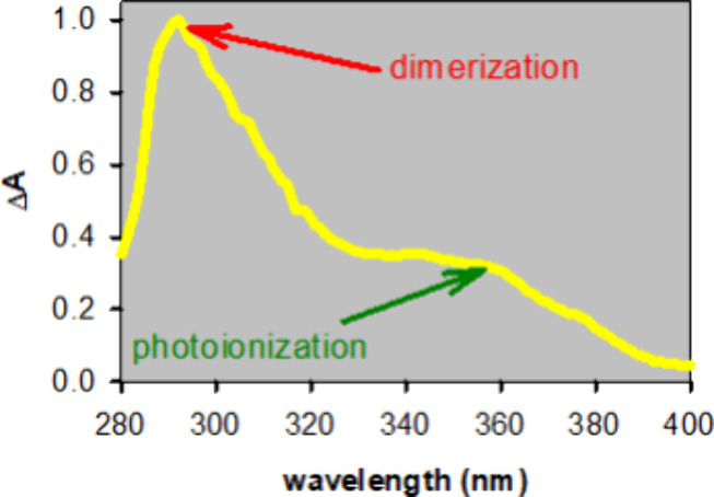
The assessment of DNA damage by means of appropriate fluorescent probes is widely spread. In the specific case of UV-induced damage, it has been suggested to use the emission of dimeric photoproducts as an internal indicator for the efficacy of spermicidal lamps. However, in the light of fundamental studies on the UV-induced processes, outlined in this review, this is not straightforward. It is by now well established that, in addition to photodimers formed via an electronic excited state, photoionization also takes place with comparable or higher quantum yields, depending on the irradiation wavelength. Among the multitude of final lesions, some have been fully characterized, but others remain unknown; some of them may emit, while others go undetected upon monitoring fluorescence, the result being strongly dependent on both the irradiation and the excitation wavelength. In contrast, the fluorescence of undamaged nucleobases associated with emission from ππ* states, localized or excitonic, appearing at wavelengths shorter than 330 nm is worthy of being explored to this end. Despite its low quantum yield, it is readily detected nowadays. Its intensity decreases due to the disappearance of the reacting nucleobases and the loss of exciton coherence provoked by the presence of lesions, independently of their type. Thus, it could potentially provide valuable information about the DNA damage induced, not only by UV radiation but also by other sanitizing or therapeutic agents.


Conflict of interest statement

The author declares no competing financial interest.

Figures

Figure 1
Figure 1
Alteration of the fluorescence of calf thymus DNA due to its damage by femtosecond laser pulses at 267 nm. Fluorescence spectra recorded exciting at 255 nm before (blue) and after (red) irradiation. Reprinted from ref (32). Copyright ACS 2010.
Figure 2
Figure 2
Dependence of the fluorescence quantum yield on the secondary DNA structure: mononucleotides (yellow), single strands (cyan), duplexes (pink), and G-quadruplexes (green). The shaded areas indicate the range of Φfl values determined for each type of structure with excitation at 255–270 nm.
Figure 3
Figure 3
Steady-state fluorescence spectra of dA20•dT20, (red), dA20 (green), and dT20 (cyan). Excitation wavelength: 335 nm. The spectral areas are proportional to the quantum yields. Adapted from ref (54). Copyright ACS 2011.
Figure 4
Figure 4
Steady-state fluorescence spectra of polymeric duplexes with simple repetitive sequence. The spectral areas are proportional to the quantum yields (a) d(GC)n•d(GC)n, Φfl = 1.5 × 10⁻⁴; (b) d(AT)n•d(AT)n, Φfl = 1.4 × 10⁻⁴; (c) dAn•dTn, Φfl = 3 × 10⁻⁴. Excitation wavelength: 267 nm. Adapted from references ( and 42). Copyright ACS 2010 and 2023.
Figure 5
Figure 5
Comparison of duplex fluorescence spectra (red) with that of the constitutive purine rich single strand (green). (a) d(CGGACAAGAAG)•d(CTTCTTGTCCG) and d(CGGACAAGAAG). Adapted from ref (51). Copyright RSC 2013. (b) d(A)20•d(T)20 and d(A)20. Adapted from ref (54). Copyright ACS 2007. Excitation wavelength: 267 nm. The intensities are normalized at the maximum.
Figure 6
Figure 6
Chemical formulas of thymine dimers: cyclobutane dimer (CPD), (6–4) photoadduct (64PP), and spore photoproduct (SP). R denotes the backbone.
Figure 7
Figure 7
Normalized steady-state fluorescence spectra. Red: (dT)20 after irradiation at 255 nm (excitation at 320 nm). Green: 8-oxodG (excitation at 255 nm). Adapted from references ( and 86). Copyright ACS 2005 and 2012.
Figure 8
Figure 8
Schematic drawing of the two adenine photodimers. Reprinted from ref (91). Copyright ACS 2016.
Figure 9
Figure 9
Chemical formulas of (a) guanosine and (b) 8-oxodG. The proton encircled in (a) is that lost upon deprotonation of the radical cation in single and double strands. R denotes the backbone.
Figure 10
Figure 10
Difference between the steady-state absorption spectra recorded before and after irradiation of d(AT)10•d(AT)10 with 266 nm laser pulses. Adapted from reference (99). Copyright RSC 2018.

References

    1. Duan X. R.; Liu L. B.; Feng F. D.; Wang S. Cationic Conjugated Polymers for Optical Detection of DNA Methylation, Lesions, and Single Nucleotide Polymorphisms. Acc. Chem. Res. 2010, 43, 260–270. 10.1021/ar9001813. - DOI - PubMed
    1. El-Yazbi A. F.; Loppnow G. R. Terbium fluorescence as a sensitive, inexpensive probe for UV-induced damage in nucleic acids. Anal. Chim. Acta 2013, 786, 116–123. 10.1016/j.aca.2013.04.068. - DOI - PubMed
    1. El-Yazbi A. F.; Loppnow G. R. A selective, inexpensive probe for UV-induced damage in nucleic acids. Can. J. Chem. 2013, 91, 320–325. 10.1139/cjc-2012-0417. - DOI - PubMed
    1. El-Yazbi A. F.; Loppnow G. R. Detecting UV-induced nucleic-acid damage. TrAC, Trends Anal. Chem. 2014, 61, 83–91. 10.1016/j.trac.2014.05.010. - DOI
    1. Condie A. G.; Yan Y.; Gerson S. L.; Wang Y. M. A Fluorescent Probe to Measure DNA Damage and Repair. PLoS One 2015, 10, e0131330.10.1371/journal.pone.0131330. - DOI - PMC - PubMed

LinkOut - more resources