. 2024 Jun 17;14(9):3739-3759.
doi: 10.7150/thno.97412. eCollection 2024.

An injectable magnesium-loaded hydrogel releases hydrogen to promote osteoporotic bone repair via ROS scavenging and immunomodulation

Affiliations

An injectable magnesium-loaded hydrogel releases hydrogen to promote osteoporotic bone repair via ROS scavenging and immunomodulation

Hang Zhou et al. Theranostics. .

Abstract

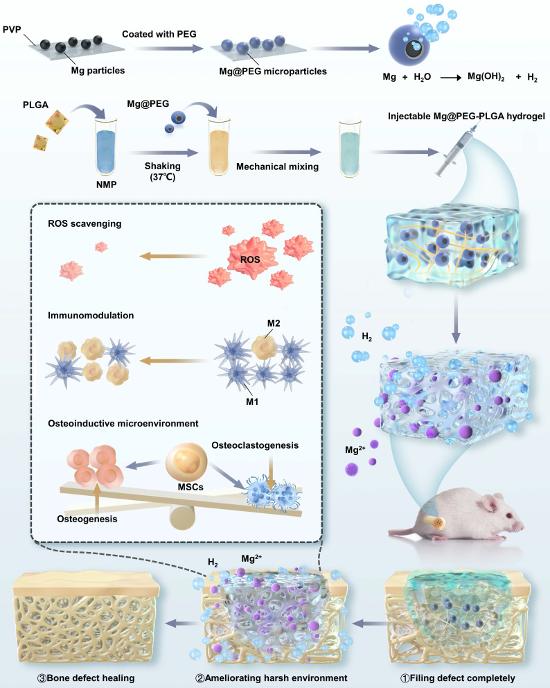
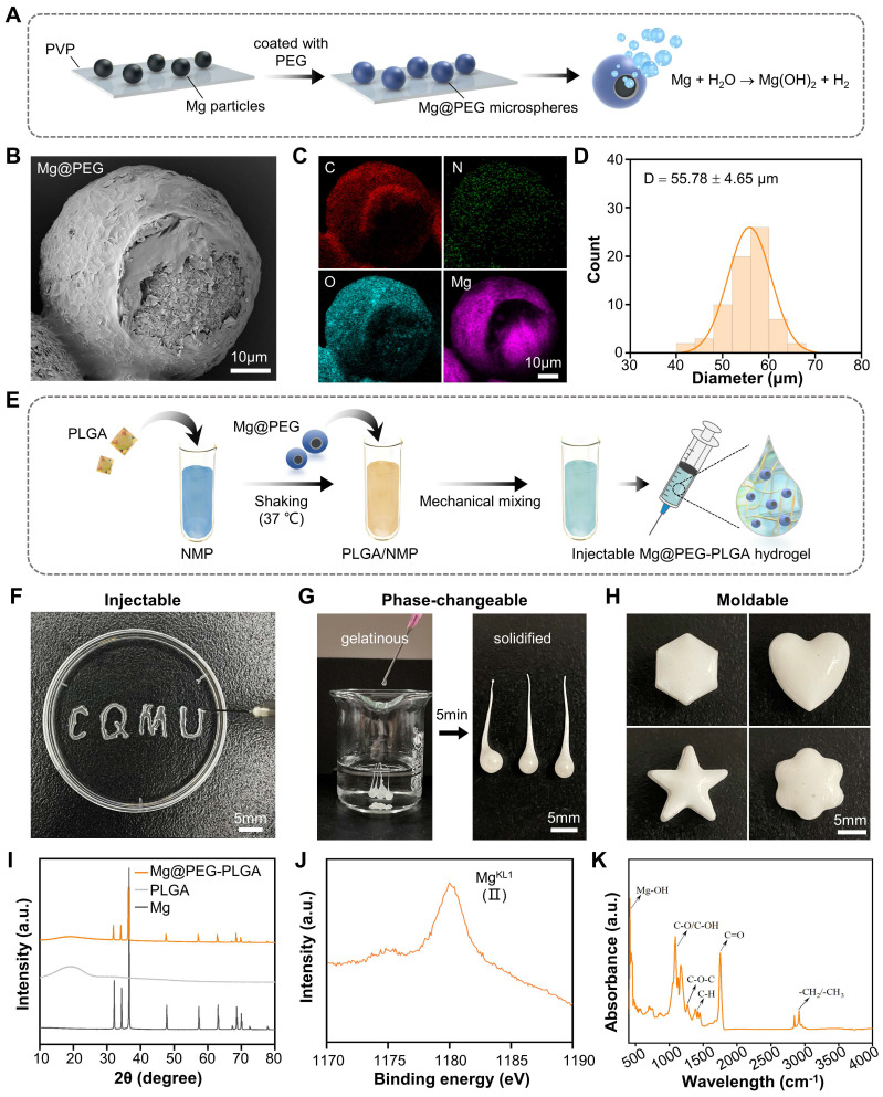
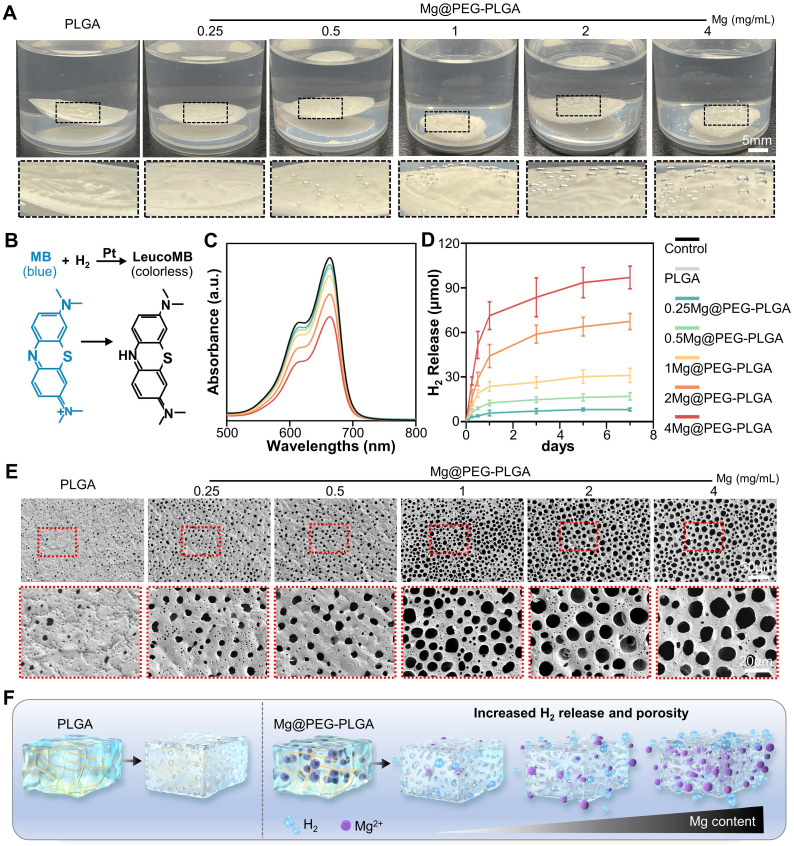
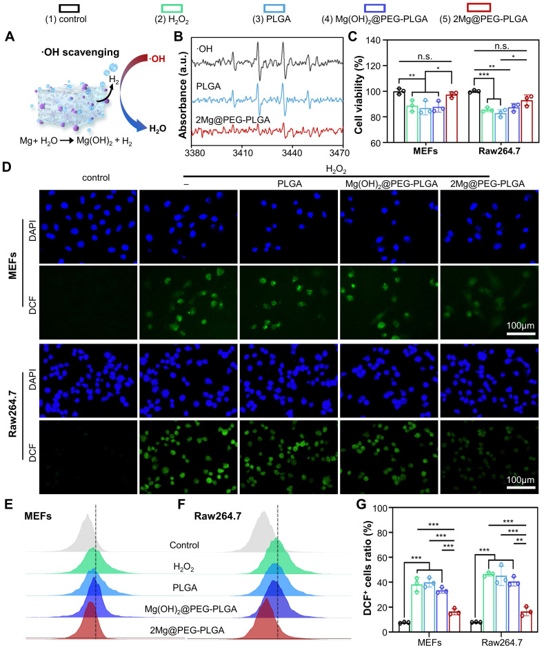
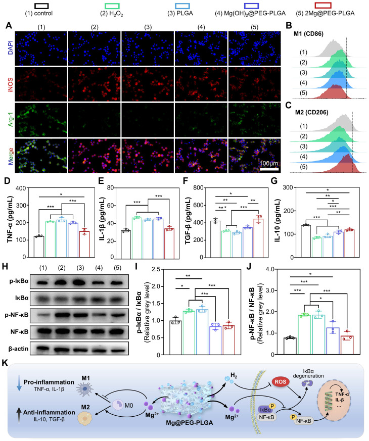
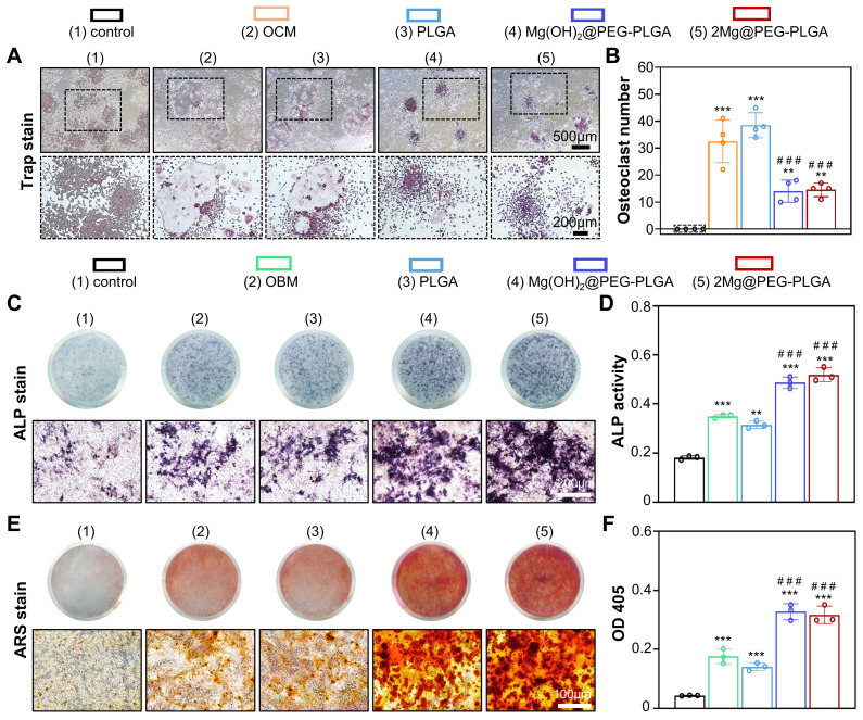
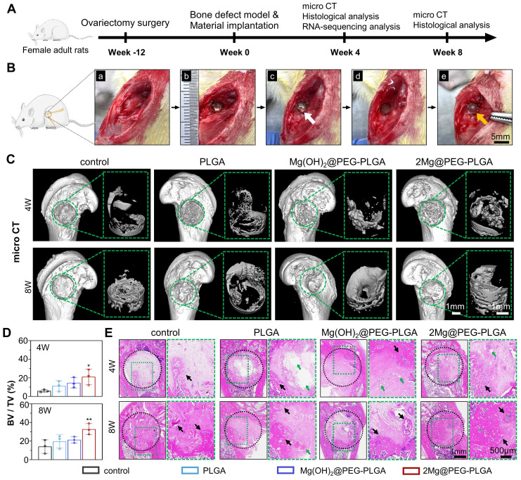
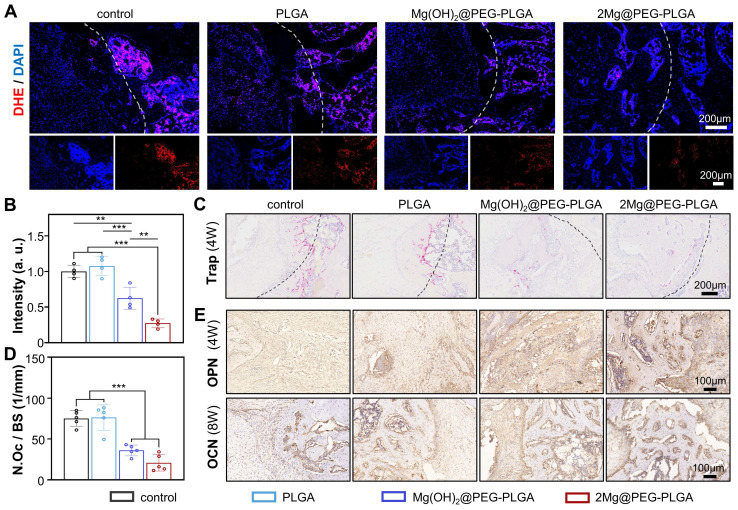
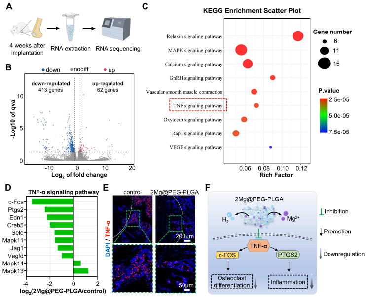
Background: The repair of osteoporotic bone defects remains challenging due to excessive reactive oxygen species (ROS), persistent inflammation, and an imbalance between osteogenesis and osteoclastogenesis. Methods: Here, an injectable H2-releasing hydrogel (magnesium@polyethylene glycol-poly(lactic-co-glycolic acid), Mg@PEG-PLGA) was developed to remodel the challenging bone environment and accelerate the repair of osteoporotic bone defects. Results: This Mg@PEG-PLGA gel shows excellent injectability, shape adaptability, and phase-transition ability, can fill irregular bone defect areas via minimally invasive injection, and can transform into a porous scaffold in situ to provide mechanical support. With the appropriate release of H2 and magnesium ions, the 2Mg@PEG-PLGA gel (loaded with 2 mg of Mg) displayed significant immunomodulatory effects through reducing intracellular ROS, guiding macrophage polarization toward the M2 phenotype, and inhibiting the IκB/NF-κB signaling pathway. Moreover, in vitro experiments showed that the 2Mg@PEG-PLGA gel inhibited osteoclastogenesis while promoting osteogenesis. Most notably, in animal experiments, the 2Mg@PEG-PLGA gel significantly promoted the repair of osteoporotic bone defects in vivo by scavenging ROS and inhibiting inflammation and osteoclastogenesis. Conclusions: Overall, our study provides critical insight into the design and development of H2-releasing magnesium-based hydrogels as potential implants for repairing osteoporotic bone defects.

Keywords: ROS scavenging; hydrogen; immunomodulation; magnesium-based hydrogel; osteoporotic bone defect.


Conflict of interest statement

Competing Interests: The authors have declared that no competing interest exists.

Figures

Scheme 1
Scheme 1
Schematic illustration of the mechanism by which the injectable Mg@PEG-PLGA hydrogel effectively promoted osteoporotic bone regeneration.
Figure 1
Figure 1
Synthesis and characteristics of the Mg@PEG-PLGA hydrogel. (A) Schematic diagram of the preparation of Mg@PEG microspheres. (B) SEM image of Mg@PEG microspheres and the corresponding (C) elemental mapping of C, O, N and Mg. (D) Size distribution of the Mg@PEG microspheres. (E) Schematic diagram of the preparation of the Mg@PEG-PLGA hydrogel. Digital images of the (F) injectability, (G) phase change ability and (H) moldability of the Mg@PEG-PLGA hydrogel. (I) XRD patterns of Mg particles, PLGA and the Mg@PEG-PLGA gel. (J) MgKL1 XPS spectrum of the solidified Mg@PEG-PLGA gel. (K) FTIR spectra of solidified Mg@PEG-PLGA gel.
Figure 2
Figure 2
H2 release and pore analysis of the Mg@PEG-PLGA hydrogels. (A) Photograph showing H2 generation from the Mg@PEG-PLGA gels in PBS. (B) Schematic illustration of H2 generation determined by the MB-Pt probe solution. (C) Absorption spectra of the MB probe solution with or without the hydrogel added (6 h). (D) Time-dependent H2 generation measured by an MB probe from Mg@PEG-PLGA hydrogels. (E) SEM images of the solidified PLGA and Mg@PEG-PLGA gels. (F) Schematic illustration of the improved H2 generation ability of the Mg@PEG-PLGA gel with increased Mg content.
Figure 3
Figure 3
ROS scavenging analysis of the 2Mg@PEG-PLGA hydrogel. (A) Schematic illustration of ·OH reacting with H2 generated from the 2Mg@PEG-PLGA hydrogel. (B) The ·OH scavenging effect of 2Mg@PEG-PLGA detected by ESR. (C) Viability of H2O2-stimulated MEFs and RAW264.7 cells without or with hydrogel treatment. (D) ROS scavenging in MEFs and RAW264.7 cells after different treatments. (E-F) FCM results showing the intracellular ROS levels in MEFs and RAW264.7 cells after different treatments and (G) the corresponding quantitative analysis. The data are expressed as the mean ± SD (n=3). n.s. indicates no significance. *p < 0.05, **p < 0.01 and ***p < 0.001.
Figure 4
Figure 4
Immunomodulatory properties of the 2Mg@PEG-PLGA hydrogel. (A) Immunofluorescence images of iNOS, Arg-1 and DAPI staining of macrophages in the different groups. FCM results of (B) M1 (CD86+) and (C) M2 (CD206+) macrophages. (D-G) Secretion levels of TNF-α, IL-1β, TGF-β and IL-10 in macrophage suspensions. (H) Representative Western blot images of p-IκBα, IκBα, p-NF-κB, NF-κB and β-actin in the indicated groups. Quantitative analyses of the (I) p-IκBα/IκBα and (J) p-NF-κB/NF-κB ratios. (K) Schematic illustration of the immunomodulatory mechanism of the 2Mg@PEG-PLGA gel. The data are expressed as the mean ± SD (n=3). *p < 0.05, **p < 0.01 and ***p < 0.001.
Figure 5
Figure 5
In vitro antiosteoclastic and pro-osteoblastic properties of the 2Mg@PEG-PLGA hydrogel. (A) TRAP staining of RAW264.7 cells cultured in OCM supplemented with or without different hydrogels for 7 days and (B) corresponding quantitative analysis of TRAP+ cells per well. (C) ALP staining and (D) ALP activity quantitative analysis of MEFs after culture with OBM with or without supplementation with different hydrogels for 7 days. (E-F) ARS staining and corresponding quantitative analysis of MEFs after culture with OBM supplemented with or without different hydrogels for 14 days. The data are expressed as the mean ± SD. **p < 0.01 and ***p < 0.001, compared with the control group; ###p<0.001, compared with the OCM (Figure 5B) or OBM (Figure 5D and Figure 5F) group.
Figure 6
Figure 6
Osteoporotic bone defect repair efficacy of the 2Mg@PEG-PLGA hydrogel. (A) Schematic timeline of the in vivo study. (B) The surgical process of in situ implantation of the 2Mg@PEG-PLGA hydrogel in osteoporotic bone defects. (a-b) The construction of bone defects (3 mm in diameter × 3 mm in depth) on the lateral epicondyle of the femur. (c-e) The implanted 2Mg@PEG-PLGA hydrogel was solidified after immersion in saline for 5 min. The white and red arrows indicate gelatinous and solidified 2Mg@PEG-PLGA gels, respectively. (C) Micro-CT 3D-reconstructed images of the distal femur of rats and the newly formed bone within the bone defect at 4 and 8 weeks. The green circle marks the bone defects. (D) BV/TV analysis of the newly formed bone within the bone defect via micro-CT. (E) HE staining of rat femurs from different groups at 4 and 8 weeks. The black circle marks the bone defects. The green arrows indicate residual materials. The black arrows indicate the newly formed bone. The data are expressed as the mean ± SD (n = 3). *p < 0.05 and **p < 0.01.
Figure 7
Figure 7
The ability of the 2Mg@PEG-PLGA hydrogel to scavenge ROS, inhibit osteoclastogenesis, and promote osteogenesis in vivo. (A) Representative images and (B) corresponding quantification of DHE staining at the bone defect site (week 4). The white dotted lines mark the bone defects. (C) TRAP staining and (D) corresponding quantification of the number of TRAP+ osteoclasts at the bone defect sites (week 4). The black dotted lines mark the bone defects. (E) Images of immunohistochemical staining for OPN and OCN in bone defects. The data are expressed as the mean ± SD; **p < 0.01 and ***p < 0.001.
Figure 8
Figure 8
Transcriptome high-throughput sequencing was used to study the effect of the 2Mg@PEG-PLGA gel on osteoporotic bone defect repair. (A) Schematic diagram of the RNA sequence analysis of extracted tissues within femoral bone defects from the control and 2Mg@PEG-PLGA groups at 4 weeks. (B) Volcano plot of genes that were differentially expressed between the control and 2Mg@PEG-PLGA groups. (C) Representative KEGG pathways associated with genes that were significantly differentially expressed between the control and 2Mg@PEG-PLGA groups. (D) Differentially expressed genes involved in the TNF-α signaling pathway. (E) IF images of TNF-α protein expression in femoral bone defects at 4 weeks. The white dotted lines mark the bone defects. Red fluorescence: TNF-α; blue fluorescence: DAPI. (F) Schematic illustration of the mechanism by which 2Mg@PEG-PLGA downregulates osteoclastogenesis and inflammation by inhibiting the TNF-α signaling pathway.

References

    1. Compston JE, McClung MR, Leslie WD. Osteoporosis. Lancet (London, England) 2019;393:364–76. - PubMed
    1. Eastell R, O'Neill TW, Hofbauer LC, Langdahl B, Reid IR, Gold DT. et al. Postmenopausal osteoporosis. Nat Rev Dis Primers. 2016;2:16069. - PubMed
    1. White VanGompel EC, Franks P, Robbins JA, Fenton JJ. Incidence and Predictors of Repeat Bone Mineral Densitometry: A Longitudinal Cohort Study. J Gen Intern Med. 2017;32:1090–6. - PMC - PubMed
    1. Zhang Y, Ma W, Zhan Y, Mao C, Shao X, Xie X. et al. Nucleic acids and analogs for bone regeneration. Bone Res. 2018;6:37. - PMC - PubMed
    1. Zeng J, Guo J, Sun Z, Deng F, Ning C, Xie Y. Osteoblastic and anti-osteoclastic activities of strontium-substituted silicocarnotite ceramics: In vitro and in vivo studies. Bioact Mater. 2020;5:435–46. - PMC - PubMed

Publication types

MeSH terms