. 2024 Jun 7:40:64-73.
doi: 10.1016/j.bioactmat.2024.05.046. eCollection 2024 Oct.

Regulating the proinflammatory response to composite biomaterials by targeting immunometabolism

Affiliations

Regulating the proinflammatory response to composite biomaterials by targeting immunometabolism

Chima V Maduka et al. Bioact Mater. .

Abstract

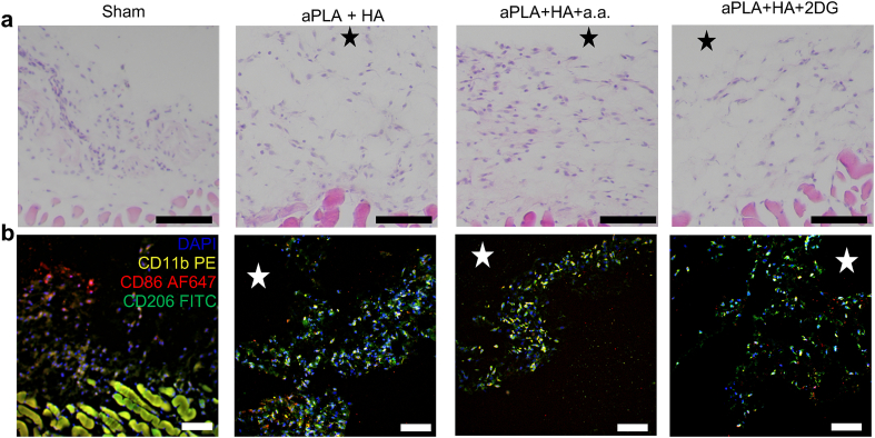
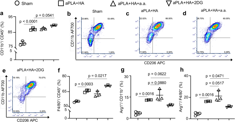
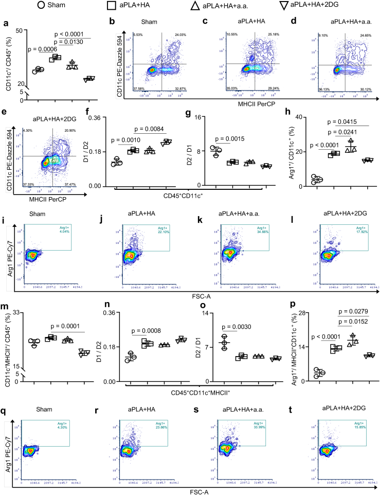
Composite biomaterials comprising polylactide (PLA) and hydroxyapatite (HA) are applied in bone, cartilage and dental regenerative medicine, where HA confers osteoconductive properties. However, after surgical implantation, adverse immune responses to these composites can occur, which have been attributed to size and morphology of HA particles. Approaches to effectively modulate these adverse immune responses have not been described. PLA degradation products have been shown to alter immune cell metabolism (immunometabolism), which drives the inflammatory response. Accordingly, to modulate the inflammatory response to composite biomaterials, inhibitors were incorporated into composites comprised of amorphous PLA (aPLA) and HA (aPLA + HA) to regulate glycolytic flux. Inhibition at specific steps in glycolysis reduced proinflammatory (CD86+CD206-) and increased pro-regenerative (CD206+) immune cell populations around implanted aPLA + HA. Notably, neutrophil and dendritic cell (DC) numbers along with proinflammatory monocyte and macrophage populations were decreased, and Arginase 1 expression among DCs was increased. Targeting immunometabolism to control the proinflammatory response to biomaterial composites, thereby creating a pro-regenerative microenvironment, is a significant advance in tissue engineering where immunomodulation enhances osseointegration and angiogenesis, which could lead to improved bone regeneration.

Keywords: Hydroxyapatite; Immunometabolism; Inflammation; Polylactide; Regenerative medicine.


Conflict of interest statement

The authors declare no competing interest.

Figures

Image 1
Graphical abstract
Fig. 1
Fig. 1
Histological evaluation of the amorphous polylactide-hydroxyapatite composite implant microenvironment with and without metabolic inhibitors suggests cellular infiltration. a, Hematoxylin and eosin staining reveals immune cellular infiltrates around implanted biomaterials (stars). b, Immunohistochemical staining using DAPI, CD11b-PE, CD206-FITC and CD86-AF647 suggests the presence of immune cells of varied immunophenotypic compositions. Scale bars, 100 μm, amorphous polylactide (aPLA), hydroxyapatite (HA), aminooxyacetic acid (a.a.), 2-deoxyglucose (2DG).
Fig. 2
Fig. 2
Glycolytic inhibition in the amorphous polylactide-hydroxyapatite composite biomaterial microenvironment modifies the numbers and inflammatory states of recruited nucleated hematopoietic cell populations. a-e, Flow cytometry quantification (a) and representative plots (b–e) of nucleated hematopoietic (CD45+) cells gated on live cells. f, Fold change of proinflammatory (H1; CD86+CD206-) cells with respect to anti-inflammatory (H2; CD206+) cells. g, Fold change of H2 with respect to H1 cells. h-k, Representative flow plots of CD86 and CD206 cells gated on CD45+ cells. l, Quantification of Arginase 1 (Arg1+) cells gated on CD45+ populations. One-way ANOVA followed by Tukey's or Newman-Keul's multiple comparison test, n = 3; amorphous polylactide (aPLA), hydroxyapatite (HA), aminooxyacetic acid (a.a.), 2-deoxyglucose (2DG).
Fig. 3
Fig. 3
Incorporation of metabolic inhibitors modulate neutrophil recruitment in the amorphous polylactide-hydroxyapatite composite biomaterial microenvironment. a-e, Quantification (a) and representative flow cytometry plots (b–e) of neutrophils (Ly6G+) cells gated on CD45+ populations. One-way ANOVA followed by Tukey's multiple comparison test, n = 3; amorphous polylactide (aPLA), hydroxyapatite (HA), aminooxyacetic acid (a.a.), 2-deoxyglucose (2DG).
Fig. 4
Fig. 4
Monocyte and macrophage populations are differentially affected by targeting different glycolytic steps in the amorphous polylactide-hydroxyapatite composite biomaterial microenvironment. a, Flow cytometry quantification of monocytes (CD11b+ cells) gated on CD45+ populations. b-e, Representative plots of monocytes (CD11b+) and macrophages (F4/80+) gated on CD45+ populations. f, Quantification of macrophages in the composite biomaterial microenvironment. g-h, Quantification of Arginase 1 (Arg1+) monocytes (g) and macrophages (h). One-way ANOVA followed by Tukey's or Newman-Keul's multiple comparison test, n = 3; amorphous polylactide (aPLA), hydroxyapatite (HA), aminooxyacetic acid (a.a.), 2-deoxyglucose (2DG).
Fig. 5
Fig. 5
Activation states of monocytes and macrophages are modulated by glycolytic inhibition in the amorphous polylactide-hydroxyapatite composite biomaterial microenvironment. a, Fold change of proinflammatory (M1; CD86+CD206-) monocytes with respect to anti-inflammatory (M2; CD206+) monocytes. b, Fold change of M2 monocytes with respect to M1 monocytes. c-f, Representative plots of CD86 and CD206 cells gated on monocyte populations (CD45+CD11b+). g, Fold change of proinflammatory (M1; CD86+CD206-) macrophages with respect to anti-inflammatory (M2; CD206+) macrophages. h, Fold change of M2 macrophages with respect to M1 macrophages. i-l, Representative plots of CD86 and CD206 cells gated on macrophage populations (CD45+F4/80b+). One-way ANOVA followed by Tukey's multiple comparison test, n = 3; amorphous polylactide (aPLA), hydroxyapatite (HA), aminooxyacetic acid (a.a.), 2-deoxyglucose (2DG).
Fig. 6
Fig. 6
Proportions and inflammatory states of dendritic cells are affected in the amorphous polylactide-hydroxyapatite composite biomaterial microenvironment. a, Flow cytometry quantification of dendritic (CD11c+) cells gated on CD45+ cells. b-e, Representative plots of dendritic (CD11c+) cells with and without MHCII gated on CD45+ cells. f, Fold change of proinflammatory (D1; CD86+CD206-) dendritic cells with respect to anti-inflammatory (D2; CD206+) dendritic cells. g, Fold change of D2 with respect to D1 dendritic cells. h-l, Quantification (h) and representative plots (i–l) of Arginase 1 (Arg1+) dendritic cells. m, Dendritic cells expressing MHCII gated on CD45+ cells. n, Fold change of proinflammatory (D1; CD86+CD206-) dendritic cells expressing MHCII with respect to anti-inflammatory (D2; CD206+) dendritic cells expressing MHCII. o, Fold change of D2 with respect to D1 dendritic cells expressing MHCII. p-t, Quantification (p) and representative plots (q–t) of Arginase 1 (Arg1+) dendritic cells expressing MHCII. One-way ANOVA followed by Tukey's or Newman-Keul's multiple comparison test, n = 3; amorphous polylactide (aPLA), hydroxyapatite (HA), aminooxyacetic acid (a.a.), 2-deoxyglucose (2DG).
Fig. 7
Fig. 7
Release kinetics of glycolytic inhibitors suggests that only small amounts are released by 12 weeks. a-b, Whereas 5.23 % of 2-deoxyglucose (2DG; a) is released by 12 weeks, released aminooxyacetic acid (a.a.; b) levels are below detection limits of the applied mass spectrometry method. Mean (SD), n = 3, amorphous polylactide (aPLA), hydroxyapatite (HA).

References

    1. Bernardo M.P., et al. PLA/Hydroxyapatite scaffolds exhibit in vitro immunological inertness and promote robust osteogenic differentiation of human mesenchymal stem cells without osteogenic stimuli. Sci. Rep. 2022;12:1–15. - PMC - PubMed
    1. Begum S.A., Krishnan P.S.G., Kanny K. Properties of poly (lactic Acid)/hydroxyapatite biocomposites for 3D printing feedstock material. J. Thermoplast. Compos. Mater. 2023
    1. Huolman R., Ashammakhi N. New multifunctional anti-osteolytic releasing bioabsorbable implant. J. Craniofac. Surg. 2007;18:295–301. - PubMed
    1. Länsman S., et al. Poly-L/D-lactide (PLDLA) 96/4 fibrous implants: histological evaluation in the subcutis of experimental design. J. Craniofac. Surg. 2006;17:1121–1128. - PubMed
    1. Ashammakhi N., Suuronen R., Tiainen J., Törmälä P., Waris T. Spotlight on naturally absorbable osteofixation devices. J. Craniofac. Surg. 2003;14:247–259. - PubMed