[Preprint]. 2024 Jun 17:2024.06.17.599413.
doi: 10.1101/2024.06.17.599413.

Liver-specific Mettl14 deletion induces nuclear heterotypia and dysregulates RNA export machinery

Affiliations

Liver-specific Mettl14 deletion induces nuclear heterotypia and dysregulates RNA export machinery

Keith A Berggren et al. bioRxiv. .

Abstract

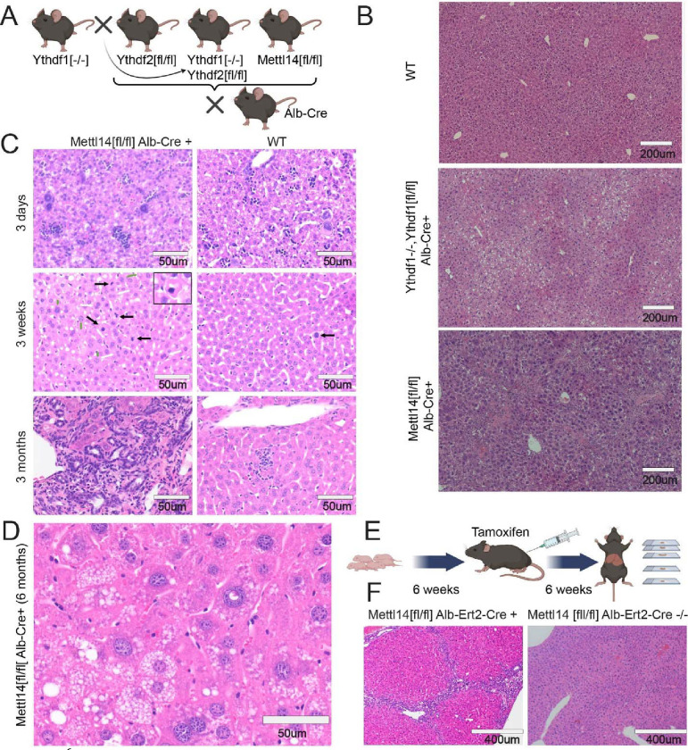
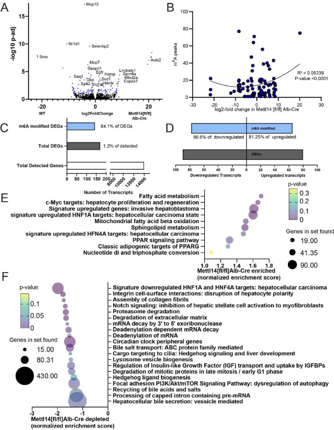
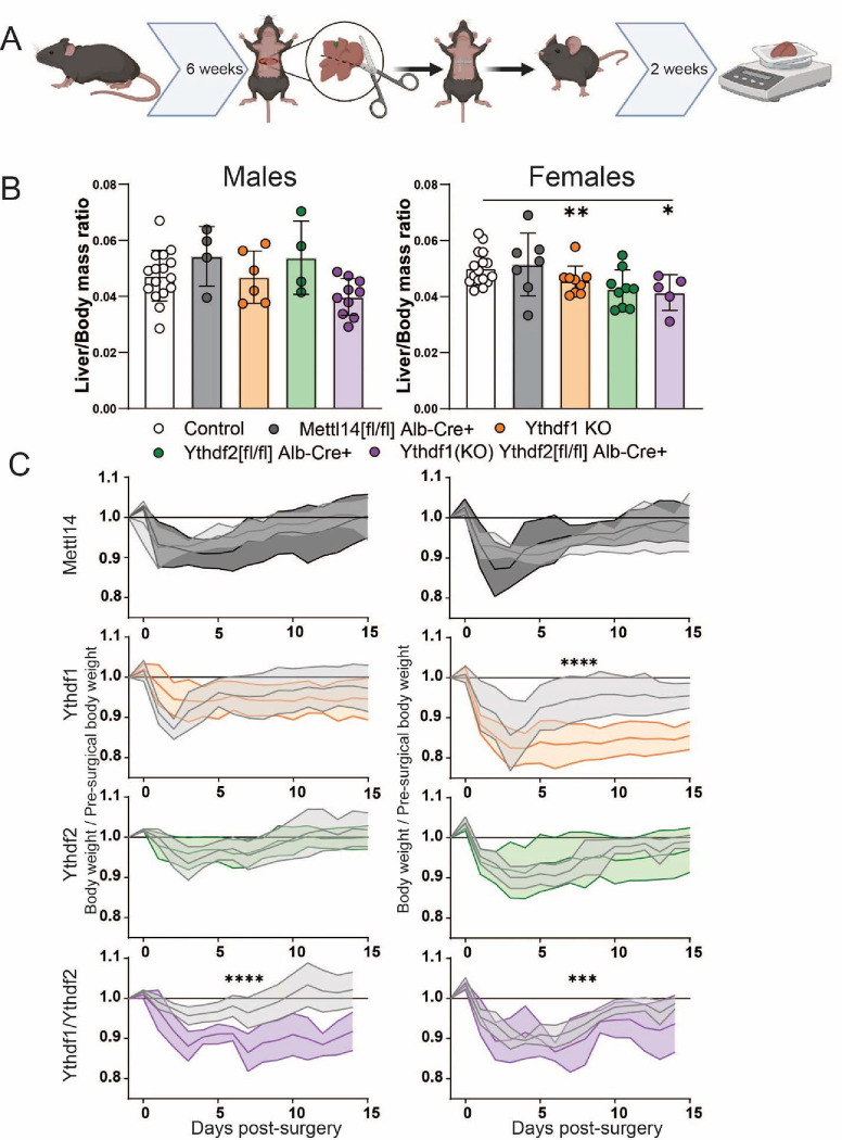
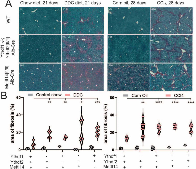
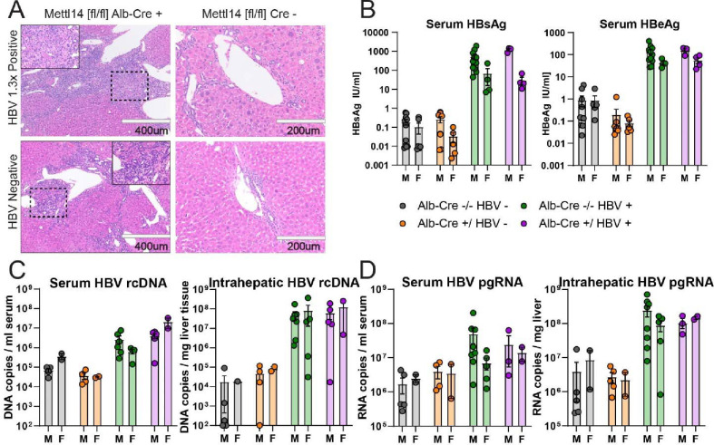
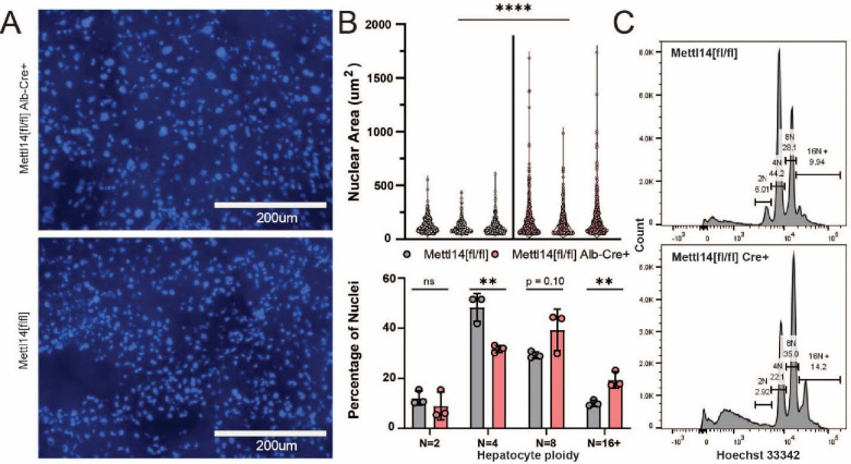
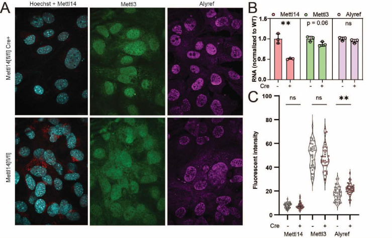
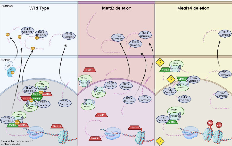
Modification of RNA with N6-methyladenosine (m6A) has gained attention in recent years as a general mechanism of gene regulation. In the liver, m6A, along with its associated machinery, has been studied as a potential biomarker of disease and cancer, with impacts on metabolism, cell cycle regulation, and pro-cancer state signaling. However these observational data have yet to be causally examined in vivo. For example, neither perturbation of the key m6A writers Mettl3 and Mettl14, nor the m6A readers Ythdf1 and Ythdf2 have been thoroughly mechanistically characterized in vivo as they have been in vitro. To understand the functions of these machineries, we developed mouse models and found that deleting Mettl14 led to progressive liver injury characterized by nuclear heterotypia, with changes in mRNA splicing, processing and export leading to increases in mRNA surveillance and recycling.


Conflict of interest statement

Competing interests All authors declare they have no competing interests.

Figures

Fig. 1.
Fig. 1.. m6A writer and reader deficiencies lead to progressive liver damage.
(A) Schematic showing crosses used to generate gene deletions in hepatocytes for this study (Created with BioRender.com). (B) Dual Ythdf1/Ythdf2 deletion in liver tissue as well as Mettl14 deletion leads to liver injury, but each with distinctly different histology. (C) Mettl14-deficient liver tissue exhibits progressive damage emerging first at 3 days and progressing in severity through 3 months of age and older. Black arrows indicate apoptotic cells with condensed nuclei, green arrows indicate enlarged nuclei, and white arrows indicate mitotic events. (D) Representative image of the advanced liver injury phenotype seen in 6-month-old Mettl14 deficient liver tissue, showing steatosis and nuclear heterotypia. (E) Schematic of tamoxifen-induction timeline where mice were injected with tamoxifen over 6 weeks before sacrifice for experiments (Created with BioRender.com). (F) Liver injury is recapitulated in this inducible Mettl14 model, showing similar levels of fibrosis and nuclear heterotypia.
Fig. 2.
Fig. 2.. Bulk RNAseq and GSEA analysis reveal key pathways of liver damage.
(A) Volcano plot of transcripts upregulated and downregulated between Mettl14[fl/fl]/Alb-Cre and Mettl14[fl/fl] male mice (n = 3) Hits with a significant adjusted p-value below 0.1 are highlighted in blue. Specific genes with particularly significant p-values or high levels of up-regulation or down-regulation, as well as those with particularly interesting functions to pathways seen in the GSEA are marked with gene names next to their data points. (B) Comparison of known m6A modified sites on transcripts with significantly upregulated or downregulated transcripts in Mettl14 deletion mice. (C) The ratio of enrichment of transcripts published to be m6A modified versus those without evidence for modification among differentially expressed genes (DEGs) seen in our data (D) and breakdown of m6A enrichment among DEGs in those upregulated and downregulated. (E) Gene set enrichment analysis (GSEA) of bulk transcriptomic data, revealing which pathways and functions are over-represented in the Mettl14 deletion mice. (F) GSEA results of which pathways and functions are downregulated in Mettl14 deletion mice.
Fig. 3.
Fig. 3.. Significantly slower liver regeneration in m6A reader-deficient mice following partial hepatectomy.
(A) Schematic of timeline for two-thirds hepatectomy surgery and recovery prior to sacrifice and sample collection (Created with BioRender.com). (B) Males (left) and females (right): comparison of liver /body mass ratios at 2 weeks post-surgery across C57BL/6 (N=16 males, 16 females) Mettl14 (N=4 males, 7 females), Ythdf1(N=6 males, 9 females), Ythdf2 (N=4 males, 9 females), and dual Ythdf1/Ythdf2 deletion mice (N=10 males, 5 females). (C) Males (left) and females (right): weight recovery curves after surgery.
Fig. 4.
Fig. 4.. m6A reader and writer deficiency increases liver fibrosis response to toxicity.
(A) Histological staining with picrosirius red of mock (chow diet) and DDC diet-treated (left) male mice reveals increased liver injury and areas of fibrosis (red). Similar staining of mock (corn oil) and CCl4-treated male mice (right) reveals the same pattern of liver injury and fibrosis advancement in m6A reader- and writer-perturbed mice. (B) Quantitative image analysis of areas of fibrosis in DDC diet- (left) and CCl4- (right) treated mice with mock comparison.
Fig. 5.
Fig. 5.. Mettl14 deficiency impacts liver injury but not HBV translation or replication.
(A) Histology of H&E-stained sections from male Mettl14[fl/fl]/Alb-Cre and Mettl14[fl/fl] control mice expressing HBV 1.3x genome transgene, and comparison with control HBV negative mice. (B) ELISA assays showing blood serum HBsAg (left) and HBeAg (right) protein levels of HBV expressing mice in comparison to control HBV negative animals to establish baseline. (C) qPCR of HBV genomic rcDNA extracted from blood serum (left) and liver tissue (right). (D) RT-qPCR of HBV pre-genomic RNA extracted from blood serum (left) and liver tissue (right).
Fig. 6.
Fig. 6.. Mettl14 deletion-related nuclear heterotypia is concurrent with increased ploidy
(A) Hoechst-3342-stained histological sections reveal an increase in nuclear size in Mettl14 deletion mice (top) relative to wild type control mice (bottom). (B) Quantitative analysis (top) of imaging data from separate animals shows consistently increased nuclear size and greater range of size in Mettl14[fl/fl]/Alb-Cre mice vs Mettl14[fl/fl] controls. Quantitative analysis of hepatocyte ploidy via flow cytometry (bottom) of Hoechst-33342 stained hepatocyte nuclei. (C) Representative histograms of Hoechst-33342 staining intensity from flow cytometry data of stained hepatocyte nuclei.
Fig. 7.
Fig. 7.. TREX complex localization changes reveal RNA trafficking machinery defects
(A) Representative images from Mettl14[fl/fl]/Cre (top) and Mettl14[fl/fl]/Cre MEFs (bottom) show differences in Mettl14 signal in red (left), Mettl3 signal in green (middle) and TREX complex marker Alyref in pink (right). (B) RT-qPCR data show gene expression of Mettl14 is significantly reduced approximately 50%, but Mettl3 and Alyref levels are unchanged. (C) Quantitative analysis of multiple replicate slides showing nuclear localized signal by co-localization with Hoechst-33342 signal demonstrates an increase in nuclear Alyref signal.
Fig. 8.
Fig. 8.. Proposed mechanisms for nuclear maintenance of TREX in Mettl14 deletion
In wild-type cells (left), Mettl3 and Mettl14 function as a complex to place m6A modifications on mRNA transcripts. In Mettl3-deficient cells (middle), export of normally m6A-modified transcripts is slowed, but transcription rates are maintained or increased, allowing alternate pathways of TREX-mediated mRNA export to function at capacity (56). In Mettl14-deficient cells, m6A-mediated mechanisms of mRNA export are impaired similarly to Mettl3 deletion, but TREX complex shuttling is also impaired through nuclear retention by mechanisms not yet understood. At the same time, global transcription rates are impacted by loss of Mettl14 chromatin binding (Created with BioRender.com).

References

    1. Berggren K. A., Schwartz R. E., Kleiner R. E., Ploss A., The impact of epitranscriptomic modifications on liver disease. Trends Endocrinol Metab 35 (2024). - PubMed
    1. Zou Y., Jiang G., Xie Y., Li H., m6A-Related Genes Contribute to Poor Prognosis of Hepatocellular Carcinoma. Comput Math Methods Med 2022 (2022). - PMC - PubMed
    1. Jiang X., Liu B., Nie Z., Duan L., Xiong Q., Jin Z., Yang C., Chen Y., The role of m6A modification in the biological functions and diseases. Signal Transduction and Targeted Therapy 2021 6:1 6, 1–16 (2021). - PMC - PubMed
    1. Yuan D., Wang S., Chen S., People’s Hospital S., Sun J., Han P., Xu B., Zhong Y., Xu Z., Zhang P., Mi P., Zhang C., Xia Y., Li S., Heikenwälder M., m6A modification-tuned sphingolipid metabolism regulates postnatal liver development. doi: 10.21203/RS.3.RS-1372810/V1 (2022). - DOI - PubMed
    1. Roundtree I. A., Evans M. E., Pan T., He C., Dynamic RNA Modifications in Gene Expression Regulation. Cell 169, 1187–1200 (2017). - PMC - PubMed

Publication types