

Cardiovascular computed tomography in cardiovascular disease: An overview of its applications from diagnosis to prediction
- PMID: 38948894
- PMCID: PMC11211902
- DOI: 10.26599/1671-5411.2024.05.002
Cardiovascular computed tomography in cardiovascular disease: An overview of its applications from diagnosis to prediction
Abstract
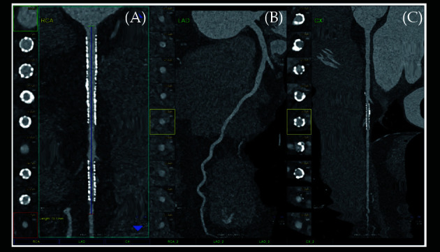
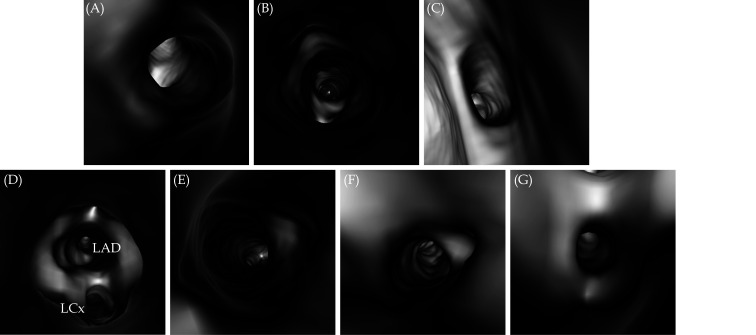
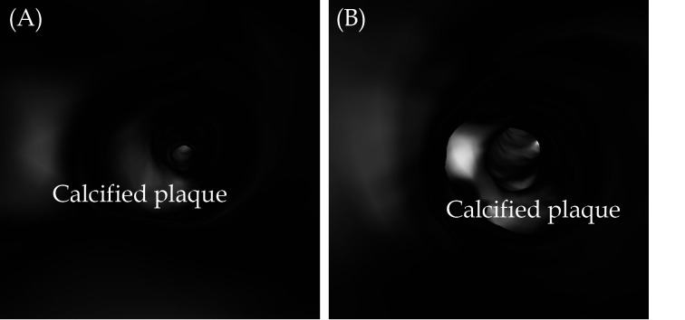
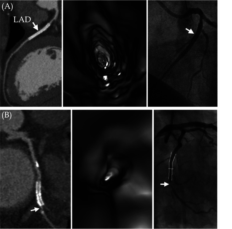
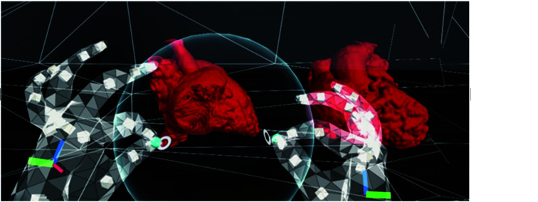
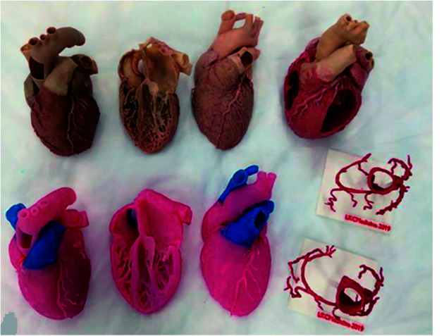
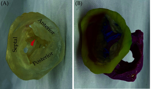
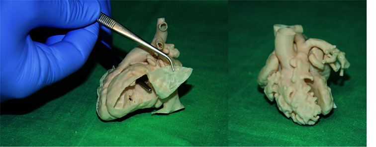
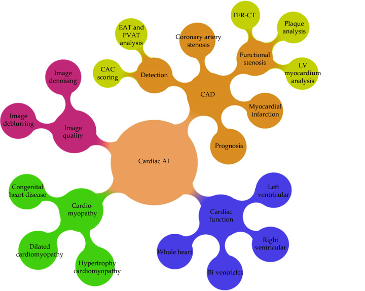
Cardiovascular computed tomography angiography (CTA) is a widely used imaging modality in the diagnosis of cardiovascular disease. Advancements in CT imaging technology have further advanced its applications from high diagnostic value to minimising radiation exposure to patients. In addition to the standard application of assessing vascular lumen changes, CTA-derived applications including 3D printed personalised models, 3D visualisations such as virtual endoscopy, virtual reality, augmented reality and mixed reality, as well as CT-derived hemodynamic flow analysis and fractional flow reserve (FFRCT) greatly enhance the diagnostic performance of CTA in cardiovascular disease. The widespread application of artificial intelligence in medicine also significantly contributes to the clinical value of CTA in cardiovascular disease. Clinical value of CTA has extended from the initial diagnosis to identification of vulnerable lesions, and prediction of disease extent, hence improving patient care and management. In this review article, as an active researcher in cardiovascular imaging for more than 20 years, I will provide an overview of cardiovascular CTA in cardiovascular disease. It is expected that this review will provide readers with an update of CTA applications, from the initial lumen assessment to recent developments utilising latest novel imaging and visualisation technologies. It will serve as a useful resource for researchers and clinicians to judiciously use the cardiovascular CT in clinical practice.
© 2024 JGC All rights reserved; www.jgc301.com.
Figures

Similar articles
-
Cardiovascular Computed Tomography in the Diagnosis of Cardiovascular Disease: Beyond Lumen Assessment.J Cardiovasc Dev Dis. 2024 Jan 12;11(1):22. doi: 10.3390/jcdd11010022. J Cardiovasc Dev Dis. 2024. PMID: 38248892 Free PMC article. Review.
-
Comparison of Coronary Computed Tomography Angiography, Fractional Flow Reserve, and Perfusion Imaging for Ischemia Diagnosis.J Am Coll Cardiol. 2019 Jan 22;73(2):161-173. doi: 10.1016/j.jacc.2018.10.056. J Am Coll Cardiol. 2019. PMID: 30654888 Clinical Trial.
-
Additional diagnostic value of new CT imaging techniques for the functional assessment of coronary artery disease: a meta-analysis.Eur Radiol. 2019 Jun;29(6):3044-3061. doi: 10.1007/s00330-018-5919-8. Epub 2019 Jan 7. Eur Radiol. 2019. PMID: 30617482
-
Diagnostic Performance of CT-Derived Fractional Flow Reserve in Australian Patients Referred for Invasive Coronary Angiography.Heart Lung Circ. 2022 Aug;31(8):1102-1109. doi: 10.1016/j.hlc.2022.03.008. Epub 2022 Apr 29. Heart Lung Circ. 2022. PMID: 35501246
-
Anatomical and Functional Computed Tomography for Diagnosing Hemodynamically Significant Coronary Artery Disease: A Meta-Analysis.JACC Cardiovasc Imaging. 2019 Jul;12(7 Pt 2):1316-1325. doi: 10.1016/j.jcmg.2018.07.022. Epub 2018 Sep 12. JACC Cardiovasc Imaging. 2019. PMID: 30219398
References
LinkOut - more resources
Full Text Sources