. 2024 Jul 15;137(14):jcs261892.
doi: 10.1242/jcs.261892. Epub 2024 Jul 22.

Distinct TORC1 signalling branches regulate Adc17 proteasome assembly chaperone expression

Affiliations

Distinct TORC1 signalling branches regulate Adc17 proteasome assembly chaperone expression

Thomas D Williams et al. J Cell Sci. .

Abstract

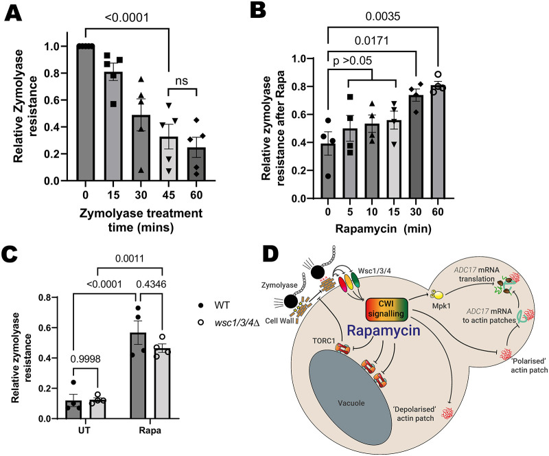
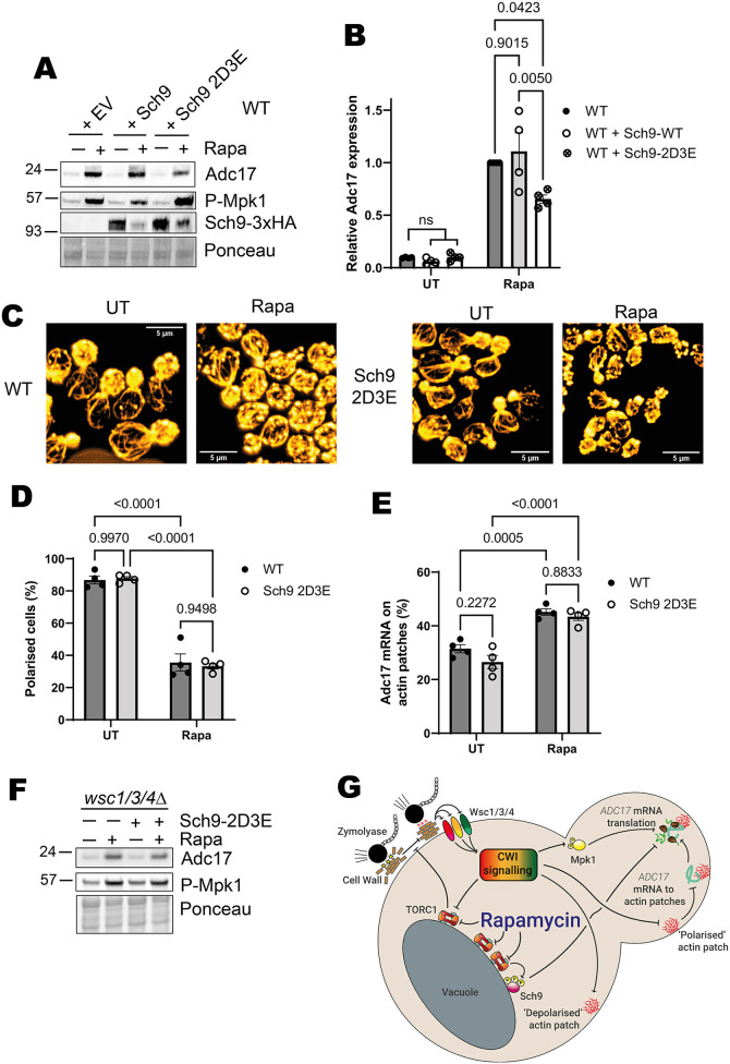
When stressed, cells need to adapt their proteome to maintain protein homeostasis. This requires increased proteasome assembly. Increased proteasome assembly is dependent on increased production of proteasome assembly chaperones. In Saccharomyces cerevisiae, inhibition of the growth-promoting kinase complex TORC1 causes increased proteasome assembly chaperone translation, including that of Adc17. This is dependent upon activation of the mitogen-activated protein kinase (MAPK) Mpk1 and relocalisation of assembly chaperone mRNA to patches of dense actin. We show here that TORC1 inhibition alters cell wall properties to induce these changes by activating the cell wall integrity pathway through the Wsc1, Wsc3 and Wsc4 sensor proteins. We demonstrate that, in isolation, these signals are insufficient to drive protein expression. We identify that the TORC1-activated S6 kinase Sch9 must be inhibited as well. This work expands our knowledge on the signalling pathways that regulate proteasome assembly chaperone production.

Keywords: Actin cytoskeleton; Mpk1 kinase; Proteasome assembly chaperone; Slt2 kinase; TORC1 signalling.


Conflict of interest statement

Competing interests The authors declare no competing or financial interests.

Figures

Fig. 1.
Fig. 1.
Mpk1 is activated and actin depolarised via the Wsc1, Wsc3 and Wsc4 cell wall integrity pathway sensors in response to rapamycin treatment. (A) Proteasome assembly chaperone translation regulation overview. Upon rapamycin treatment, TORC1 is inhibited, leading to activation of Mpk1 and ADC17 mRNA relocalisation to cortical actin patches, together driving increased Adc17 production. (B) Drop assay for WT and CWI sensor mutants on YEPD and YEPD plus rapamycin after 3 days of growth. Representative of three experiments. (C) Mpk1 activation (P-Mpk1) and Adc17 levels following 4 h rapamycin treatment in WT and wsc1Δ cells. Representative of three experiments. (D) Drop assay of WT and wsc1/3/4Δ cells on YEPD and YEPD plus rapamycin after 3 days of growth. Representative of three experiments. (E) Mpk1 activation (P-Mpk1) and Adc17 levels following 4 h rapamycin treatment in WT and wsc1/3/4Δ cells. Representative of three experiments. (F) Quantification of the blots shown in E (mean±s.e.m., n=3, two-way ANOVA followed by a Tukey's multiple comparisons test). UT, untreated; Rapa, rapamycin treated.
Fig. 2.
Fig. 2.
Actin is depolarised via the CWI pathway sensors in response to rapamycin treatment. (A) Effect of 1 h rapamycin treatment on actin polarisation in WT and wsc1/3/4Δ cells. Representative of five experiments. Cellular actin is labelled with rhodamine–phalloidin. Scale bars: 5 µm. (B) Quantification of images from A (mean±s.e.m., n=5, two-way ANOVA followed by a Tukey's multiple comparisons test). Budding cells are classified as unpolarised if there are six or more circular actin patches in the mother cell and polarised if this is not the case. (C) Updated scheme from Fig. 1A, showing role of Wsc1, Wsc3 and Wsc4 and CWI signalling in ADC17 translation. UT, untreated; Rapa, rapamycin treated.
Fig. 3.
Fig. 3.
Rapamycin treatment activates the CWL pathway by altering cell wall properties. (A) Time courses of zymolyase resistance in WT cells growing exponentially in YEPD (mean with s.e.m., n=5, one-way ANOVA followed by a Tukey's multiple comparisons test). (B) WT cell resistance to 45 min zymolyase treatment after treatment with rapamycin (mean±s.e.m., n=4, one-way ANOVA followed by a Tukey's multiple comparisons test). (C) Resistance to 45 min zymolyase treatment by WT and wsc1/3/4Δ cells that were either untreated or after 1 h rapamycin treatment (mean±s.e.m., n=4, two-way ANOVA followed by a Tukey's multiple comparisons test). (D) Updated scheme from Fig. 1A, showing role of cell wall alterations, which increase zymolyase resistance in ADC17 translation. UT, untreated; Rapa, rapamycin treated.
Fig. 4.
Fig. 4.
Cell wall stress activates the CWI pathway, Adc17 protein expression and TORC1 inhibition. (A) Mpk1 activation (P-Mpk1) and Adc17 levels in WT and mpk1Δ cells after 4 h Calcofluor White treatment. Representative of three experiments. (B) Effect of 1 h Calcofluor White treatment on actin polarisation in WT cells. Representative of four experiments. Cellular actin is labelled with rhodamine–phalloidin. Scale bars: 5 µm. (C) Quantification of images from B (mean±s.e.m., n=4, unpaired two-tailed t-test). Budding cells are classified as unpolarised if there are six or more circular actin patches in the mother cell and polarised if this is not the case. (D) Quantification of ADC17 mRNA localised to actin patches in untreated or 1 h Calcofluor White-treated WT cells (mean±s.e.m., n=5, unpaired two-tailed t-test). (E) TORC1 activity (P-Rps6) and Rps6 levels in WT cells after 4 h Calcofluor White treatment. Representative of three experiments. UT, untreated; CFW, Calcofluor White treated.
Fig. 5.
Fig. 5.
Mpk1 activation and ADC17 mRNA recruitment to CAPs is insufficient for Adc17 translation induction. (A) Scheme showing Mpk1 activation through inducible expression of the Mpk1 upstream kinase Mkk1 with the constitutively active ‘DD’ mutations (bottom) and ADC17 mRNA tethering to actin patches using the actin patch protein Abp1 tagged with an anti-GFP nanobody (top), which binds GFP-tagged PP7 coat protein (PCP), which in turn binds ADC17 mRNA with PP7 stem loops inserted into the 3′ UTR. (B) Mkk1-DD-3xHA and Adc17 levels, and Mpk1 and TORC1 activation (P-Mpk1 and P-Rps6, respectively) following 4 h rapamycin treatment or galactose induction in the cells with actin patch-tethered ADC17 mRNA (Adc17-24xSL Abp1-αGFP+PCP-GFP as described in Fig. 3A). Representative of at least 3 experiments. (C) Adc17 expression and Mpk1 and TORC1 activity (P-Mpk1 and P-Rps6, respectively) untreated and following 4 h rapamycin treatment in WT and ede1Δ cells with ADC17 mRNA tethered to actin patches. Representative of at least 3 experiments.
Fig. 6.
Fig. 6.
Sch9 inhibition is required for rapamycin-induced Adc17 expression by a CWI-independent mechanism. (A) Mpk1 activation (P-Mpk1) and Adc17 levels following 4 h rapamycin treatment in WT cells containing either empty vector (EV), WT Sch9 or the Sch9-2D3E constitutively active mutant. Representative of four experiments. (B) Quantification of blots shown in A (mean±s.e.m., n=4, two-way ANOVA followed by a Tukey's multiple comparisons test). (C) Effect of 1 h rapamycin treatment on actin polarisation in WT and Sch9-2D3E expressing cells. Representative of four experiments. Cellular actin is labelled with rhodamine-phalloidin. Scale bars: 5 µm. (D) Quantification of cellular actin polarisation from images shown in C (mean±s.e.m., n=4, two-way ANOVA followed by a Tukey's multiple comparisons test). Budding cells are classified as unpolarised if there are 6 or more circular actin patches in the mother cell and polarised if not. (E) Quantification of ADC17 mRNAs localised to actin patches in WT and Sch9-2D3E expressing cells from C (mean±s.e.m., n=4, two-way ANOVA followed by a Tukey's multiple comparisons test). (F) Effect of introducing constitutively active Sch9-2D3E into wsc1/3/4Δ cells on the response to rapamycin. Representative of three experiments. (G) Updated scheme from Fig. 1A, showing additional role of Sch9 in Adc17 protein translation. UT, untreated; Rapa, rapamycin treated.
Fig. 7.
Fig. 7.
Absence of Chc1 chronically activates the proteasome assembly pathway through Mpk1 activation and Sch9 inhibition. (A) Mpk1 activation (P-Mpk1) and Adc17 levels following rapamycin treatment in WT and chc1Δ cells. Representative of four experiments. (B) Sch9 levels in WT and chc1Δ cells. Representative of three experiments. (C) Mpk1 activity (P-Mpk1), TORC1 activity (P-Rps6) and Adc17 levels in WT and chc1Δ cells containing either empty vector (EV) or expressing Sch9-2D3E. Representative of four experiments. (D) Quantification of Adc17 levels from C (mean±s.e.m., n=4, one-way ANOVA followed by a Tukey's multiple comparisons test). (E) Actin polarisation in WT and chc1Δ cells. Representative of four experiments. Cellular actin is labelled with rhodamine-phalloidin. Scale bars: 5 µm. (F) Quantification of actin polarisation from images shown in D (mean±s.e.m., n=4, unpaired two-tailed t-test). Budding cells are classified as unpolarised if there are 6 or more circular actin patches in the mother cell and polarised if not. (G) Quantification of ADC17 mRNAs localised to actin patches in untreated WT and chc1Δ cells (mean±s.e.m., n=4, unpaired two-tailed t-test). (H) Scheme showing the proposed signalling control of Adc17 expression.

References

    1. Agrotis, A., Lamoliatte, F., Williams, T. D., Black, A., Horberry, R. and Rousseau, A. (2023). Multiple phosphorylation of the Cdc48/p97 cofactor protein Shp1/p47 occurs upon cell stress in budding yeast. Life Sci Alliance 6, e202201642. 10.26508/lsa.202201642 - DOI - PMC - PubMed
    1. Black, A., Williams, T. D., Soubigou, F., Joshua, I. M., Zhou, H., Lamoliatte, F. and Rousseau, A. (2023). The ribosome-associated chaperone Zuo1 controls translation upon TORC1 inhibition. EMBO J. 42, e113240. 10.15252/embj.2022113240 - DOI - PMC - PubMed
    1. Dupres, V., Alsteens, D., Wilk, S., Hansen, B., Heinisch, J. J. and Dufrêne, Y. F. (2009). The yeast Wsc1 cell surface sensor behaves like a nanospring in vivo. Nat. Chem. Biol. 5, 857-862. 10.1038/nchembio.220 - DOI - PubMed
    1. Dupres, V., Heinisch, J. J. and Dufrêne, Y. F. (2011). Atomic force microscopy demonstrates that disulfide bridges are required for clustering of the yeast cell wall integrity sensor Wsc1. Langmuir 27, 15129-15134. 10.1021/la203679s - DOI - PubMed
    1. Elorza, M. V., Rico, H. and Sentandreu, R. (1983). Calcofluor white alters the assembly of chitin fibrils in Saccharomyces cerevisiae and Candida albicans cells. J. Gen. Microbiol. 129, 1577-1582. - PubMed

Publication types

MeSH terms

Substances

LinkOut - more resources