. 2024 Oct 2;22(10):973-988.
doi: 10.1158/1541-7786.MCR-23-1003.

NAPRT Silencing in FH-Deficient Renal Cell Carcinoma Confers Therapeutic Vulnerabilities via NAD+ Depletion

Affiliations

NAPRT Silencing in FH-Deficient Renal Cell Carcinoma Confers Therapeutic Vulnerabilities via NAD+ Depletion

Katelyn J Noronha et al. Mol Cancer Res. .

Abstract

Hereditary leiomyomatosis and renal cell carcinoma (HLRCC) is caused by loss of function mutations in fumarate hydratase (FH) and results in an aggressive subtype of renal cell carcinoma with limited treatment options. Loss of FH leads to accumulation of fumarate, an oncometabolite that disrupts multiple cellular processes and drives tumor progression. High levels of fumarate inhibit alpha ketoglutarate-dependent dioxygenases, including the ten-eleven translocation (TET) enzymes, and can lead to global DNA hypermethylation. Here, we report patterns of hypermethylation in FH-mutant cell lines and tumor samples are associated with the silencing of nicotinate phosphoribosyl transferase (NAPRT), a rate-limiting enzyme in the Preiss-Handler pathway of NAD+ biosynthesis, in a subset of HLRCC cases. NAPRT is hypermethylated at a CpG island in the promoter in cell line models and patient samples, resulting in loss of NAPRT expression. We find that FH-deficient RCC models with loss of NAPRT expression, as well as other oncometabolite-producing cancer models that silence NAPRT, are extremely sensitive to nicotinamide phosphoribosyl transferase inhibitors (NAMPTi). NAPRT silencing was also associated with synergistic tumor cell killing with PARP inhibitors and NAMPTis, which was associated with effects on PAR-mediated DNA repair. Overall, our findings indicate that NAPRT silencing can be targeted in oncometabolite-producing cancers and elucidates how oncometabolite-associated hypermethylation can impact diverse cellular processes and lead to therapeutically relevant vulnerabilities in cancer cells. Implications: NAPRT is a novel biomarker for targeting NAD+ metabolism in FH-deficient HLRCCs with NAMPTis alone and targeting DNA repair processes with the combination of NAMPTis and PARP inhibitors.


Conflict of interest statement

K.J. Noronha reports grants from NIH (R01CA21543-05, R.S. Bindra; NRSA F31, K.J. Noronha) and Department of Defense (DOD; W81XWH-22-1-0549, B. Shuch and R.S. Bindra) during the conduct of the study and personal fees from AtlasXomics, Inc., outside the submitted work. K.N. Lucas reports grants from NIH (R01CA21543-05, R.S. Bindra) and DOD (W81XWH-22-1-0549, B. Shuch and R.S. Bindra) during the conduct of the study and other support from York Analytical Laboratories outside the submitted work. S. Friedman reports grants from NIH (R01CA21543-05, R.S. Bindra) and DOD (W81XWH-22-1-0549, B. Shuch and R.S. Bindra) during the conduct of the study. M.A. Murray reports grants from NIH NRSA F31 outside the submitted work. S. Liu reports grants from Yale College First-Year Research Fellowship during the conduct of the study. J. Spurrier reports employment with Alphina Therapeutics. M. Raponi reports a patent for Anti-NAPRT Antibodies and Methods of Use pending to Alphina Therapeutics. J.C. Vasquez reports grants from NIH K08, Robert Wood Johnson Harold Amos Medical Faculty Development Program, Doris Duke Charitable Foundation, and American Cancer Society Institutional Research Grant during the conduct of the study. No disclosures were reported by the other authors.

Figures

Figure 1.
Figure 1.
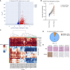
FH-deficient RCC models and patient tumors exhibit DNA hypermethylation of specific genes. A, Volcano plot of differential DNA methylation in YUNK1 shNT vs. YUNK1 shFH cell lines. B, Unsupervised clustering of FH-deficient RCC cell line models by methylation β values of NAPRT-associated probes. C, Boxplot showing methylation β values of probes from DMGs in relation to CpG island features. Statistics (Kruskal–Wallis H test) reported in Supplementary Table S3. D, Schematic of NAPRT promoter. E, Methylation array β values arranged by probe chromosomal location across cell lines models. F, Protein expression of FH and NAPRT in FH-deficient RCC models detected by western blot (left). Quantification of relative NAPRT signal is normalized to YUNK1 parental (right, n = 3). G and H, Western blot of NAPRT expression after treatment with 5-aza-2′-deoxycytidine for 7 days in UOK262 (G) and NCCFH1 (H). Quantification of NAPRT signal (right) shown, normalized to highest intensity band (n = 3). HKP, house-keeping protein. One-way ANOVA: **, P ≤ 0.01; ***, P ≤ 0.001; ****, P ≤ 0.0001.
Figure 2.
Figure 2.
NAPRT silencing in FH-deficient RCC models is associated with promoter hypermethylation. A, Volcano plot of DMGs in FH-deficient RCC normal (n = 12) versus tumor tissue (n = 20; ref. 13). B, Average methylation β value of all NAPRT gene methylation probes in normal and FH-deficient RCC tumor tissue (13). Matched normal and tumor samples are connected. C, Unsupervised clustering of FH-deficient RCC tumor samples and normal tissue samples (13) by β values of NAPRT associated probes. D, Summary of NAPRT IHC scoring in samples from patients with FH-deficient HLRCC. E, Representative hematoxylin and eosin (H&E; top) and NAPRT IHC (bottom) images in adjacent normal tissue and FH-deficient tumors from patients with HLRCC. Scale bar, 1 mm.
Figure 3.
Figure 3.
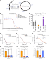
NAPRT loss confers sensitivity to NAMPTis in FH-deficient RCC models. A, Schematic of NAD+ biosynthesis pathways, Triple arrow in the de novo synthesis pathway represents five additional enzymatic conversion steps in the pathway. B, YUNK1 cells treated with increasing concentrations of FK866 with or without the addition of 10-μmol/L of NA. C, Total NAD+ levels in YUNK1 and YUNK1 shFH cells at baseline and upon treatment with 25-nmol/L FK866 for 24 hours alone or with 10-μmol/L NA. D–F, FH-deficient and NAPRT-silenced NCCFH1 (D), UOK262 (E), and UOK268 (F) cell lines treated with increasing concentrations of FK866 after 8 (D), 6 (E), or 8 (F) days with or without 10-μmol/L NA. G–I, Total NAD+ quantification in NCCFH1 (G), UOK262 (H), and UOK268 (I) after 24-hour treatment with 25-nmol/L FK866 alone or with 10-μmol/L NA. Two-way ANOVA, *, P = 0.0151; ****, P ≤ 0.0001. Trp, tryptophan; QA, quinolinic acid; QPRT, Quinolinate phosphoribosyl transferase; NAMN, nicotinic acid mononucleotide; NAPRT, nicotinate phosphoribosyltransferase; NA, nicotinic acid; NMNAT, Nicotinamide mononucleotide adenylyl transferase; NAAD, nicotinic acid adenine dinucleotide; NADS, NAD+ synthetase; PARPs, poly(ADP-ribose) polymerases, NAM, nicotinamide; NAMPT, nicotinamide phosphoribosyltransferase; NMN, nicotinamide mononucleotide; NMNAT, nicotinamide mononucleotide adenylyl transferase.
Figure 4.
Figure 4.
NAMPTi and PARPi synergize in NAPRT-deficient RCC models. A, Mapped synergy plots of YUNK1 parental (left) and YUNK1 shFH (right) cell lines treated with combinations of increasing doses of FK866 and olaparib for 6 days. B–D, Mapped synergy plots of UOK262 (B), NCCFH1 (C), and UOK268 (D) cell lines treated with increasing doses of FK866 and olaparib for 6 days. All synergies performed with 10-μmol/L NA and all synergy scores calculated with the BLISS model.
Figure 5.
Figure 5.
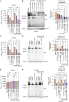
Combination of NAMPTi and PARPi reduces NAD+ and PAR chain formation. A, Total NAD+ quantification in NCCFH1 cell line after 24-hour treatment with FK866 alone or with 0.6-μmol/L olaparib. B, PAR chains detected by western blot (left) in NCCFH1 cells treated for 24 hours at indicated concentrations of FK866 and olaparib and quantified (right, n = 3). All samples were also treated with 10-μmol/L NA. Arrow indicates the molecular weight for PARP1-associated PARylation. C, Total NAD+ quantification in UOK262 cell line after 24-hour treatment with FK866 alone or in combination with 0.6-μmol/L olaparib. D, PAR chains detected by western blot (left) in UOK262 cells treated for 24 hours with the indicated combinations of 1.0-nmol/L FK866, 0.6-μmol/L olaparib, and 10-μmol/L NA and quantified (right, n = 3). E, Total NAD+ quantification in UOK268 cell line after 24-hour treatment with FK866 alone or in combination with 0.6-μmol/L olaparib. F, PAR chains detected by western blot (left) in UOK268 cells treated for 24 hours with the indicated combinations of 1.0-nmol/L FK866, 0.6-μmol/L olaparib, and 10-μmol/L NA and quantified (right, n = 3). All NAD+ assays performed in the presence of 10 μmol/L NA. HKP, house-keeping protein. Two-way ANOVA: *, P ≤ 0.05; **, P ≤ 0.01; ***, P ≤ 0.001; ****, P ≤ 0.0001.
Figure 6.
Figure 6.
NAMPTi and PARPi combination increases PARP retention at the chromatin. A, Subcellular fractionation of NCCFH1 cells after treatment with combinations of 25-nmol/L FK866, 10-μmol/L olaparib, 10-μmol/L BMN673, and 0.01% MMS. B, Quantification of nuclear soluble PARP1 signal relative to Sirt6 signal (top) and chromatin-bound PARP1 signal relative to H3 signal (bottom) in NCCFH1 cells. All lanes were normalized to DMSO within the same blot (n = 3). C, Subcellular fractionation of UOK268 cells after treatment with combinations of 25-nmol/L FK866, 10-umol/L olaparib, 10-umol/L BMN673, and 0.01% MMS. D, Quantification of nuclear soluble PARP1 signal relative to Sirt6 signal (top) and chromatin-bound PARP1 signal relative to H3 signal (bottom) in UOK268 cells. All lanes were normalized to DMSO within the same blot (n = 3). Cells were treated with FK866 and 10-μmol/L NA for 24 hours and then with PARPis and/or MMS for 30 minutes before harvesting. Sirt6 is a nuclear-localized protein. H3 is a histone protein localized to chromatin. One-way ANOVA, **, P = 0.009; ***, P ≤ 0.001. Statistics for significant comparisons are shown (B and D), others are nonsignificant.
Figure 7.
Figure 7.
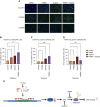
Combination of PARPi and NAMPTi increases DNA damage in NAPRT-silenced models. A, Representative images of NCCFH1 (top), UOK262 (middle), and UOK268 (bottom) with indicated 25-nmol/L FK866 and/or 5-μmol/L olaparib treatment for 24 hours. All cells were treated with 10-μmol/L NA. Scale bar, 50 μm. B–D. Quantification of the proportion of γH2AX foci positive cells in NCCFH1 (B), UOK262 (C), and UOK268 (D) cells. Thresholds for counting foci positive cells were set by the number of γH2AX foci in DMSO-treated cells. One-way ANOVA, **, P ≤ 0.01; ***, P ≤ 0.001; ****, P ≤ 0.0001. E, Schematic describing current understanding of NAPRT silencing in 75% of FH-deficient cell lines and 90% of patient samples and associated drug sensitivity in FH-deficient cancers.

References

    1. Zheng L, Zhu Z-R, Sneh T, Zhang W-T, Wang Z-Y, Wu G-Y, et al. . Circulating succinate-modifying metabolites accurately classify and reflect the status of fumarate hydratase–deficient renal cell carcinoma. J Clin Invest 2023;133:e165028. - PMC - PubMed
    1. Srinivasan R, Gurram S, Harthy MA, Singer EA, Sidana A, Shuch BM, et al. . Results from a phase II study of bevacizumab and erlotinib in subjects with advanced hereditary leiomyomatosis and renal cell cancer (HLRCC) or sporadic papillary renal cell cancer. J Clin Oncol 2020;38(15_Suppl):5004.
    1. Carril-Ajuria L, Colomba E, Cerbone L, Romero-Ferreiro C, Crouzet L, Laguerre B, et al. . Response to systemic therapy in fumarate hydratase-deficient renal cell carcinoma. Eur J Cancer 2021;151:106–14. - PubMed
    1. Yang H, Ye D, Guan KL, Xiong Y. IDH1 and IDH2 mutations in tumorigenesis: mechanistic insights and clinical perspectives. Clin Cancer Res 2012;18:5562–71. - PMC - PubMed
    1. Sciacovelli M, Gonçalves E, Johnson TI, Zecchini VR, da Costa AS, Gaude E, et al. . Fumarate is an epigenetic modifier that elicits epithelial-to-mesenchymal transition. Nature 2016;537:544–7. - PMC - PubMed

Publication types

MeSH terms

Supplementary concepts