Review
. 2024 Aug 2;16(15):1583-1599.
doi: 10.1080/17568919.2024.2366146. Epub 2024 Jul 1.

Regulation of post-translational modification of PD-L1 and associated opportunities for novel small-molecule therapeutics

Affiliations
Review

Regulation of post-translational modification of PD-L1 and associated opportunities for novel small-molecule therapeutics

Jinglin Tang et al. Future Med Chem. .

Abstract

PD-L1 is overexpressed on the surface of tumor cells and binds to PD-1, resulting in tumor immune escape. Therapeutic strategies to target the PD-1/PD-L1 pathway involve blocking the binding. Immune checkpoint inhibitors have limited efficacy against tumors because PD-L1 is also present in the cytoplasm. PD-L1 of post-translational modifications (PTMs) have uncovered numerous mechanisms contributing to carcinogenesis and have identified potential therapeutic targets. Therefore, small molecule inhibitors can block crucial carcinogenic signaling pathways, making them a potential therapeutic option. To better develop small molecule inhibitors, we have summarized the PTMs of PD-L1. This review discusses the regulatory mechanisms of small molecule inhibitors in carcinogenesis and explore their potential applications, proposing a novel approach for tumor immunotherapy based on PD-L1 PTM.

Keywords: N-linked glycosylation; PD-L1; PTMs; acetylation; methylation; palmitoylation; phosphorylation; ubiquitination.

Plain language summary

[Box: see text].


Conflict of interest statement

The authors have no competing interests or relevant affiliations with any organization or entity with the subject matter or materials discussed in the manuscript. This includes employment, consultancies, honoraria, stock ownership or options, expert testimony, grants or patents received or pending, or royalties.

Figures

None
Graphical abstract
Figure 1.
Figure 1.
The schematic diagram for two signals of T-cell activation. When a T-cell encounters an Ag, it initially binds to the MHC using its TCR. Subsequently, a second signal is generated when the costimulatory molecule on APCs interacts with the corresponding receptor or ligand on the surface of T-cells such as CD28 binding to CD80. Both signals collaborate to initiate T-cell function. Ag: Antigen; APC: Antigen-presenting cell; MHC: Major histocompatibility complex; TCR: T-cell receptor.
Figure 2.
Figure 2.
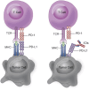
The schematic diagram for tumor immune escape and principles of immune checkpoint inhibitors therapy. T-cells serve as ‘sentinels’ capable of identifying tumor cells within the body and executing cytotoxic attacks. Tumor cells will produce the PD-L1 protein in response to the presence of the PD-1 protein on T-cells, which binds to the PD-1 protein and transmits inhibitory signals, thereby impeding normal T-cell function, inducing T-cell apoptosis and facilitating immune evasion. ICIs block the binding of PD-1 to PD-L1, thereby enabling T-cell functionality. ICI: Immune checkpoint inhibitor.
Figure 3.
Figure 3.
Chemical structure of PD-L1 inhibitor, PD-L1 glycosylation inhibitors and agonist.
Figure 4.
Figure 4.
Chemical structures of small molecule PD-L1 polyubiquitination, deubiquitination and phosphorylation inhibitors.
Figure 5.
Figure 5.
Regulations of PD-L1 by proteolysis targeting chimera. The AbTAC of PD-L1 is a specific antibody that binds to both PD-L1 and the E3 ubiquitin ligase complex RNF-43, thereby inducing the lysosomal degradation pathway for PD-L1. Peptide PROTAC targets palmitoacyltransferase DHHC3, which stabilizes PD-L1, leading to its degradation through the lysosomal pathway. Another peptide-PROTAC, named FOXM1-Protac, degrades PD-L1 by inhibiting the degradation of FOXM1. PROTAC: Proteolysis targeting chimera.
Figure 6.
Figure 6.
Regulations of PD-L1 though polyubiquitination, deubiquitination. PD-L1 can be degraded through binding to E3 ubiquitin ligase complexes, such as cullin3 SPOP, STUB1, β-Trcp, HUWEI, HRD1, ARIH1, MARCH8 (blue circle), followed by proteasomal degradation. The ubiquitination of PD-L1 can also be inhibited by deubiquitinating enzymes such as CSN5, USP22, OTUB1, USP9X (orange circle). Currently, some small molecule inhibitors like curcumin, C-15 and WP1130 have been discovered to inhibit the PD-L1 deubiquitination pathway and degrade PD-L1. Several upstream factors and enzymes including CDK4, CMTM4/6, GSK3 and ERAD are involved in regulating the ubiquitination of PD-L1.
Figure 7.
Figure 7.
Chemical structure of palmitoacyltransferase inhibitors.
Figure 8.
Figure 8.
Regulations of PD-L1 by post-translational modifications. The schematic illustrates the molecular functional domain of the PD-L1 protein and its various PTMs. PD-L1 consists of two extracellular Ig-like domains, a TM region and an ICD. The activity and stability of PD-L1 are regulated by a range of PTMs, including N-glycosylation (G, red circles), phosphorylation (P, yellow circles), methylation (M, purple squares), palmitoylation (PM, five-pointed yellow stars) and acetylation (Ac, green circles). Glycosylation and palmitoylation are crucial for maintaining the stability of PD-L1. Ubiquitination can facilitate the degradation of PD-L1. Phosphorylation can modulate levels of PD-L1 by indirectly influencing glycosylation and ubiquitination. ICD: Intracellular domain; PTM: Post-translational modification; TM: Transmembrane.

References

    1. Grakoui A, Bromley SK, Sumen C, et al. The immunological synapse: a molecular machine controlling T cell activation. Science. 1999;285:221–227. doi: 10.1126/science.285.5425.221 - DOI - PubMed
    1. Monks CRF, Freiberg BA, Kupfer H, et al. Three-dimensional segregation of supramolecular activation clusters in T-cells. Nature. 1998;395:82–86. doi: 10.1038/25764 - DOI - PubMed
    1. Saito T, Yokosuka T, Hashimoto-Tane A, et al. Dynamic regulation of T-cell activation and co-stimulation through TCR-microclusters. FEBS Lett. 2010;584:4865–4871. doi: 10.1016/j.febslet.2010.11.036 - DOI - PubMed
    1. Cadranel J, Canellas A, Matton L, et al. Pulmonary complications of immune checkpoint inhibitors in patients with nonsmall cell lung cancer. Eur Respir Rev. 2019;28:190058. doi: 10.1183/16000617.0058-2019 - DOI - PMC - PubMed
    1. Isaacsson Velho P, Antonarakis ES, Isaacsson Velho P, et al. PD-1/PD-L1 pathway inhibitors in advanced prostate cancer. Expert Rev Clinical Pharmacology. 2018;11:475–486. doi: 10.1080/17512433.2018.1464388 - DOI - PMC - PubMed

MeSH terms