. 2024 Oct 10;144(15):1633-1645.
doi: 10.1182/blood.2023023349.

Use of HSC-targeted LNP to generate a mouse model of lethal α-thalassemia and treatment via lentiviral gene therapy

Affiliations

Use of HSC-targeted LNP to generate a mouse model of lethal α-thalassemia and treatment via lentiviral gene therapy

Maxwell E Chappell et al. Blood. .

Abstract

α-Thalassemia (AT) is one of the most commonly occurring inherited hematological diseases. However, few treatments are available, and allogeneic bone marrow transplantation is the only available therapeutic option for patients with severe AT. Research into AT has remained limited because of a lack of adult mouse models, with severe AT typically resulting in in utero lethality. By using a lipid nanoparticle (LNP) targeting the receptor CD117 and delivering a Cre messenger RNA (mRNACreLNPCD117), we were able to delete floxed α-globin genes at high efficiency in hematopoietic stem cells (HSC) ex vivo. These cells were then engrafted in the absence or presence of a novel α-globin-expressing lentiviral vector (ALS20αI). Myeloablated mice infused with mRNACreLNPCD117-treated HSC showed a complete knock out (KO) of α-globin genes. They showed a phenotype characterized by the synthesis of hemoglobin H (HbH; also known as β-tetramers or β4), aberrant erythropoiesis, and abnormal organ morphology, culminating in lethality ∼8 weeks after engraftment. Mice infused with mRNACreLNPCD117-treated HSC with at least 1 copy of ALS20αI survived long term with normalization of erythropoiesis, decreased production of HbH, and amelioration of the abnormal organ morphology. Furthermore, we tested ALS20αI in erythroid progenitors derived from α-globin-KO CD34+ cells and cells isolated from patients with both deletional and nondeletional HbH disease, demonstrating improvement in α-globin/β-globin mRNA ratio and reduction in the formation of HbH by high-performance liquid chromatography. Our results demonstrate the broad applicability of LNP for disease modeling, characterization of a novel mouse model of severe AT, and the efficacy of ALS20αI for treating AT.


Conflict of interest statement

Conflict-of-interest disclosure: H.P. and D.W. are scientific founders and hold equity in Capstan Therapeutics. Y.K.T. and B.L.M. are employees and hold equity in Acuitas Therapeutics. D.W. receives research support from BioNTech. F.D.B. is founder of Biocept; reports intellectual property licensed to Novartis; and serves as a consultant for Sana Biotechnology, Poseida Therapeutics, Encoded Therapeutics, and Johnson & Johnson. In accordance with the University of Pennsylvania policies and procedures and our ethical obligations as researchers, H.P., and D.W. are named on several patents that describe the use of nucleoside-modified mRNA and targeted LNP as platforms to deliver therapeutic proteins and vaccines. The vector ALS20αI is protected in the patent “Generation of a lentiviral vector for the cure of alpha-thalassemia” (The Children's Hospital of Philadelphia, M.E.C., and S.R.). The remaining authors declare no competing financial interests.

Figures

None
Graphical abstract
Figure 1.
Figure 1.
Characterization of WT, Hba-a1Fl/Fl/Hba-a2KO/KO + mRNACre-LNPCD117 (AG-KO), and HSC Hba-a1Fl/Fl/Hba-a2KO/KO + mRNACre-LNPCD117 + ALS20αI (AG-KOALS20αI) transplant recipients. (A) Scheme showing the Cre-mediated excision of Hba-a1 after administration of mRNACre-LNPCD117. Although administration of mRNACreLNPCD117 offers impressive in vivo recombination, we opted for in vitro treatment and engraftment of isolated lin cells to ensure the near total gene deletion. (B) Agarose gels showing the deletion of the Hba-a1F) genes in cells collected and kept in vitro after lin cell collection. Amplification after recombination results in a 496-base pair (bp) product, whereas the absence of recombination (no cells exposed to LNP) does not produce an amplification because of size constraints. The deletion was observed after treatment with mRNACre-LNPCD117 and keeping the cells in culture, as indicated. Only relevant lanes are shown; the full gel is shown in supplemental Figure 37. The black line separates 2 different sections of the gel. (C) Levels of endogenous genomic mouse Hba (Hba-a1 + Hba-a2), after treatment with mRNACre-LNPCD117 measured by droplet digital polymerase chain reaction (ddPCR). The ddPCR assay amplifies any remaining intact genomic α-globin gene, either Hba-a1 or Hba-a2. The residual genomic α-globin amplification in AG-KO level mice is compared with nonrecombined WT control (4 copies of the α-globin genes) and mice carrying 3 or 2 copies of the α-globin genes, shown as a percentage. The WT is set to 100% by default. (D) Cation-exchange HPLC chromatograms of WT, AG-KO, and AG-KOALS20αI mice that received transplantation were analyzed. Absolute counts of erythroblasts in the BM (E) and the spleen (F) include proerythroblasts (I, in purple), basophilic erythroblasts (II, in blue), polychromatic erythroblasts (III, in yellow), orthochromatic erythroblasts and reticulocytes (IV, in green), and RBC (V, in red). Significance was assessed by ordinary 2-way analysis of variance (ANOVA) using Tukey multiple comparisons test (n = 4-6; test described in supplemental Table 1). (G) Oxygen equilibrium curves of WT and AG-KO mice, as well as AG-KOALS20αI with VCN in the range of 1.1 to 2.9, values were extrapolated using the method described in supplementary Materials, in “Oxygen binding affinity.”
Figure 2.
Figure 2.
Evaluation of gene expression and spleen-to-body weight ratios in WT, AG-KO, and AG-KOALS20αIrecipients. (A) EPO gene expression was measured in kidney tissue of WT (n = 12), AG-KO (n = 14), and AG-KOALS20αI (n = 7) animals. (B) Hepcidin (HAMP) gene expression was measured in liver tissue of WT (n = 12), AG-KO (n = 14), and AG-KOALS20αI (n = 6) animals. (C) Ratio of the spleen-to-body weight measured in WT (n = 2), AG-KO (n = 6), and AG-KOALS20αI (n = 7). The mean and standard error of the mean are indicated. Significant differences between groups were measured using a Kruskal-Wallis test with Dunns multiple comparisons test: ∗P ≤ .05; ∗∗P ≤ .01; ∗∗∗P ≤ .001; ∗∗∗∗P ≤ .0001.
Figure 3.
Figure 3.
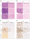
Histology of the spleen and liver, and immunohistochemistry of lung and brain tissue from WT, AG-KO, and AG-KOALS20αIrecipient animals. (A) Representative spleens from WT, AG-KO, and AG-KOALS20αI mouse chimeras stained with hematoxylin and eosin (H&E) and Perls Prussian blue, shown at ×4 original magnification. The spleens of WT animals exhibit sites of high iron staining corresponding to the retention of iron in the macrophages. (B) Representative livers from WT, AG-KO, and AG-KOALS20αI recipient animals stained with H&E and Perls Prussian blue, shown at ×10 original magnification. Arrows indicate sites of extramedullary hematopoiesis. (C) Brain tissues from WT, AG-KO, and AG-KOALS20αI mouse chimeras were stained using antifibrinogen and anti–von Willebrand factor (VWF) immunohistochemistry. Numbered arrows indicate sites of possible vessel occlusion. (D) Lung tissue from WT, AG-KO, and AG-KOALS20αI mouse chimeras was stained using antifibrinogen and anti-VWF immunohistochemistry. Numbered arrows indicate sites of possible vessel occlusion. Arrows 1 and 5 indicate sites of fibrinogen staining, whereas arrows 2-4 and 6 indicate sites of high VWF staining.
Figure 4.
Figure 4.
Schematic diagrams of lentiviral vectors expressing the human α-globin gene. Vectors differ in the inclusion and orientation of core sequences of the DNase I hypersensitive sites (HS) of the β-globin locus control region (LCR) but maintain the same short β-globin promoter, 3′ enhancer, and untranslated regions (UTRs). These vectors also use a 3′ self-inactivating LTR (sinLTR) that includes an Ankyrin insulator element and HBA2 genomic sequence. All vectors include HS2, HS3, and HS4 of the β-globin LCR. In addition, ALS19αI includes HS1 (in 3′→5′ orientation), whereas ALS20αI includes HS1 (in 3′→5′ orientation).
Figure 5.
Figure 5.
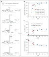
Evaluation of the effect of ALS20αI transduction on α-globin–KO cells. (A) Chromatograms from cation-exchange HPLC of hemolysates from nontransduced and ALS20αI–transduced HUDEP-αKO cells (clone 9). In nontransduced HUDEP-αKO cells, only a single peak corresponding to HbH (β4) is visible, because of the lack of all α-globin chains. After transduction, HbA (α2β2) and HbF (α2γ2) peaks become visible, whereas HbH decreases. (B) Correlation between VCN and percent HbA + HbF + HbA2 tetramers in HUDEP-αKO clones and α-globin-KO CD34+–derived erythroid cells. (C) Correlation between VCN and percent α-globin chains (top); correlation between VCN and percent single β-globin-like chains (β + γ + δ; bottom) detected by reversed-phase HPLC in hemolysates from terminally differentiated primary α-KO CD34+ cells or immortalized HUDEP-αKO erythroblast untreated or treated with ALS20αI. Gray bars represent values in healthy human erythroblasts. The values normalize at VCN of ∼4 for both α-globin–KO CD34+ and HUDEP-αKO cells, indicating that 1 copy of ALS20αI can generate as many α-globin chains as 1 endogenous α-globin gene.
Figure 6.
Figure 6.
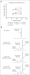
Stable engraftment and expression of human α-globin protein in primary, secondary, and tertiary transplanted mice. (A) The VCN was measured via ddPCR analysis of genomic DNA (gDNA) from AG-KOALS20αI mice. gDNA for the first 2 time points was isolated from the peripheral blood, whereas gDNA from the final time point was isolated from the BM. Each point represents a single mouse. (B) Cation-exchange HPLC analysis of RBC lysate collected from primary, secondary, and tertiary transplanted AG-KOALS20αI mice. This analysis indicated that ALS20αI transduced long-term HSC, and its expression is stable over time.
Figure 7.
Figure 7.
Evaluation of the effect of ALS20αI transduction on HbH and α-globin mRNA levels in mutant cells. (A) The percentage of total HbH relative to VCN measured by cation-exchange HPLC in erythroblasts differentiated from HUDEP-αKO clones, α-globin–KO CD34+ cells, and cells from patients with HbH. Each symbol represents 1 biological replicate: Patients 1 (n = 12), 3 (n = 3), 4 (n = 5), and 5 (n = 5). Genotypes are summarized in Table 2. In HUDEP-αKO clone 9, n = 7, in clone 7, n = 2, and in clone 62, n = 2. In α-globin–KO CD34+, n = 2. (B) The HBA:(HBB + HBG + HBD) mRNA relative to VCN in a patient with the –α3.7/− −SEA deletion (n = 8, patient 1 in Table 2), in a patient with homozygous Agrinio (427 T>A) mutation (n = 5, patient 5 in Table 2), and in α-globin–KO CD34+–derived erythroid cells. These analyses also suggested that 1 copy of ALS20αI produced as many human α-globin chains as those of a single endogenous α-globin gene.

Comment in

References

    1. Higgs DR, Gibbons RJ. The molecular basis of alpha-thalassemia: a model for understanding human molecular genetics. Hematol Oncol Clin North Am. 2010;24(6):1033–1054. - PubMed
    1. Taher AT, Weatherall DJ, Cappellini MD. Thalassaemia. Lancet. 2018;391(10116):155–167. - PubMed
    1. Galanello R, Cao A. Alpha-thalassemia. Genet Med. 2011;13(2):83–88. - PubMed
    1. Hughes JR, Cheng JF, Ventress N, et al. Annotation of cis-regulatory elements by identification, subclassification, and functional assessment of multispecies conserved sequences. Proc Natl Acad Sci U S A. 2005;102(28):9830–9835. - PMC - PubMed
    1. Sollaino MC, Paglietti ME, Loi D, Congiu R, Podda R, Galanello R. Homozygous deletion of the major alpha-globin regulatory element (MCS-R2) responsible for a severe case of hemoglobin H disease. Blood. 2010;116(12):2193–2194. - PubMed

Publication types