. 2024 Jun 17;134(12):e179559.
doi: 10.1172/JCI179559.

CIAO1 loss of function causes a neuromuscular disorder with compromise of nucleocytoplasmic Fe-S enzymes

Affiliations

CIAO1 loss of function causes a neuromuscular disorder with compromise of nucleocytoplasmic Fe-S enzymes

Nunziata Maio et al. J Clin Invest. .

Abstract

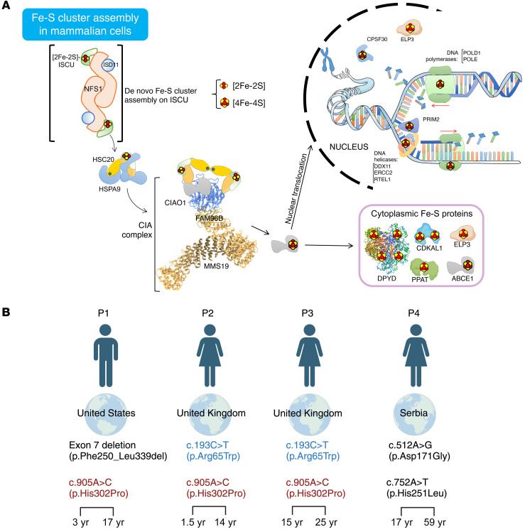
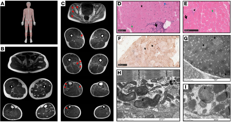
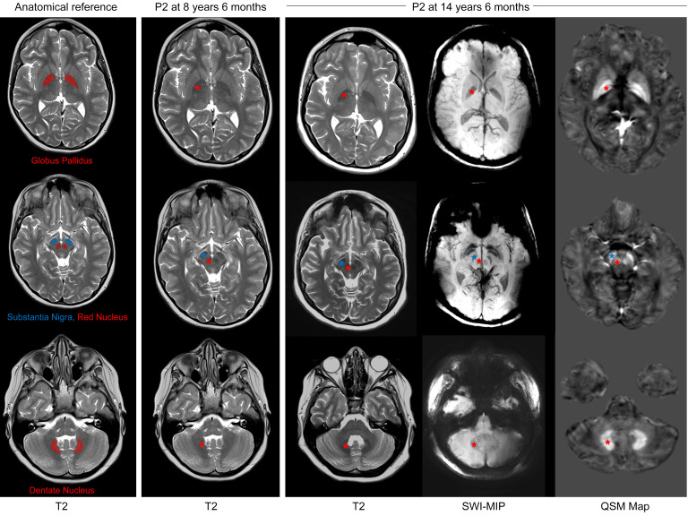
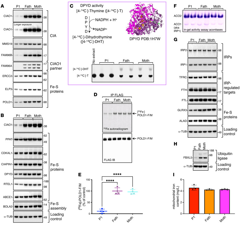
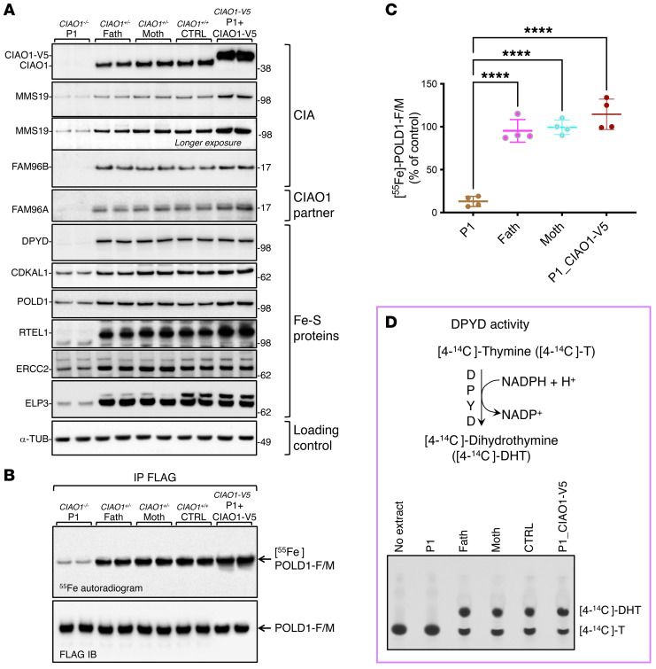
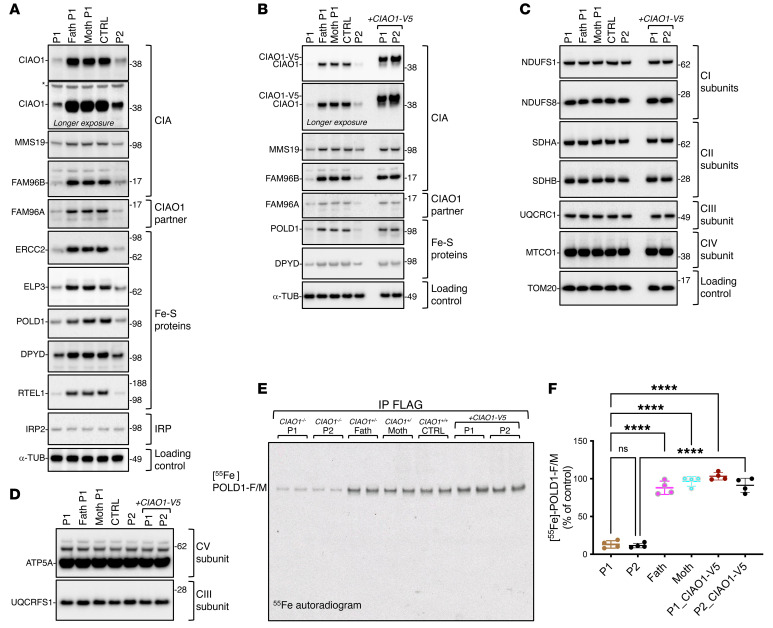
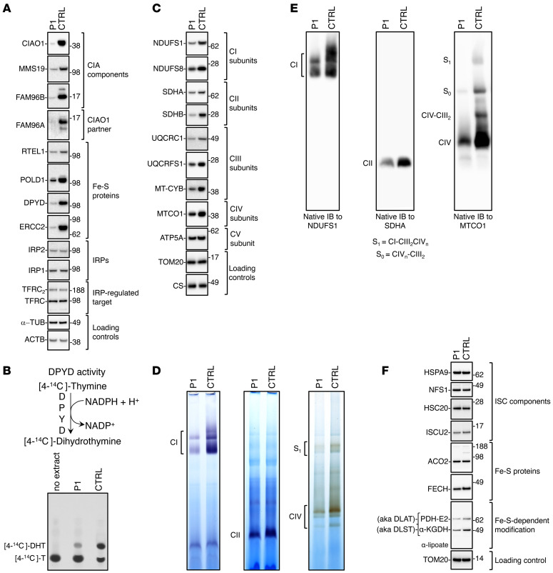
Cytoplasmic and nuclear iron-sulfur (Fe-S) enzymes that are essential for genome maintenance and replication depend on the cytoplasmic Fe-S assembly (CIA) machinery for cluster acquisition. The core of the CIA machinery consists of a complex of CIAO1, MMS19 and FAM96B. The physiological consequences of loss of function in the components of the CIA pathway have thus far remained uncharacterized. Our study revealed that patients with biallelic loss of function in CIAO1 developed proximal and axial muscle weakness, fluctuating creatine kinase elevation, and respiratory insufficiency. In addition, they presented with CNS symptoms including learning difficulties and neurobehavioral comorbidities, along with iron deposition in deep brain nuclei, mild normocytic to macrocytic anemia, and gastrointestinal symptoms. Mutational analysis revealed reduced stability of the variants compared with WT CIAO1. Functional assays demonstrated failure of the variants identified in patients to recruit Fe-S recipient proteins, resulting in compromised activities of DNA helicases, polymerases, and repair enzymes that rely on the CIA complex to acquire their Fe-S cofactors. Lentivirus-mediated restoration of CIAO1 expression reversed all patient-derived cellular abnormalities. Our study identifies CIAO1 as a human disease gene and provides insights into the broader implications of the cytosolic Fe-S assembly pathway in human health and disease.

Keywords: DNA repair; Genetic diseases; Metabolism; Muscle biology.


Figures

Figure 1
Figure 1. Identification of biallelic CIAO1 variants in 4 independent patients with a neuromuscular condition of undefined etiology.
(A) Proposed model for the biogenesis of ISCs in mammalian cells. De novo assembly of ISCs occurs upon the main scaffold protein ISCU by the coordinated action of a multiprotein complex, which consists of the cysteine desulfurase NFS1 and the accessory protein ISD11. The HSC20-HSPA9 cochaperone-chaperone complex interacts with ISCU to facilitate cluster transfer to recipient proteins. The functional unit of HSC20 is a dimer (15). A subset of recipient Fe-S proteins acquire their clusters directly from the HSC20-HSPA9-ISCU1 complex (15). In the cytoplasm, binding of HSC20 to the LYR motif of CIAO1 recruits the CIA-targeting complex, which is known to form a platform to which Fe-S recipients involved in DNA metabolism dock to acquire their clusters (13, 14). The Fe-S proteins shown in the model were all identified as HSC20 interacting partners (15) (i.e., NUBP2, GLRX3, CIAPIN1, ABCE1, ERCC2, POLD1, PRIM2, PPAT, ELP3, CPSF30, DDX11, etc.). (B) Diagram showing the country of origin of the 4 patients with biallelic CIAO1 variants. Recurring variants are shown in red and blue. The age when symptoms were first recognized (on the left) and the age at the latest clinical assessment (on the right) are shown at the bottom. Figure 1B was created with BioRender.com.
Figure 2
Figure 2. Muscle MRI, histopathology, and ultrastructural findings.
(A) Anatomical reference. MRI scan positions are indicated with red lines on a human reference image. Figure 2A was created with BioRender.com. (B) Normal muscle MRI cross-sectional images showing anatomy of pelvic (top), thigh (middle), and lower leg (bottom) muscles. AL, adductor longus; AM, adductor magnus; BF, biceps femoris; EDL, extensor digitorum longus; Gmax, gluteus maximus; Gmed, gluteus medius; Gmin, gluteus minimus; Gr, gracilis; Il, iliacus; LG, lateral gastrocnemius; MG, medial gastrocnemius; PL, peroneus longus; RF, rectus femoris; Sa, sartorius; SM, semimembranosus; ST, semitendinosus; VI, vastus intermedius; TP, tibialis posterior; VL, vastus lateralis; VM, vastus medialis. (C) Axial muscle MRI images of P1 at age 17 years (proximal to distal) at pelvic, thigh, and calf levels showing diffuse fatty transformation (red arrows) greater in proximal muscles and more pronounced in the posterior thighs. (D) Quadriceps muscle biopsy from P2 at 6 years of age. H&E staining showed abnormal variation in fiber size, internal nuclei (small arrow), slight hypercontraction of fibers (green arrow), a focal area of cellularity possibly associated with necrosis (large black arrow), and slightly basophilic fibers (blue arrow). Scale bar: 100 μm. (E) Vastus lateralis muscle biopsy from P3 at 15 years of age. H&E staining shows abnormal variation in fiber size, necrotic fibers (*), increased number of internal nuclei (black arrow), increase in endomysial connective tissue, and endomysial cellularity (green arrow). Scale bar: 250 μm. (F) Combined cytochrome oxidase (COX) and succinic dehydrogenase (SDH) stains of quadriceps muscle from P2 at 6 years of age shows prominent mitochondria in several type 1 myofibers (arrows). Scale bar: 100 μm. (G) EM image of vastus medialis muscle biopsy from P1 at age 5 years and 10 months shows scattered clusters of morphologically abnormal mitochondria (arrows). Scale bar: 2 μm. (H) EM image of quadriceps muscle biopsy of P2 at 6 years of age shows morphologically abnormal, large mitochondria with whorled cristae (arrow). Scale bar: 500 nm. (I) Additional EM image from P1 shows large mitochondria with disoriented cristae with concentric arrangements (arrow). Scale bar: 500 nm.
Figure 3
Figure 3. Brain MRI of P2 demonstrating evolving increased iron deposition in deep nuclei of the brain.
Brain MRI of P2 performed at age 8 years 6 months shows normal anatomy and susceptibility signals. Brain MRI acquired at age 14 years 6 months shows increased, atypical-for-age susceptibility of bilateral globus pallidus (interna and externa with laminar sparing, upper row), substantia nigra (middle row), red nucleus (middle row), and dentate nucleus (lower row). The increased mineralization is evidenced as hypointense signal on T2 and SWI, and hyperintensity on QSM. The areas of interest are denoted by asterisks, with their color coding corresponding to the regions specified in the anatomical reference on the left. Of note, mild, diffuse cerebral and cerebellar volume reduction was also apparent when compared with the earlier scan. The left panel is displayed for anatomical reference.
Figure 4
Figure 4. The CIAO1 variants identified in P1 cause protein instability and compromised biogenesis of multiple Fe-S clients of the CIA complex.
(A and B) Levels of the CIA components and Fe-S proteins in P1- and parent-derived fibroblasts (“Fath” and “Moth” correspond to father and mother of P1, respectively). Levels of FAM96A are also shown, along with the cytosolic iron and ISC chaperone BOLA2 (46). α-Tubulin (α-TUB) was included as a loading control and is presented again in panel G. To avoid reprobing of the same blotting membrane, the same lysates were run on adjacent wells on the gel shown in Supplemental Figure 4A, and α-tubulin was probed only once for the set of samples. (C) Top left corner shows the reaction catalyzed by DPYD. Top right corner is a ribbon representation of the crystal structure of DPYD (Protein Data Bank [PDB] ID: 1H7W), which assembles into a dimer containing a total of 8 [4Fe-4S] clusters. Bottom section shows DPYD-mediated conversion of [4-14C]-thymine to [4-14C]-dihydrothymine in lysates derived from P1 or control cells assayed by TLC and autoradiography. The reaction mix containing [4-14C]-T alone (no extract) was loaded to visualize the substrate (4-14C-thymine). (D) 55Fe incorporation into POLD1-FLAG/MYC expressed for 16 hours in P1 and parental fibroblasts. Anti-FLAG IB shows equal amounts of POLD1-F/M immunoprecipitated (A–D, n = 4 biological replicates). (E) Quantification by scintillation counter of 55Fe incorporated into POLD1-F/M. [55Fe]-POLD1-F/M levels in control cells (father of P1) were quantified and set to 100%. Values are expressed as a percentage of control and are given as the mean ± SEM. ****P < 0.0001, by 1-way ANOVA Šidák’s multiple-comparison test for P1 versus the father and P1 versus the mother. n = 4 biological replicates. (F) In-gel activity assays of cytosolic (ACO1) and mitochondrial (ACO2) aconitases in fibroblasts from P1 compared with control cells. (G) IBs to detect IRP1 and IRP2, TFRC, FTH, FTL, GLRX3, and ALAD on lysates from P1- and parent-derived fibroblasts. (H) Levels of FBXL5 in P1 and parental cells (F and G, n = 3 biological replicates). (I) Iron content in P1- and parent-derived mitochondria as assessed by ICP-MS (n = 3 biological replicates). No statistically significant difference was detected between experimental groups by 1-way ANOVA Šidák’s multiple-comparison test.
Figure 5
Figure 5. Lentivirus-mediated transduction of V5-tagged WT CIAO1 in patient-derived cells reversed all the abnormalities caused by impaired function of the CIA machinery.
(A) IBs to detect CIAO1, MMS19, FAM96B, FAM96A, and recipient Fe-S proteins (DPYD, CDKAL1, POLD1, RTEL1, ERCC2, and ELP3) on lysates from P1-derived (CIAO1–/–), parent-derived (CIAO1+/–), and control-derived (CTRL, CIAO1+/+) fibroblasts and from P1-derived fibroblasts after lentivirus-mediated restoration of WT CIAO1 (CIAO1-V5) (n = 4 biological replicates). (B) Representative 55Fe incorporation into POLD1-FLAG/MYC transiently expressed in P1-derived, parent-derived, and control-derived (FC2-derived) fibroblasts and in P1-derived fibroblasts after lentivirus-mediated restoration of WT CIAO1 (CIAO1-V5). Anti-FLAG IB shows that equal amounts of POLD1-F/M were immunoprecipitated (n = 4 biological replicates). (C) Quantification of radioactive iron incorporated into POLD1-F/M as assessed by scintillation counter. [55Fe]-POLD1-F/M levels in control cells (father of P1) were quantified and set to 100%. Values are expressed as a percentage of control and are given as the mean ± SEM. ****P < 0.0001 by 1-way ANOVA Šidák’s multiple-comparison test for P1 versus the father, P1 versus the mother, and P1 versus P1_CIAO1-V5. n = 4 biological replicates. (D) Top section: schematic representation of the reaction catalyzed by the cytosolic Fe-S enzyme DPYD. Bottom section: DPYD-mediated conversion of [4-14C]-thymine to [4-14C]-dihydrothymine in lysates derived from P1, parental, or control fibroblasts (representing a fibroblast cell line harboring 2 WT copies of CIAO1, CTRL), and in P1- derived fibroblasts stably expressing CIAO1-V5, as indicated, assayed by TLC and autoradiography. The reaction mix containing the substrate of the reaction, [4-14C]-T without cell extract, was loaded as a negative control (no extract) to visualize the substrate (4-14C-thymine) by TLC (n = 3 biological replicates).
Figure 6
Figure 6. A second cell line derived from P2 demonstrates abnormal characteristics similar to those seen in P1 cells, and these defects in P2-derived cells are entirely reversed when the WT CIAO1 gene is reintroduced.
(A)SDS IBs were used to detect CIA components, FAM96A, and Fe-S recipient proteins (ERCC2, ELP3, POLD1, DPYD, and RTEL1) in fibroblasts from P1, his parents, a control cell line with 2 wild-type CIAO1 copies (CTRL), and P2. α-Tubulin was used as a loading control (n = 3 biological replicates). (B) SDS IBs also detected CIA components, FAM96A, and Fe-S recipient proteins (POLD1, DPYD) in the same cell lines as in (A). Lysates from P1 and P2 cell lines transduced with V5-tagged wild-type CIAO1 showed full restoration of CIA components and Fe-S recipient levels (n = 3 biological replicates). (C) SDS IBs to detect subunits of the mitochondrial respiratory chain complexes I (NDUFS1, NDUFS8), II (SDHA, SDHB), III (UQCRC1), and IV (MTCO1) in lysates from the cell lines presented in B. Levels of the mitochondrial marker TOM20 are shown as a reference for the loading control (n = 3 biological replicates). (D) SDS IBs to detect the mitochondrial respiratory chain subunits of complex V (CV) (ATP5A) and complex III (CIII) (UQCRFS1) in lysates from the cell lines presented in B and C (n = 3 biological replicates). (E) Representative 55Fe incorporation into POLD1-FLAG/MYC expressed in cell lines as presented the same cell lines presented in panels B and C (n = 4 biological replicates). (F) Quantification of radioactive iron incorporated into POLD1-F/M as assessed by scintillation counter. [55Fe]-POLD1-F/M levels in control cells (father of P1) were quantified and set to 100%. Values are expressed as a percentage of the control and shown as the mean ± SEM. ****P < 0.0001, by 1-way ANOVA Šidák’s multiple-comparison test for P1 versus the father, P1 versus the mother; P1 versus P1_CIAO1-V5 and P2 versus P2_CIAO1-V5. P1 versus P2 was not statistically significant (NS, P = 0.9991). n = 4 biological replicates.
Figure 7
Figure 7. Mitochondrial dysfunction and compromised biogenesis of Fe-S recipients of the CIA complex in muscle from P1.
(A) SDS IBs to detect the CIA components (CIAO1, MMS19, FAM96B) and FAM96A in P1 and control (CIAO1+/+) muscle tissue specimens. Levels of cytoplasmic and nuclear Fe-S proteins (RTEL1, POLD1, DPYD, ERCC2), of the IRPs IRP1 and IRP2, and of the IRP-regulated target TFRC were also assessed (TFRC2 designates dimeric TFRC). β-Actin (ACTB) and α-tubulin were included as references for even loading between samples. (B) Top panel illustrates the reaction catalyzed by the cytosolic Fe-S enzyme DPYD. Blot shows DPYD-mediated conversion of [4-14C]-thymine to [4-14C]-dihydrothymine in lysates derived from P1-derived and control-derived (CIAO1+/+; CTRL) muscle tissue specimens, assayed by TLC and autoradiography. The reaction mix containing the substrate of the reaction [4-14C]-T without cell extract was loaded as a negative control (no extract) to visualize the substrate (4-14C-thymine). (C) SDS IBs of lysates from isolated mitochondria to detect subunits of mitochondrial respiratory complex I (NDUFS1 and NDUFS8), complex II (SDHA, SDHB), complex III (UQCRC1, UQCRFS1, MT-CYB), complex IV (MTOC1), and complex V (ATP5A) in P1- and control-derived muscle tissue specimens. Levels of TOM20 and CS were included as a reference for the loading control. (D) In-gel activity assays of mitochondrial respiratory complexes I, -II, and -IV in P1- and control-derived muscle tissue specimens. (E) Native IBs of subunits of complex I (NDUFS1), complex II (SDHA), and complex IV (MTCO1) to assess the overall levels of fully assembled respiratory complexes. (F) SDS IBs of lysates from isolated mitochondria to detect components of the de novo ISC biogenesis pathway proteins HSPA9, NFS1, HSC20, and ISCU, the mitochondrial Fe-S enzymes ACO2 and FECH, and lipoylated PDH and α-KGDH complexes using an anti-lipoate antibody. Lipoylation is a posttranslational modification that depends on the Fe-S enzyme lipoic acid synthase LIAS (AF, n = 2 biological replicates).

Update of

References

    1. Maio N, Rouault TA. Outlining the complex pathway of mammalian Fe-S cluster biogenesis. Trends Biochem Sci. 2020;45(5):411–426. doi: 10.1016/j.tibs.2020.02.001. - DOI - PMC - PubMed
    1. Lill R. Mechanisms of intracellular compartmentalization and disease. Eur J Cell Biol. 2015;94(7-9):277–279. doi: 10.1016/j.ejcb.2015.06.008. - DOI - PubMed
    1. Fox NG, et al. Structure of the human frataxin-bound iron-sulfur cluster assembly complex provides insight into its activation mechanism. Nat Commun. 2019;10(1):2210. doi: 10.1038/s41467-019-09989-y. - DOI - PMC - PubMed
    1. Maio N, et al. Mammalian iron-sulfur cluster biogenesis: Recent insights into the roles of frataxin, acyl carrier protein and ATPase-mediated transfer to recipient proteins. Curr Opin Chem Biol. 2020;55:34–44. doi: 10.1016/j.cbpa.2019.11.014. - DOI - PMC - PubMed
    1. Maio N, et al. Cochaperone binding to LYR motifs confers specificity of iron sulfur cluster delivery. Cell Metab. 2014;19(3):445–457. doi: 10.1016/j.cmet.2014.01.015. - DOI - PMC - PubMed