. 2024 Jun 29;15(1):5514.
doi: 10.1038/s41467-024-49635-w.

HIV-1 Vpr combats the PU.1-driven antiviral response in primary human macrophages

Affiliations

HIV-1 Vpr combats the PU.1-driven antiviral response in primary human macrophages

Maria C Virgilio et al. Nat Commun. .

Abstract

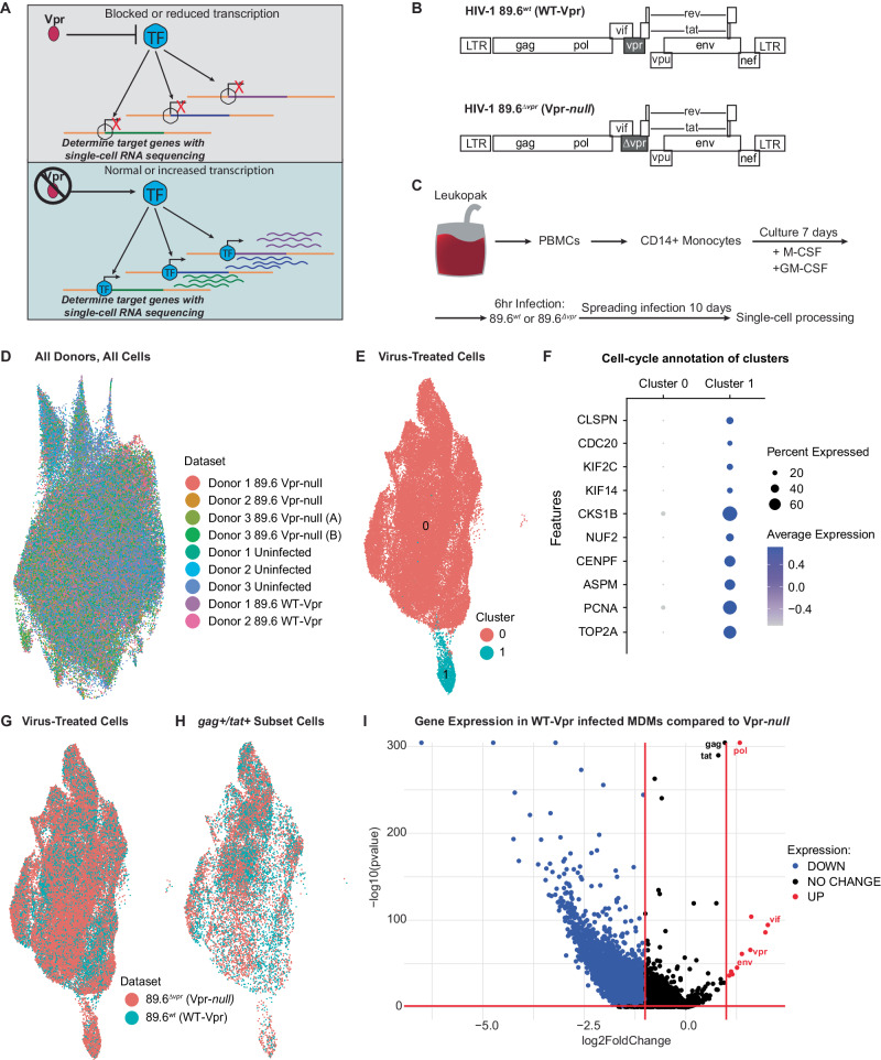
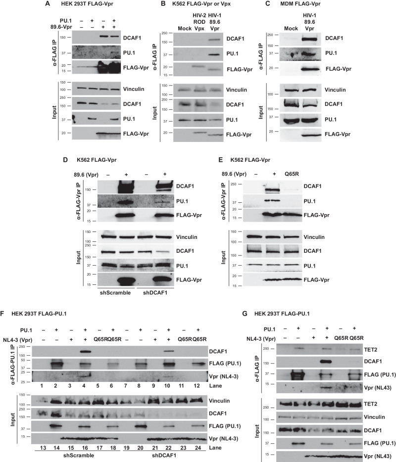
HIV-1 Vpr promotes efficient spread of HIV-1 from macrophages to T cells by transcriptionally downmodulating restriction factors that target HIV-1 Envelope protein (Env). Here we find that Vpr induces broad transcriptomic changes by targeting PU.1, a transcription factor necessary for expression of host innate immune response genes, including those that target Env. Consistent with this, we find silencing PU.1 in infected macrophages lacking Vpr rescues Env. Vpr downmodulates PU.1 through a proteasomal degradation pathway that depends on physical interactions with PU.1 and DCAF1, a component of the Cul4A E3 ubiquitin ligase. The capacity for Vpr to target PU.1 is highly conserved across primate lentiviruses. In addition to impacting infected cells, we find that Vpr suppresses expression of innate immune response genes in uninfected bystander cells, and that virion-associated Vpr can degrade PU.1. Together, we demonstrate Vpr counteracts PU.1 in macrophages to blunt antiviral immune responses and promote viral spread.


Conflict of interest statement

The authors declare no competing interests.

Figures

Fig. 1
Fig. 1. Single-cell RNA sequencing of HIV-1 infected MDMs reveals Vpr-dependent transcriptional changes.
A Schematic diagram illustrating the objective of single-cell RNA sequencing; the identification of Vpr-targeted transcription factor(s) in HIV-infected primary macrophages; TF transcription factor. B Genome maps for full-length 89.6wt HIV-1 (top) and the same viral genome with a premature stop-codon in vpr (bottom). C Experimental setup for the generation of the scRNA-seq datasets. D UMAP representation of LIGER-integrated scRNA-seq data from MDM samples treated as shown in C and listed in Dataset. E, G, H UMAP representations of LIGER-integrated scRNA-seq data from MDMs treated as indicated. E Colors indicate individual clusters. Cluster 0 = pro-inflammatory macrophages, Cluster 1 = cycling cells. F Dot plot representation of cell-cycle genes used to determine clusters in E. The size of the dot equates to the percentage of cells within the population expressing the feature, and the color indicates the average expression of the feature across all cells in each cluster. G, H Cells are colored according to whether they were exposed to 89.6 WT-Vpr virus (blue) or Vpr-null virus (pink). H Bona fide infected cells were identified based on expression of HIV tat and gag. I Volcano plot of differentially expressed genes from HIV 89.6wt verses 89.6vpr infected MDMs from H as determined by two-sided Wilcoxin Rank Sum. Significance determined as >1 log2 fold change and false discovery rate adjusted p-value of p < 0.05 (red-bars). Blue colored genes indicate genes less highly expressed in HIV 89.6wt verses 89.6vpr infected MDMs, red colored genes indicate genes more highly expressed, and black colored genes indicate no significant difference between datasets.
Fig. 2
Fig. 2. Vpr downmodulates PU.1-dependent transcription.
A PU.1 motifs identified by HOMER as present in the promoters of Vpr-downmodulated genes (Fig. 1I, Blue). B Violin plots displaying RNA abundance of the indicated transcription factor genes in MDMs infected with the indicated virus. C Volcano plot as in Fig. 1I except that genes containing a PU.1 or PU.1-IRF binding motif in their promoter region are highlighted in green (see also Supplementary Fig. 2). D Biological processes associated with the PU.1 targeted genes from C. Size of circles indicates the relative number of GO terms associated with the process. FDR adjusted q-values associated with GO terms are indicated by the color. Bolded rings are associated with biological processes listed. E Violin plots displaying RNA abundance of the indicated genes in MDMs infected with the indicated virus. HIV-1 89.6wt (WT); HIV-1 89.6vpr (∆Vpr). False-discovery rate corrected two-sided Wilcoxon rank-sum p-values are shown above the conditions being compared.
Fig. 3
Fig. 3. Vpr counteracts the innate immune response to HIV infection.
A Selected biological processes associated with infection and inflammation (selected from GO terms represented in Fig. 2D). Pathways were identified as associated with PU.1 regulated genes downmodulated in MDMs infected with 89.6wt or 89.6∆vpr virus as determined by expression of gag and tat transcripts (highlighted in Fig. 2C). The −log10 FDR-adjusted p-values (q-values) are plotted for each gene ontology term. Blue bars represent terms associated with TLR signaling; Black bars represent related gene ontology terms that are similar, but not directly associated with TLR signaling. B Violin plots summarizing single-cell RNA transcripts expressed by MDMs treated with the indicated virus and cultured for 10 days. Gag + /tat+ are the subset of cells expressing HIV genes within in each culture. SPI1 is the gene that codes for PU.1. MRC1 is the gene that codes for mannose receptor. C Violin plots summarizing single-cell RNA transcripts expressed by primary human macrophages as in B. False-discovery rate corrected two-sided Wilcoxon rank-sum p-values are shown above the conditions being compared. D, G Representative immunofluorescent images of MDMs from a single donor infected with either 89.6wt or 89.6∆vpr (MDMs from n = 2 independent donors). HIV-infected cells identified by Gag staining. E, H Quantification of ISG15- or IFITM3-corrected total cell fluorescence in Gag+ cells, or F, I Gag- cells divided by the number of nuclei in the cell area. The number of cells quantified for each condition is indicated. Error bars represent standard error of the mean, n = the number of cells quantified. P values were determined using an unpaired two-sided t test. **p = 0.0096; ****p < 0.0001. Source data are provided as a Source Data file.
Fig. 4
Fig. 4. PU.1 levels decrease in the presence of Vpr.
A NL4-3 ∆GPE-GFP viral genome map. B Representative flow cytometry histogram of PU.1 expression in infected (GFP+) or uninfected bystander (GFP-) MDMs infected with NL4-3 ∆GPE-GFP with or without vpr and collected on day 7 post infection. C Summary graph showing the percentage of infected (GFP+) cells that do not express PU.1 as determined by flow cytometry as depicted in Fig. 4B. The mean ±standard deviation from n = 4 independent donors is shown for each time point. P values were determined using a two-sided, one-way analysis of variance (ANOVA) with Tukey’s multiple comparisons test. ***p < 0.001; ****p < 0.0001. D Lentiviral map of vectors encoding 3xFLAG-encoding vpr or vpx genes. E PU.1 expression plasmid map for full-length, human PU.1. F Representative flow cytometric plots of HEK 293T cells transiently transfected with PU.1 and the indicated FLAG-tagged viral protein. G Summary graph showing PU.1+ expression in transfected (FLAG+) cells. Each point represents the mean of three technical replicates. The mean ± standard deviation of four independent experiments is shown. P values were determined using one-way ANOVA compared to control; ****p < 0.0001. H Immunoblot analysis of PU.1 in K562 cells transduced with the indicated lentivirus expressing 3xFLAG-tagged viral proteins. Results are representative of those from three independent experiments. I Summary graph of SPI1 (the gene encoding PU.1) expression in HEK 293T cells co-transfected with PU.1 and an 89.6 expression vector with or without an intact vpr open reading frame. SPI1 levels were assessed from purified RNA using RT quantitative real time PCR. Results represent the mean fold change compared with wild type ±standard deviation for samples performed in triplicate for two independent experiments. Source data are provided as a Source Data file.
Fig. 5
Fig. 5. Vpr-mediated reduction of PU.1 is conserved in HIV-2 and SIV molecular clones.
A Genomic maps for HIV-1, HIV-2, and select SIV genomes. HIV-1, SIVcpz, and SIVgor genomes contain vpr and vpu genes but not vpx. HIV-2, SIVrcm, and SIVsmm contain vpx and vpr. SIVagm contains vpr. B, D Representative flow cytometric plots of HEK 293T cells transiently transfected with expression plasmids for PU.1 and the indicated FLAG-tagged viral protein. C, E Summary graph of data from B and D, respectively. The percentage of PU.1+ cells per transfected (FLAG+) cells is shown. Each point represents the average of three technical replicates. The mean ± standard deviation is shown for n = 6 (C) or n = 11 (E) independent experiments, respectively. Part E was additionally normalized to HIV-2RODVpx for each experiment. ****p < 0.0001 using two-sided one-way ANOVA compared to control with Tukey’s multiple comparisons test. Source data are provided as a Source Data file.
Fig. 6
Fig. 6. Vpr forms a DCAF-1-dependent complex with PU.1.
A Western blot analysis of lysates from HEK 293T cells co-transfected with the indicated expression constructs and immunoprecipitated with an antibody directed against the FLAG epitope. B Western blot analysis of lysates from K562 cells transduced with lentiviruses and treated as in part A. C Western blot analysis of lysates from MDMs transduced with lentivirus and treated as in part A. For parts AC, results are representative of three independent experiments. D Western blot analysis of lysates from K562 cells stably expressing either control, non-targeting shRNA (shScramble) or an shRNA targeting DCAF1 (shDCAF1), then transduced with FLAG-tagged 89.6-Vpr expression lentivirus and immunoprecipitated using an antibody directed against the FLAG epitope. E Western blot analysis of lysates from K562 cells transduced with FLAG-tagged 89.6-VprWT or 89.6-VprQ65R expressing lentiviruses and immunoprecipitated using an antibody directed against the FLAG epitope. F Western blot of lysates from HEK 293T cells stably transduced with virus expressing a non-targeting shRNA (shScramble) or one targeting DCAF1 (shDCAF1) and immunoprecipitated with an antibody directed against the FLAG epitope. G Western blot analysis of lysates from HEK 293T cells co-transfected with the indicated expression constructs and immunoprecipitated with an antibody directed against the FLAG epitope. Results are representative of three independent experiments. Source data are provided as a Source Data file.
Fig. 7
Fig. 7. An intact DCAF-1 interaction domain is required for Vpr and DCAF1 to degrade PU.1.
A Immunoblot analysis of lysates from MDMs treated for five hours with the indicated virions. B Summary graph of PU.1 protein normalized to vinculin from MDMs incubated for five hours with the indicated viruses from A. Each point and matched color is representative of an independent donor. Statistical significance was determined using a mixed-effects analysis with Tukey’s multiple comparisons test. *p < 0.05. C Immunoblot analysis of lysates from HEK 293T cells transfected with the indicated expression construct and treatment as indicated with 10 µM MG132 or vehicle (Veh) control (DMSO). A GFP-expressing plasmid was included where indicated as control for transfection efficiency, n = 2. D Immunoblot analysis of lysates from MDMs preincubated for two hours with vehicle (Veh) or MG132 as indicated and then treated for five hours with the indicated virus as in part A. E Summary graph of PU.1 protein normalized to vinculin from MDMs treated for five hours with the indicated viruses from D. Statistical significance was determined using a mixed-effects analysis with Šidák’s multiple comparisons test. **p = 0.0073. Each point and matched color is representative of an independent donor. F Working model of PU.1 and TET2 interacting with Vpr and DCAF1 in macrophages. Source data are provided as a Source Data file.
Fig. 8
Fig. 8. Reducing PU.1 increases Env output in HIV-1 infected MDMs.
A Genome map for full-length 89.6wt HIV-1 modified to replace the vpr ORF with a U6-promoter followed by either a non-targeting shRNA control (shScramble) or an shRNA targeting the PU.1 transcripts (shSPI1). B Immunoblot from K562 cells stably expressing the shRNAs from A, n = 2. C Immunoblot analysis from two independent MDM donors. Lysates collected from MDMs infected with the virus from A expressing the indicated shRNA. White lines indicate the location where the digital image of the stained membrane was cropped to remove irrelevant samples. D Proposed model of the PU.1-mediated antiviral response disrupted by HIV-Vpr. HIV (−) Vpr indicates an infection of primary macrophages with HIV that does not express Vpr. PU.1 protein is maintained and available to regulate anti-viral response genes with co-factors such as IRF or TET2. HIV (+) Vpr indicates an infection with HIV that expresses Vpr. PU.1 protein is less available to regulate anti-viral response genes, contributing to innate immune evasion. Source data are provided as a Source Data file.

Update of

References

    1. Balliet JW, et al. Distinct effects in primary macrophages and lymphocytes of the human immunodeficiency virus type 1 accessory genes vpr, vpu, and nef: mutational analysis of a primary HIV-1 isolate. Virology. 1994;200:623–631. doi: 10.1006/viro.1994.1225. - DOI - PubMed
    1. Lu YL, Bennett RP, Wills JW, Gorelick R, Ratner L. A leucine triplet repeat sequence (LXX)4 in p6gag is important for Vpr incorporation into human immunodeficiency virus type 1 particles. J. Virol. 1995;69:6873–6879. doi: 10.1128/jvi.69.11.6873-6879.1995. - DOI - PMC - PubMed
    1. Eckstein DA, et al. HIV-1 Vpr enhances viral burden by facilitating infection of tissue macrophages but not nondividing CD4+ T cells. J. Exp. Med. 2001;194:1407–1419. doi: 10.1084/jem.194.10.1407. - DOI - PMC - PubMed
    1. Connor RI, Chen BK, Choe S, Landau NR. Vpr is required for efficient replication of human immunodeficiency virus type-1 in mononuclear phagocytes. Virology. 1995;206:935–944. doi: 10.1006/viro.1995.1016. - DOI - PubMed
    1. Dedera D, Hu W, Vander Heyden N, Ratner L. Viral protein R of human immunodeficiency virus types 1 and 2 is dispensable for replication and cytopathogenicity in lymphoid cells. J. Virol. 1989;63:3205–3208. doi: 10.1128/jvi.63.7.3205-3208.1989. - DOI - PMC - PubMed

MeSH terms

Substances