. 2025 Jan;21(1):120-130.
doi: 10.1038/s41589-024-01664-8. Epub 2024 Jul 1.

Transition transferases prime bacterial capsule polymerization

Affiliations

Transition transferases prime bacterial capsule polymerization

Christa Litschko et al. Nat Chem Biol. 2025 Jan.

Abstract

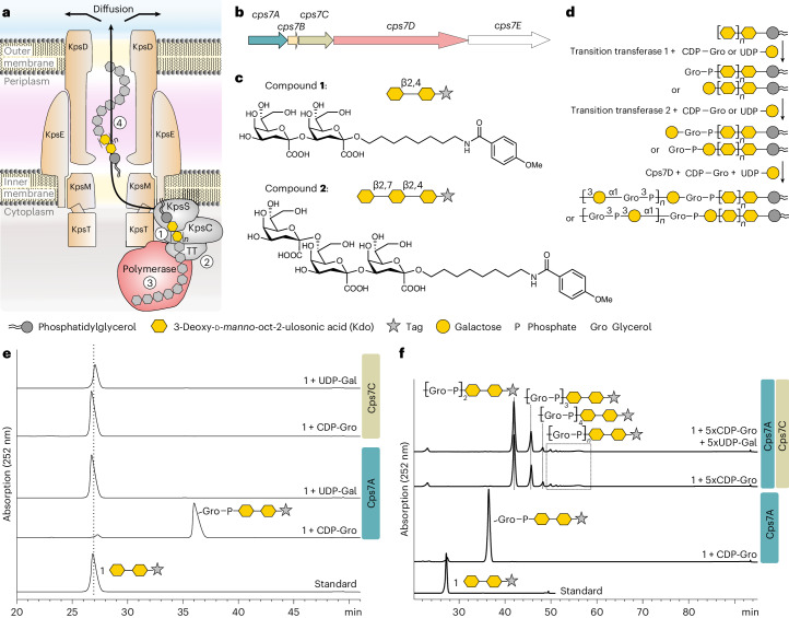
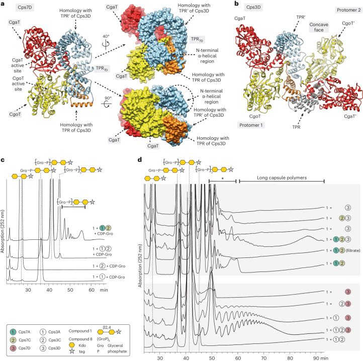
Capsules are long-chain carbohydrate polymers that envelop the surfaces of many bacteria, protecting them from host immune responses. Capsule biosynthesis enzymes are potential drug targets and valuable biotechnological tools for generating vaccine antigens. Despite their importance, it remains unknown how structurally variable capsule polymers of Gram-negative pathogens are linked to the conserved glycolipid anchoring these virulence factors to the bacterial membrane. Using Actinobacillus pleuropneumoniae as an example, we demonstrate that CpsA and CpsC generate a poly(glycerol-3-phosphate) linker to connect the glycolipid with capsules containing poly(galactosylglycerol-phosphate) backbones. We reconstruct the entire capsule biosynthesis pathway in A. pleuropneumoniae serotypes 3 and 7, solve the X-ray crystal structure of the capsule polymerase CpsD, identify its tetratricopeptide repeat domain as essential for elongating poly(glycerol-3-phosphate) and show that CpsA and CpsC stimulate CpsD to produce longer polymers. We identify the CpsA and CpsC product as a wall teichoic acid homolog, demonstrating similarity between the biosynthesis of Gram-positive wall teichoic acid and Gram-negative capsules.


Conflict of interest statement

Competing interests: A.B., M.B., T.F. and C.L. have submitted the following patent application: Fiebig T, Litschko C, Gerardy-Schahn R, Berger M, Bethe A. Means and methods for producing phosphate containing capsular polysaccharides (WO2019020735A1). The remaining authors declare no competing interests.

Figures

Fig. 1
Fig. 1. Identification of transition transferases.
a, The current model for capsule assembly in ABC transporter-dependent systems comprises (1) the generation of a conserved glycolipid consisting of β-linked Kdo and phosphatidylglycerol, (2) the addition of a priming capsule polymer repeating unit by putative transition transferases (TT), (3) the elongation of said repeating unit by the capsule polymerase and (4) export of the polymer. b, Capsule gene cluster of App7, showing the capsule polymerase (Cps7D, pink), the enzyme providing CDP-Gro (Cps7B, yellow), putative transition transferases (Cps7A, turquoise; Cps7C, green) and a gene of unknown function (Cps7E, white). c, Chemical and symbolic structures of compounds 1 and 2. d, Current working model for the in vivo synthesis of the capsule polymer in App7. The conserved glycolipid is extended by a putative transition transferase 1 that adds glycerol-phosphate or galactose. A second transition transferase 2 transfers galactose or glycerol-phosphate, completing the priming repeating unit, which serves as an acceptor for the capsule polymerase Cps7D, a two-domain enzyme that uses UDP-Gal and CDP-Gro as substrates for the generation of the capsule polymer. e,f, HPLC–AEC-based activity assay for the analysis of transition transferase activity of Cps7A (e) and Cps7C (f). Compound 1 was used as standard. Detection was performed at 252 nm. Nucleotide substrates and products are also detectable at this wavelength but elute early in the gradient (app. after 8–14 min; see Supplementary Fig. 3h for an example). e, Cps7A and Cps7C were incubated with compound 1 and either UDP-Gal or CDP-Gro. f, Cps7C utilizes CDP-Gro to generate a negatively charged polymer.
Fig. 2
Fig. 2. Comprehensive 1D and 2D NMR analysis of Cps7A and Cps7C reaction products.
ac, 31P NMR analysis of compound 3 (a), compound 4 (b) and compound 5 (c). d, Chemical structure of compound 3 (left), compound 4 (middle) and compound 5 (right). e,f, A combination of 2D 1H–13C HSQC (e) and 1H–31P HMBC (f) analysis was used to analyze the linkages between Kdo, phosphate and glycerol, demonstrating that Cps7A transfers Gro3P onto the 7-OH of the nonreducing end Kdo, thereby creating a new phosphodiester linkage. Cps7C adds additional Gro3P moieties onto 1-OH of the terminal glycerol phosphate (see Supplementary Table 3 for chemical shifts).
Fig. 3
Fig. 3. The presence of Cps7A promotes the generation of long chains by Cps7D.
HPLC–AEC analysis of Cps7A/C/D reactions. Species eluting between 48 and 60 min (marked with asterisk) can comprise products of Cps7C and (short) products of Cps7D (see also Extended Data Fig. 4d). Long capsule polymers synthesized in Cps7A/(C)/D reactions have retention times between 60 and 90 min (see label at the top of a and b). a, Compound 3 was incubated with Cps7A or mutants of Cps7A, and Cps7C/D. The generation of long chains could only be observed in the presence of all three enzymes Cps7A/C/D. Interestingly, Cps7A could be substituted by inactive amino acid exchange mutants without losing the stimulating effect, demonstrating that the presence but not the activity of Cps7A is required once the product of Cps7A has been built. b, Compound 1 was elongated with Cps7A/C (r1), the enzymes were removed by filtration and the filtrate (r2) was used as substrate in subsequent reactions. Interestingly, long chains were produced in the presence of inactive Cps7C mutants and even in the absence of Cps7C, suggesting that, after the products of Cps7C have been assembled, the enzyme is not required for stimulating the assembly of long polymers.
Fig. 4
Fig. 4. The overall structure of Cps3D.
a, Each protomer of the homodimer is composed of a TPR domain (orange), the Gro3P-transferase CgoT (yellow) and the α-1,2-galactosyltransferase CgaT (red). The active sites of the two enzymes in each protomer face the concave surface of the homodimer. b, Middle: structure of CgoT. The N-terminal and C-terminal domains comprise residues 361–539 and 720–736 (CgoT-NT, orange) and residues 540–719 (CgoT-CT, gray), respectively. Left and right: CgoT active site showing the donor substrate CDP-Gro and selected loops and amino acid residues c, Middle: structure of CgaT. The N-terminal and C-terminal domains comprise residues 747–947 and 1,120–1,136 (CgaT-NT, salmon) and residues 948–1,119 (CgaT-CT, gray), respectively. Left and right: CgoT active site showing UDP-Gal and selected loops and amino acid residues. d, Structure of the TPR (dark orange) and TPR′ (salmon) domains. The predicted α4HB (α1B (residues 10–24), α2B (residues 27–41), α3B (residues 45–59) and α4B (residues 62–75)) and αL (residues 80–110) reveals a charge distribution with an intense positive character. e, Structure of Cps3D with possible location of the α4HB and αL structural elements shown as cartoon representation.
Fig. 5
Fig. 5. Differences and similarities between the App3 and App7 biosynthesis systems.
a,b, AlphaFold model of Cps7D (a) as a cartoon representation (left) and surface representation (right) with rotations as indicated. Crystal structure of Cps3D as a cartoon representation (b). Color code: red, CgaT; yellow, CgoT; orange, homologous TPR repeats from monomeric Cps7DAF (α16–α20) and protomer 1 of the Cps3D dimer (α11–α15); light blue, homologous TPR repeats from monomeric Cps7DAF (α1–α15) and protomer 2 of the Cps3D dimer (α1′–α15′). c, Cps3A and Cps3C were incubated with compound 1 and CDP-Gro. Cps3A used compound 1 as acceptor and transferred one Gro3P moiety. Cps3C transferred Gro3P onto the product of Cps3A. A Cps7A/C reaction (from Supplementary Fig. 3f) is shown as a reference. d, The presence of Cps3A/C stimulates Cps7D to produce long polymers, and the presence of Cps7A/C stimulates Cps3D to produce larger products, indicating a high conservation of the stimulating effect and its components in both systems. Species eluting between app. 48 and 60 min (marked with asterisk) can comprise products of CpsC and (short) products of CpsD (see also Extended Data Fig. 4d). Long capsule polymers synthesized in CpsA/(C)/D reactions have retention times between 60 and 90 min (see label at the top of d).
Fig. 6
Fig. 6. The role of the TPR domain in Cps3D and Cps7D.
a, N-terminal truncations of Cps7D (top) and Cps3D (bottom) generated herein. See also Extended Data Fig. 8 and Supplementary Fig. 12. b,c, Compound 1 was elongated with CpsA and CpsC, the enzymes were removed by filtration and the filtrate was used as acceptor with proteins as indicated. None of the TPR truncations was able to elongate poly(Gro3P). However, Cps3D94–1138, in which only α4HB was truncated, retained this ability (b) and even produced long polymers when Cps3A/C or Cps7A/C were present (c). d,e, Elongation of the products of CpsA/C by the enzymes as indicated and subsequent analysis using PAGE (d) and HPLC-AEC (e). TPR truncations were unable to elongate poly(Gro3P). Species eluting between app. 48 and 60 min (marked with asterisk) can comprise products of CpsC and (short) products of CpsD (panel c and e; see also Extended Data Fig. 4d). wt, wildtype; hyd., hydrolyzed. Source data
Extended Data Fig. 1
Extended Data Fig. 1. Capsule gene clusters containing transition transferases analyzed in this study.
The organization of the cluster differs between a, E. coli b, Neisseria meningitidis and c, Actinobacillus pleuropneumoniae. In all strains, the cluster is divided in conserved regions (grey boxes) and capsule-specific regions (white boxes), encoding enzymes involved in the generation of poly(Kdo) (green), proteins involved in polymer export (blue), and enzymes involved in capsule polymer synthesis. Characterized (solid) or predicted (cross-hatched) capsule polymerases are shown in pink–,,,. Enzymes involved in nucleotide substrate provision or polymer modification are shown in yellow,,, or violet,,, respectively. Putative transition transferases characterized herein (shown in orange) are CslA (CCP19790.1, NmL), CsaD (CAM07516.1, NmA), CsxB/CsxC (ATG32052.1, ATG32051.1, NmX), KfiB (TEZ99055.1, E. coli K5), Cps1A/Cps1C (AWG96007.1, AWG96005.1, App1), Cps7A/Cps7C (ACE62294.1, ACE62292.1, App7).
Extended Data Fig. 2
Extended Data Fig. 2. Putative transition transferases tested in this study.
a, Overview of strain, capsule polymer repeating unit, putative transition transferase, and putative donor substrates for transition transferase candidates. Nm, Neisseria meningitidis; App, Actinobacillus pleuropneumoniae; E. coli, Escherichia coli. b-g, HPLC-AEC-based activity assays. Compounds 1 or 2 were incubated with the donor substrates and putative transition transferases as indicated. As UDP-ManNAc (panel c) is commercially unavailable, UDP-GlcNAc was supplied together with the epimerase CsaA, which has been used in a previous study to convert UDP-GlcNAc to UDP-ManNAc in situ. Only Cps7A was able to utilize compound 1 (Fig. 1e) and compound 2 (panel g), leading to a species eluting at a later retention time. The inactivity of the other enzymes could be due to construct design, buffer conditions or choice of substrates. These enzymes could still be active transition transferases in the respective strains in vivo.
Extended Data Fig. 3
Extended Data Fig. 3. Characterization of Cps7A and Cps7C.
a, AlphaFold model of Cps7A (Cps7AAF, B3GYR3; ACE62294.1; 380 residues) comprising two Rossmann-fold domains (beige, salmon), as observed in glycosyltransferase(GT)-B enzymes,, and a C-terminal α-helix (orange). Catalytically important amino acid residues identified in this study (Supplementary Fig. 3a,b) are located in a cleft between these two domains. b, Structural alignment of Cps7AAF with the crystal structure of TagF, the WTA type I polymerase from Staphylococcus epidermidis, an enzyme with Gro3P transferase activity (PDB code 3L7L; 15% sequence identity). In TagF, H444 likely functions as an active-site base, while H584 seems to play an essential role in catalysis by forming a complex with the pyrophosphate group of CDP-Gro. H198/H199 of Cps7AAF superimpose with H584 of TagF, while H444 of TagF superimposes with Y94 of Cps7AAF. Interestingly, H444 is part of the WHG motif in TagF and TagF-like Gro3P polymerases. In GT-B fold hexose-phosphate transferases and other GT-B fold enzymes, variations of the WHG motif comprise a QHG or a QYA motif, respectively. c, AlphaFold model of Cps7C (B3GYR1; ACE62292.1; 378 residues), comprising the N-terminal tetratricopeptide repeat (TPR) domain (blue, residues 1-190 and 368-378) and the C-terminal nucleotidyltransferase (NTase) domain (beige, residues 203-365). TPRs commonly mediate protein–protein interactions and the assembly of multi-protein complexes, but also the recognition of polymers. Interestingly, the NTase domain of Cps7C displays homology with FKRP, an enzyme that modifies the phosphor-core M3 structure of the O-glycan of human α-Dystroglycan. Using CDP-ribitol as donor, FKRP transfers the second ribitol-phosphate moiety of a tandem ribitol-phosphate and thus, like Cps7C, utilizes a polyol-phosphate as acceptor and a CDP-polyol as donor. FKRP (PDB code 6KAN, chain D) contains two aspartate residues important for catalytic activity, which align with D247 and D293 of Cps7C according to PHYRE2 homology modelling and are crucial for activity (Supplementary Fig. 3e).
Extended Data Fig. 4
Extended Data Fig. 4. The reaction products of Cps7C are acceptor substrates for the α-1,1-galactosyltransferase (CgaT) domain of Cps7D.
a, Cps7D consists of three regions from the N- to the C-terminus: (i) a domain rich in tetratricopeptide repeats (TPR), (ii) the Gro3P transferase CgoT (Capsule glycerol-3-phosphate Transferase), (iii) a GT-B fold α-1,1-galactosyltransferase CgaT (Capsule α-1,1-galactosyl Transferase), and an N-terminal α-helical bundle (α4HB). Cps7D requires UDP-Gal and CDP-Gro as substrates for polymerization. b, The reaction products of a Cps7A/C reaction were incubated with either UDP-Gal or CDP-Gro, and Cps7D-H743A or Cps7D-R1123A, in which either CgaT or CgoT remained active. Only CgaT could use poly(Gro3P) as acceptor, indicated by a shift of all species to earlier retention times due to the addition of a neutral galactose (r2). c, Compounds 3, 4, and 5 were incubated with Cps7D and UDP-Gal. While only 15% of compound 3 (the product of Cps7A) were used up by Cps7D over-night (r1), compounds 4–5 (products of Cps7C) were completely converted (r2, r4). The addition of one equivalent of CDP-Gro to reaction r2, or in other words, the addition of a complete repeating unit to compound 4 (r3), led to a species eluting at precisely the same time as compound 4. d, Compound 5 was analyzed before (r2) and after (r3) incubation with Cps7D, CDP-Gro and UDP-Gal and products were visualized by PAGE (left) and HPLC-AEC (right), demonstrating that poly(Gro3P) can prime Cps7D. A reaction in the absence of compound 5 (r1) was performed as control for PAGE to document the extent of Cps7D’s previously published de novo activity,. A reaction in the presence of compound 1 was performed as standard for HPLC-AEC. Due to the complex composition of the polymer (tag, linker, Kdo, poly(Gro3P), (Gal-Gro3P)n), it is difficult to predict its electrophoretic mobility. Source data
Extended Data Fig. 5
Extended Data Fig. 5. Kdo is not required for poly(Gro3P) to be recognized as acceptor by Cps7D.
a, Chemical structure of the non-tagged Gro3P-pentamer (Gro3P)5 (compound 8). b, Because compound 8 is not readily detected by UV, products were visualized by Alcian blue / silver staining after separation using PAGE. Cps7D was used at a nanomolar concentration to avoid its previously shown de novo activity, (production of long chains at high enzyme concentrations even without any acceptor present). Samples were incubated with different protein combinations as indicated and with or without compound 8 (+/−). Compound 8 is no suitable acceptor substrate for Cps7A (lane 1), but for the Gro3P polymerase Cps7C (lane 3) and the capsule polymerase Cps7D (lane 5). The presence of Cps7A stimulates Cps7D to produce polymer, both in the presence and absence of compound 8 (compare lanes 5 and 9, and lanes 6 and 10). Cps7A has a stimulating effect on Cps7C product formation (compare lanes 3 and 13). The presence of Cps7C has no detectable effect on product formation by Cps7A/D (compare lanes 7/8 with lanes 9/10). Cps7C alone also seems to increase polymer synthesis by Cps7D (lanes 11 and 12). Source data
Extended Data Fig. 6
Extended Data Fig. 6. Structural basis of CgoT substrate specificity and catalysis in Cps3D.
a-b, Ribbons representation (a) and electrostatic surface potential representation (b) of CgoT. The active site cleft is located between the two Rossmann domains (CgoT-NT; salmon, CgoT-CT; grey) and contains the essential residues for CgoT activity. The N-terminal and C-terminal domain cores are composed by seven-stranded and six-stranded parallel β-sheets, respectively, surrounded by α-helices (a). c, d, The structural comparison of CgoT with (i) the glycerol-phosphate polymerase TagF from Staphylococcus epidermidis in complex with CDP-glycerol (Supplementary Fig. 10a, PDB code 3L7K; root mean square deviation (r.m.s.d.) value between chains of 1.206 Å for 212 pruned atoms, and of 3.843 Å for all 355 pairs) and (ii) the capsule ribitol-5-phosphate transferase CroT from H. influenzae in complex with CMP (PDB code 8A0C; r.m.s.d. value between chains of 1.189 Å for 112 pruned atoms, and of 5.576 Å for all 292 pairs) revealed the active site architecture. The active site comprises α-helices 28-30, loop 1 (β5-α22, red), loop2 (α22-β10, light blue), and loop 3 (β11-α30, orange) (panels a, c, d). Exploiting the fact that TagF from S. epidermidis utilizes the same donor substrate as CgoT, we performed docking calculations revealing the location of CDP-glycerol in the active site (c, d). Key residues conserved between the two enzymes include F391, H479, G480, T481, P482, R542, A573, R607, S647, S648 and D652. Active site residues H479 and H609 of CgoT and their homologs in TagF, and TagF-like capsule polymerases, are crucial for enzyme activity (Supplementary Fig. 11). CgoT transfers Gro3P to the O4 position of the terminal galactose residue of the nascent chain, whereas TagF and Bcs3 transfer Gro3P and ribitol-5-phosphate to the O1 and O3 position of glycerol and ribose, respectively. By homology, we propose that CgoT follows a simple displacement mechanism similar to that observed in TagF and Bcs3. We suggest that H479 acts as the putative base that contributes to the deprotonation of the O4 of the acceptor galactose residue, facilitating the nucleophilic attack of galactose to the β-PO4 of CDP-glycerol,.
Extended Data Fig. 7
Extended Data Fig. 7. Structural basis for CgaT substrate specificity and catalysis in Cps3D.
a,b, Ribbons representation (a) and electrostatic surface potential representation (b) of CgaT. The N-terminal (orange) and C-terminal (grey) domain cores are composed by seven-stranded and six-stranded parallel β-sheets, respectively, surrounded by α-helices (a). The active site is located in a deep fissure at the interface of the two Rossmann folds, suggesting an important inter-domain flexibility. c, d, Structural comparison of CgaT with the glycosyltransferase BshA from Staphylococcus aureus in complex with UDP and N-acetylglucosamine (PDB code 6N1X; r.m.s.d. value between chains of 10.137 Å across all 339 pairs; Supplementary Fig. 10), support the location of the active site, which is defined by loop 1 (β20-α46; pink), loop 2 (β21-α48; red), loop 3 (β22-α49; light green), loop 4 (β23-α50; light blue), loop 5 (β24-α51; orange) and by helices α36, α51 and α52. Molecular docking calculations placed the donor UDP-Gal into the active site of CgaT. Residues R982 and K987 of the active site are highly conserved in TagF-like capsule polymerases, crucial for Cps3D activity (Supplementary Fig. 11) and frequently involved in coordinating the phosphate moieties in retaining GT-B fold enzymes,. E1059 and E1067 are part of the EX7E motif found (among others) in the GT4 family of retaining glycosyltransferases, which includes Cps3D homologs Cps2D and Cps7D (Cps3D is not yet listed in Carboyhdrate-active enzyme database CAZy), as well as BshA, PimA and TarM. CgaT catalyzes the transfer of a galactose residue to the 2 OH position of the non-reducing end Gro3P of the nascent capsule polymer,. By homology, we suggest that CgaT is a retaining enzyme most likely following a SNi catalytic mechanism, involving an oxocarbenium ion transition state,.
Extended Data Fig. 8
Extended Data Fig. 8. Comparison of secondary structural elements of Cps3D and Cps7D based on a superposition of the dimeric Cps3D crystal structure with the monomeric Alphafold model of Cps7D.
Coloured secondary structural elements are labelled according to the crystal structure and the predicted N-terminus (α1B-α4B, αL) of Cps3D. Compared to Cps3D, Cps7DAF has an extended TPR domain that comprises regions from both protomers of the Cps3D dimer, which is the molecular reason for the monomeric state of Cps7D. The first 15 α-helices of Cps7DAF’s TPR domain superimpose with the corresponding α-helices from the TPR domain of the neighbouring protomer 2 of the Cps3D dimer (α1’-α15’, light blue). In contrast, α16-α20 of Cps7D’s TPR domain superimpose with α11-α15 (orange) of the equivalent protomer 1 of the Cps3D dimer. An arrow labeled ‘TPR shift’ indicates that α1’-α15’ (light blue) and α11-α15 (orange) belong to two different protomers of the Cps3D structure. Surface representation of full-length Cps3D/Cps7DAF and their truncations are presented in Fig. 5 and Supplementary Fig. 14. The TPR(‘) domain is required for the elongation of poly(Gro3P) (Fig. 6) and in close vicinity to CgaT(NT) (Fig. 5a, b), which transfers the first galactose onto poly(Gro3P). TPR(‘) and CgaT(NT) interact through many structural elements, that are more closely described in Supplementary Fig 14.
Extended Data Fig. 9
Extended Data Fig. 9. Comparison between Gram-negative group 2 capsule biosynthesis and Gram-positive WTA biosynthesis.
a, Schematic showing the synthesis of Gram-negative capsule polymer at the cytoplasmic side of the inner membrane. KpsS/C generate the conserved glycolipid, which is linked to the capsule polymer (assembled by CpsD) via a poly(Gro3P)-linker assembled by CpsA/C. b, Schematic showing the biosynthesis of Gram-positive WTA of type I (reviewed in Brown et al. and Sewell et al., pathways for type II-IV are uncharacterized). In analogy to poly(Kdo), a conserved priming glycolipid is assembled on the cytoplasmic side of the bacterial cell membrane, but instead of (lyso)phosphatidylglycerol, membrane anchored undecaprenyl phosphate is used as lipid carrier. The enzymes TagO/TarO and TagA/TarA create a ManNac(β1-4)GlcNAc discaccharide, which serves as acceptor for the priming glycerophosphotransferase TagB,. In analogy to Cps7A, TagB transfers the first GroP residue. The resulting product is conserved and used by all WTA I biosynthesis pathways investigated so far. Subsequently, the biosynthesis pathways diverge depending on the pathogen. In B. subtilis 168, the WTA type I polymerase TagF generates a chain of up to 40–60 Gro3P moieties. Unlike the product of CpsC (a), this structure represents the final WTA I polymer and is not further elongated by a polymerase like CpsD. In contrast, and in better analogy to the reactions catalyzed by CpsC and CpsD, the glycerophosphotransferase TarF from S. aureus only extends the product of TagB by one or a few Gro3P moieties, before the WTA I polymerase TarL adds the structurally distinct WTA I polymer (poly(ribitol-phosphate)), similar to CpsD in App (although CpsD generates a WTA type II structure).
Extended Data Fig. 10
Extended Data Fig. 10. Revised working model for the biosynthesis of WTA-like group 2 capsule polymers.
a, Characterized capsule structures of strains harbouring CpsA and CpsC homologs. b, Revised working model for group 2 capsule biosynthesis for WTA-like polymers: (1) KpsS and KpsC assemble the conserved glycolipid consisting of (lyso)phosphtidylglycerol and poly(Kdo), which is hypothesized to be an export signal and might already be associated with the ABC-transporter. (2) CpsA transfers the first Gro3P residue onto poly(Kdo), creating a primer for (3) the assembly of the WTA-type I-like poly(Gro3P) linker catalysed by CpsC. (4) The TPR domain enables CpsD to utilize poly(Gro3P) as primer for capsule polymerization and is required for processive elongation. (5) CpsA and CpsC stimulate CpsD to produce more and longer products. (6) The concave surface of CpsD and the predicted location of α4HB (see Fig. 4) above the concave surface suggest an orientation in which the active centres of the polymerase (CpsD) face the cytoplasmic membrane. Similar to the dimeric Hib capsule polymerase Bcs3, the distance between the two active centres (app. 90 Å) might allow an enzymatic assembly in which each of the two chains generated by the Cps3D dimer is exported by one ABC transporter complex (diameter of 60–70 Å,). It is of note that interactions between components of the App biosynthesis system have not been experimentally proven whereas studies in E. coli present evidence for the formation of a multi-protein complex,. Moreover, the processive finalization of a polymer by a multi-enzyme system coupled to ATP-dependent translocation is hypothesized as efficient scenario for capsule expression.

References

    1. Whitfield, C., Wear, S. S. & Sande, C. Assembly of bacterial capsular polysaccharides and exopolysaccharides. Annu. Rev. Microbiol.74, 521–543 (2020). - PubMed
    1. Sande, C. & Whitfield, C. Capsules and extracellular polysaccharides in Escherichia coli and Salmonella. EcoSal9, eESP00332020 (2021). - PMC - PubMed
    1. Willis, L. M. et al. Conserved glycolipid termini in capsular polysaccharides synthesized by ATP-binding cassette transporter-dependent pathways in Gram-negative pathogens. Proc. Natl Acad. Sci. USA110, 7868–7873 (2013). - PMC - PubMed
    1. Willis, L. M. & Whitfield, C. KpsC and KpsS are retaining 3-deoxy-d-manno-oct-2-ulosonic acid (Kdo) transferases involved in synthesis of bacterial capsules. Proc. Natl Acad. Sci. USA110, 20753–20758 (2013). - PMC - PubMed
    1. Lanz, N. D. et al. Characterization of the β-KDO transferase KpsS, the initiating enzyme in the biosynthesis of the lipid acceptor for Escherichia coli polysialic acid. Biochemistry10.1021/acs.biochem.1c00088 (2021). - PubMed

Publication types

MeSH terms

LinkOut - more resources