. 2024 Nov;31(11):1422-1438.
doi: 10.1038/s41418-024-01336-z. Epub 2024 Jun 29.

TRIM65 deficiency alleviates renal fibrosis through NUDT21-mediated alternative polyadenylation

Affiliations

TRIM65 deficiency alleviates renal fibrosis through NUDT21-mediated alternative polyadenylation

Sisi Wei et al. Cell Death Differ. 2024 Nov.

Abstract

Chronic kidney disease (CKD) is a major global health concern and the third leading cause of premature death. Renal fibrosis is the primary process driving the progression of CKD, but the mechanisms behind it are not fully understood, making treatment options limited. Here, we find that the E3 ligase TRIM65 is a positive regulator of renal fibrosis. Deletion of TRIM65 results in a reduction of pathological lesions and renal fibrosis in mouse models of kidney fibrosis induced by unilateral ureteral obstruction (UUO)- and folic acid. Through screening with a yeast-hybrid system, we identify a new interactor of TRIM65, the mammalian cleavage factor I subunit CFIm25 (NUDT21), which plays a crucial role in fibrosis through alternative polyadenylation (APA). TRIM65 interacts with NUDT21 to induce K48-linked polyubiquitination of lysine 56 and proteasomal degradation, leading to the inhibition of TGF-β1-mediated SMAD and ERK1/2 signaling pathways. The degradation of NUDT21 subsequently altered the length and sequence content of the 3'UTR (3'UTR-APA) of several pro-fibrotic genes including Col1a1, Fn-1, Tgfbr1, Wnt5a, and Fzd2. Furthermore, reducing NUDT21 expression via hydrodynamic renal pelvis injection of adeno-associated virus 9 (AAV9) exacerbated UUO-induced renal fibrosis in the normal mouse kidneys and blocked the protective effect of TRIM65 deletion. These findings suggest that TRIM65 promotes renal fibrosis by regulating NUDT21-mediated APA and highlight TRIM65 as a potential target for reducing renal fibrosis in CKD patients.


Conflict of interest statement

The authors declare no competing interests.

Figures
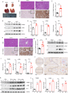
Fig. 1
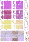
Fig. 1. Upregulation of TRIM65 in various experimental models of renal fibrosis.
A The appearances of post-operative gross kidney in the Sham and UUO mice; B Representative photomicrographs of H&E and IHC staining of mice kidneys in Sham groups and UUO groups, scale bars are 100 μm; C The intensity of TRIM65 IHC staining in kidneys of Sham and UUO mice was computed using ImageJ software (n = 6 per group); D Expression of TRIM65, α-SMA, and FN-1 in kidneys of WT mice from Sham and UUO groups assessed by western blot, with GAPDH for standardization; E Quantification of the western blot bands was carried out using Gel-Pro Analyzer software; F Representative images of H&E and IHC staining of WT mouse kidneys in Control and FAN groups, scale bars are 100 μm; G The intensity of TRIM65 IHC staining in kidneys of control and FAN mice was computed using ImageJ software (n = 6 per group); H Western blot assessment of TRIM65, α-SMA, and FN-1 expression in kidneys of WT mice from Control and FAN groups, standardized by GAPDH; I Quantification of the western blot bands was carried out using Gel-Pro Analyzer software; J Representative IHC staining of TRIM65 on renal cancer adjacent tissues; K Expression levels of TRIM65, α-SMA, and FN-1 in HK-2 cells stimulated by TFG-β1 at 0 h, 24 h, and 48 h, assessed by western blot; L Quantification of the western blot bands was carried out using Gel-Pro Analyzer software. All results are expressed as the mean ± SD. ** P < 0.01, ***P < 0.001 by Student’s t test.
Fig. 2
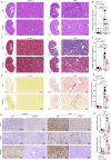
Fig. 2. TRIM65 deletion alleviates UUO-induced renal injury and fibrosis.
A Assessment of renal damage in four groups of mice (Sham group of WT mice, Sham group of Trim65-/- mice, UUO group of WT mice, and UUO group of Trim65-/- mice) via H&E staining; B Statistical graph of H&E staining showing the semi-quantitative analysis of the tubulointerstitial injury results (n = 6 per group); C Evaluation of renal fibrosis levels in four groups of mice using Masson staining; D Quantitative assessment of Masson areas in the kidneys of UUO mice or Sham mice (n = 6 per group); E Examination of renal fibrosis levels in four groups of mice through Sirius Red staining; F Quantitative assessment of Sirius Red areas in the kidneys of UUO mice or Sham mice (n = 6 per group); G Representative photomicrographs of kidney sections stained for FN-1, α-SMA, and TRIM65 in four groups of mice (n = 6 per group); H Statistical analysis of IHC staining results for FN-1 and α-SMA. All results are expressed as the mean ± SD. **P < 0.01, ***P < 0.001 by one-way ANOVA followed by Tukey’s test.
Fig. 3
Fig. 3. Removal of TRIM65 mitigates folic acid-induced renal fibrosis.
A Evaluation of renal damage in four cohorts of mice (Control group of WT mice, Control group of Trim65-/- mice, FAN group of WT mice, and FAN group of Trim65-/- mice) via H&E staining; B Statistical analysis of H&E staining results (n = 6 per group); C Assessment of renal fibrosis levels in four groups of mice using Masson staining; D Statistical analysis of Masson staining results (n = 6 per group); E Examination of renal fibrosis levels in four groups of mice through Sirius Red staining; F Statistical analysis of Sirius Red staining results (n = 6 per group); G IHC staining assessing the expression and distribution of renal FN-1, α-SMA, and TRIM65 in four groups of mice (n = 6 per group); H Statistical analysis of IHC staining results for FN-1 and α-SMA. All results are expressed as the mean ± SD. *P < 0.05, **P < 0.01, ***P < 0.001 by one-way ANOVA followed by Tukey’s test.
Fig. 4
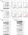
Fig. 4. TRIM65 aggravates TGF-β1-induced fibrosis in renal tubular epithelial cells.
A Western blot analysis depicting the impact of TRIM65 knockdown in HK-2 cells on the protein levels of FN-1, VIM, α-SMA, and TRIM65 under TGF-β1 stimulation, with GAPDH utilized for standardization; B Quantification of the western blot bands was carried out using Gel-Pro Analyzer software; C Western blot examination of the influence of upregulating TRIM65 in HK-2 cells on the levels of FN-1, VIM, α-SMA, and TRIM65 expression under TGF-β1 stimulation, with GAPDH used for standardization; D Quantification of the western blot bands was carried out using Gel-Pro Analyzer software; E Western blot analysis of the phosphorylation levels of Smad2, Smad3, P65, ERK1/2, and IKKβ under TGF-β1 stimulation after intervening in TRIM65 expression in HK-2 cells, with GAPDH used for standardization; F Western blot analysis of the phosphorylation levels of Smad2, Smad3, P65, ERK1/2, and IKKβ under TGF-β1 stimulation after upregulating TRIM65 expression in HK-2 cells, with GAPDH used for standardization. Data presented as mean ± SD. *P < 0.05, **P < 0.01, ***P < 0.001 by one-way ANOVA followed by Tukey’s test. Each experiment independently repeated at least three times.
Fig. 5
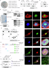
Fig. 5. Interaction between TRIM65 and NUDT21.
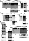
A The binding of TRIM65 and NUDT21 was identified through yeast two-hybrid system, P53 and the SV40 large T-antigen (T) or Lamin C66-230 protein (Lam) were selected as positive and negative controls, respectively; B, C Co-immunoprecipitation (Co-IP) analysis and protein blotting conducted on HEK293T cells co-transfected with Flag-TRIM65 and HA-NUDT21; D GST or GST-TRIM65 proteins purified and incubated with lysates from HEK293T cells transfected with HA-NUDT21, following GST-pulldown experiment, eluted proteins and cell lysates analyzed via protein blotting, and purity of GST-TRIM65 was assessed through Coomassie blue staining; E Co-IP analysis to examine endogenous interaction between TRIM65 and NUDT21 on HK-2 cells or ACHN cells stimulated with TGF-β1 for 1 h; F Representative confocal microscopy images showing co-localization of TRIM65 and NUDT21 in HK-2 cells; G Co-IP analysis and protein blotting on HEK293T cells transfected with GFP-TRIM65(WT), GFP-TRIM65 deletion mutants, and HA-NUDT21 to study their interaction; H Schematic representation of plasmid generation for continuous deletion mutants of TRIM65 fused with GFP and the interaction with NUDT21; I Co-IP analysis and protein blotting on HEK293T cells transfected with Flag-TRIM65(WT) and HA-NUDT21 deletion mutants to investigate their interaction; J Schematic representation of plasmid generation for continuous deletion mutants of NUDT21 and the interaction with TRIM65.
Fig. 6
Fig. 6. TRIM65 catalyzes K48-linked polyubiquitination of NUDT21.
A HEK293T cells were transfected with Flag-TRIM65, GFP-NUDT21, and HA-Ub or its mutants (HA-K6R, HA-K11R, HA-K27R, HA-K29R, HA-K33R, HA-K48R, and HA-K63R). At 24 h post transfection, cells were processed for IP with anti-GFP antibodies. Whole cell lysates and precipitated proteins were analyzed by western blot with the indicated antibodies; B HK-2 cells were transfected with Flag-TRIM65 and HA-Ub or HA-Ub-K48R to perform the ubiquitination analysis; C Using the specific antibody for Ub, Ub-K48 or Ub-K63 to analysis NUDT21 ubiquitination in HEK293T cells transfected with plasmids expressing NUDT21, Flag-TRIM65, and HA-Ub; D Detection of the NUDT21 ubiquitination in HEK293T cells transfected with plasmids expressing NUDT21, Flag-TRIM65 or Flag-TRIM65-M, and HA-Ub; E Analysis of the endogenous NUDT21 ubiquitination in HK-2 cells transfected with control siRNA (NC) or TRIM65-specific siRNA (si-TRIM65) after 24 h of TGF-β1 (10 ng/mL) stimulation; F Screening the site of polyubiquitination of NUDT21 by TRIM65 in HEK293T cells transfected with plasmids encoding GFP-tagged NUDT21 or its point site mutants, Flag-TRIM65, and HA-ubiquitin (WT). Each experiment was independently repeated at least three times.
Fig. 7
Fig. 7. TRIM65 facilitates the proteasomal degradation of NUDT21.
A Western blot assessing the impact of Flag-TRIM65 on GFP-NUDT21 (WT) and GFP-NUDT21 (K56R) stability after transfection with varying amounts of Flag-TRIM65; B The degradation of NUDT21 protein in HK-2 cells after CHX treatment in three groups (co-transfection of Flag-TRIM65 and GFP-NUDT21, co-transfection of Flag-TRIM65 and GFP-NUDT21-K56R, and co-transfection of Flag-Vector and GFP-NUDT21); C The graph plots the half-life of NUDT21 influenced by TRIM65. Statistical analysis of NUDT21 protein grayscale values in HK-2 cells at various time points for the three mentioned groups by Gel-Pro Analyzer software, Data presented as mean ± SD. *P < 0.05, **P < 0.01, ***P < 0.001 by one-way ANOVA followed by Tukey’s test; D HK-2 cells overexpressing TRIM65 were treated with the proteasome inhibitor MG132, lysosome inhibitor chloroquine, and autophagy inhibitor 3-MA for 8 h. Western blot was conducted to assess the expression of NUDT21 protein, GAPDH served as the reference gene; E Quantification of the western blot bands was carried out using Gel-Pro Analyzer software, data were presented as mean ± SD. *P < 0.05, **P < 0.01 vs untreated sample by one-way ANOVA followed by Tukey’s test; ###P < 0.001 vs DMSO group by Student’s t test. F HEK293T cells transfected with Flag-TRIM65 were treated with the proteasome inhibitor MG132 for 0, 2, and 4 h. Western blot was performed to measure the expression of NUDT21; G Illustration of pPAS and dPAS, along with two primer sets designed for targeting the 3’-UTR translation region (P1) and the distal region (P2); H RT-qPCR examining the dPAS of COL1A1, FN-1, TGFBR1, WNT5A, and FZD2 in TRIM65-knockdown HK-2 cells stimulated with TGF-β1 for 24 h; I RT-qPCR assessing the dPAS of COL1A1, FN-1, TGFBR1, WNT5A, and FZD2 in HK-2 cells overexpressing TRIM65 stimulated with TGF-β1 for 24 h. J RT-qPCR detecting the dPAS of Col1a1, Tgfbr1, Wnt5a, and Fzd2 in UUO kidney of Trim65-/- mice. The results in the figures represent the log2 ratio of long transcripts in Lenti-TRIM65/Lenti-Con, si-TRIM65/si-Con, and Trim65-/-/WT (percentage of long transcripts). *P < 0.05, **P < 0.01, compared to 0 through one-sample t-test.
Fig. 8
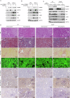
Fig. 8. TRIM65 targets NUDT21 to regulate renal fibrosis.
A Western blot was used to detect the expression of FN-1, α-SMA, and VIM in HK-2 cells overexpressing TRIM65 and NUDT21 protein while stimulated with TGF-β1. B Western blot was used to detect the expression of FN-1, α-SMA, and VIM in HK-2 cells with TRIM65 and NUDT21 protein knocked down while stimulated with TGF-β1. C Western blot was used to detect the expression of TRIM65, NUDT21, KIM-1, FN-1, α-SMA, and VIM in the kidney tissues of 4 groups of C57BL/6 J mice (AAV-si-NUDT21 + WT, AAV-si-NUDT21+Trim65-/-, AAV-si-Control+WT, AAV-si-Control+Trim65-/-) after UUO. GAPDH was used as a housekeeping gene. D Representative images of Sirius Red, Masson’s Trichrome, and IHC staining of renal tissue from mice.

References

    1. Romagnani P, Remuzzi G, Glassock R, Levin A, Jager KJ, Tonelli M, et al. Chronic kidney disease. Nat Rev Dis Prim. 2017;3:17088. - PubMed
    1. Carney EF. The impact of chronic kidney disease on global health. Nat Rev Nephrol. 2020;16:251. - PubMed
    1. Glassock RJ, Warnock DG, Delanaye P. The global burden of chronic kidney disease: estimates, variability and pitfalls. Nat Rev Nephrol. 2017;13:104–14. - PubMed
    1. Mutsaers HA, Olinga P. Editorial: organ fibrosis: triggers, pathways, and cellular plasticity. Front Med. 2016;3:55. - PMC - PubMed
    1. Huang R, Fu P, Ma L. Kidney fibrosis: from mechanisms to therapeutic medicines. Signal Transduct Target Ther. 2023;8:129. - PMC - PubMed

MeSH terms

Substances

LinkOut - more resources