Review
. 2024 Jul 1;22(1):386.
doi: 10.1186/s12951-024-02648-5.

Recent advances in gene delivery nanoplatforms based on spherical nucleic acids

Affiliations
Review

Recent advances in gene delivery nanoplatforms based on spherical nucleic acids

Nazila Valatabar et al. J Nanobiotechnology. .

Abstract

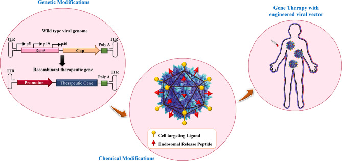
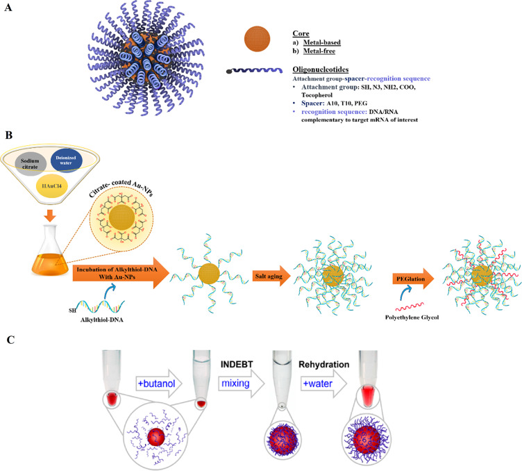
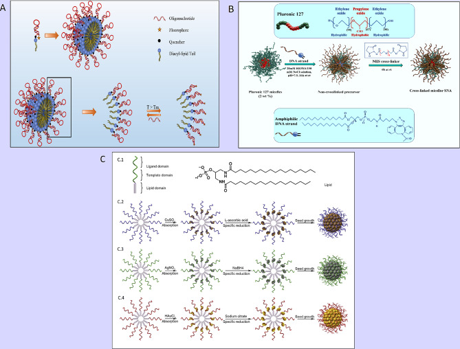
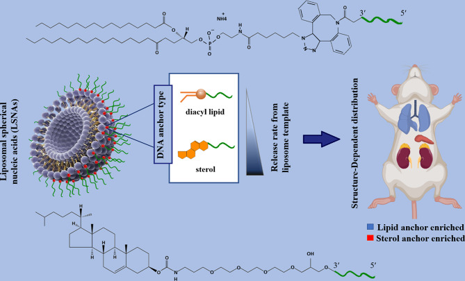
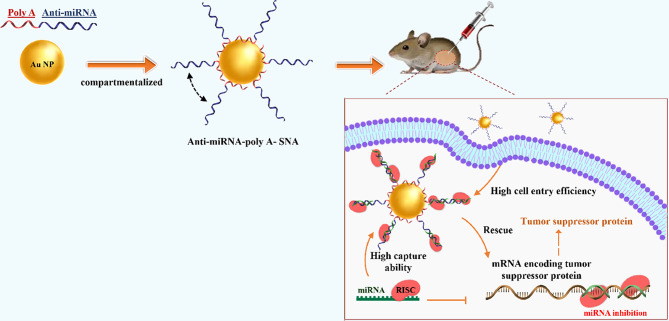
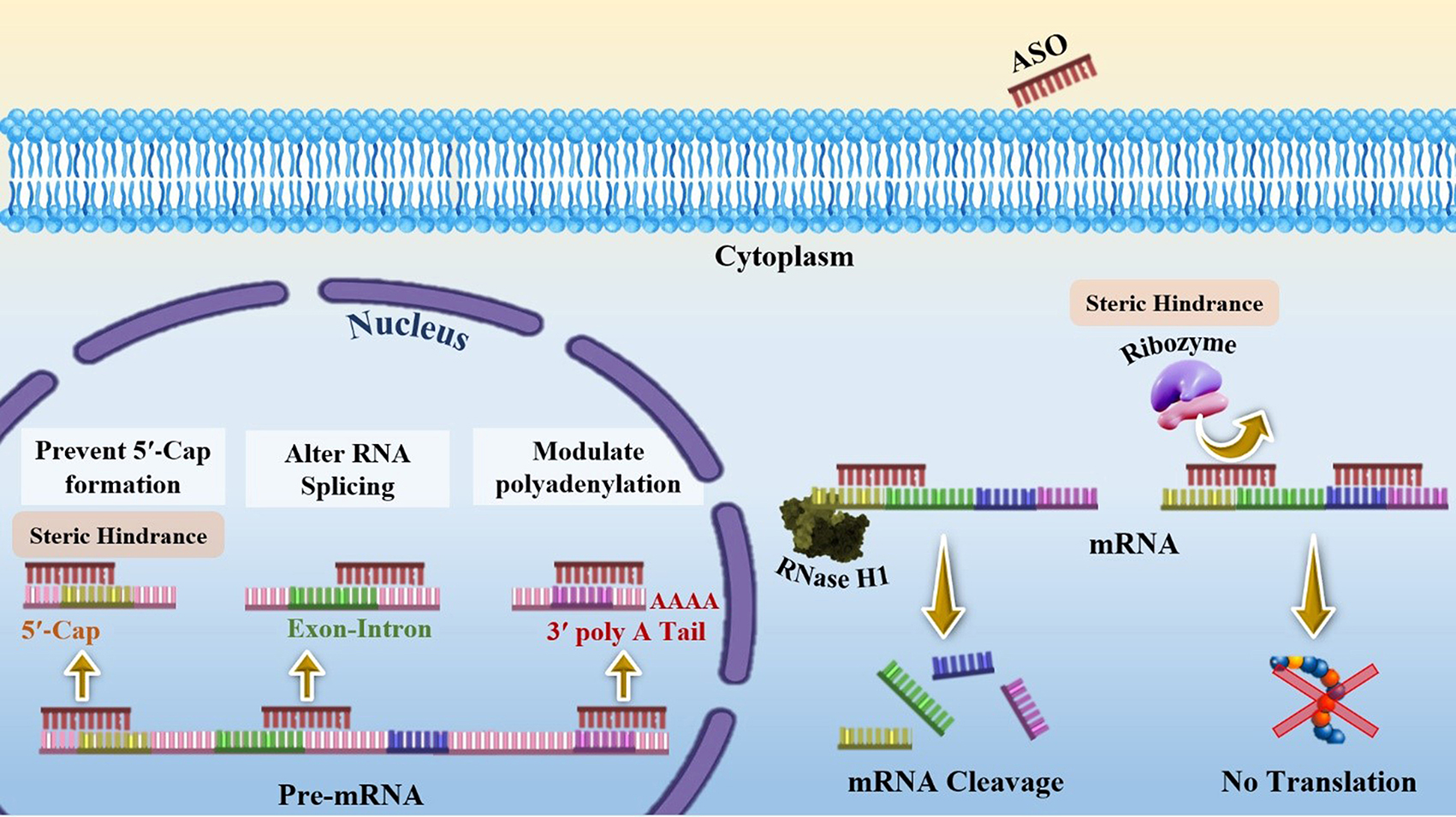
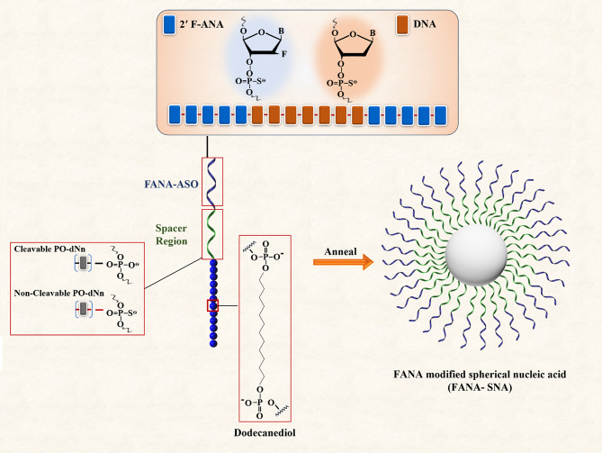
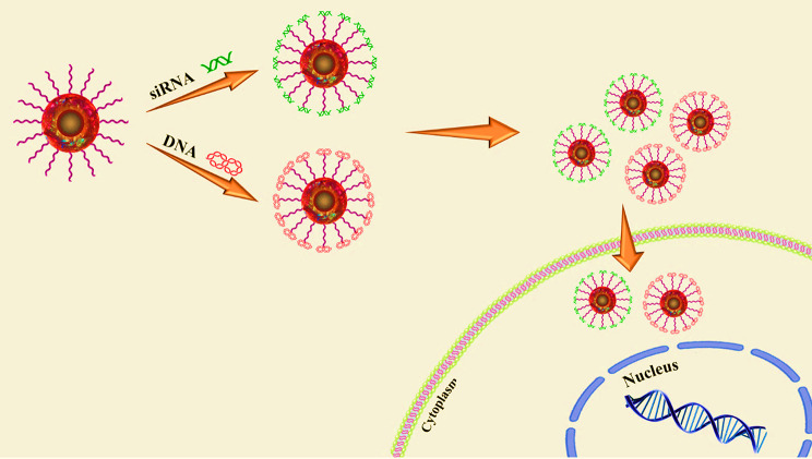
Gene therapy is a therapeutic option for mitigating diseases that do not respond well to pharmacological therapy. This type of therapy allows for correcting altered and defective genes by transferring nucleic acids to target cells. Notably, achieving a desirable outcome is possible by successfully delivering genetic materials into the cell. In-vivo gene transfer strategies use two major classes of vectors, namely viral and nonviral. Both of these systems have distinct pros and cons, and the choice of a delivery system depends on therapeutic objectives and other considerations. Safe and efficient gene transfer is the main feature of any delivery system. Spherical nucleic acids (SNAs) are nanotechnology-based gene delivery systems (i.e., non-viral vectors). They are three-dimensional structures consisting of a hollow or solid spherical core nanoparticle that is functionalized with a dense and highly organized layer of oligonucleotides. The unique structural features of SNAs confer them a high potency in internalization into various types of tissue and cells, a high stability against nucleases, and efficay in penetrating through various biological barriers (such as the skin, blood-brain barrier, and blood-tumor barrier). SNAs also show negligible toxicity and trigger minimal immune response reactions. During the last two decades, all these favorable physicochemical and biological attributes have made them attractive vehicles for drug and nucleic acid delivery. This article discusses the unique structural properties, types of SNAs, and also optimization mechanisms of SNAs. We also focus on recent advances in the synthesis of gene delivery nanoplatforms based on the SNAs.


Conflict of interest statement

The authors declare no competing interests.

Figures

Fig. 1
Fig. 1
Viral vectors designing strategies for gene therapy. This figure was redrawn with permission from ref [8]
Fig. 2
Fig. 2
Schematic display of (A) A Spherical Nucleic Acid (SNA) nanoconjugate [37]. (B) SNAs synthetizing procedure based on Turkevich–Frens (chemical method) [41]. (C) SNAs synthetizing procedure based on instant dehydration in butanol (INDEBT) method [42]. This figure was redrawn with permission from the mentioned references
Fig. 3
Fig. 3
(A) Schematic display of a molecular beacon micelle flare (MBMF). Hairpin-shaped DNA–diacyl lipid segments self-assemble into a sphere micellar flare nanostructure, in which the hairpin-shaped DNA molecular beacon can lead to an ON/OFF switching by binding to targets, changing temperature, or degradation. This figure was redrawn with permission from ref [53]. (B) Schematic illustration of thermo-responsive cross-linked micellar SNAs assembling from a Pluronic F127 block copolymer core and amphiphilic DNA in a temperature-dependent condition. This figure was redrawn with permission from ref [34]. (C) Preparation of Metal-Conjugated ssDNA Micelles (C.1) with the monomer of single-stranded lipid- DNA. Right: 5ʹ end » 3ʹend: domains (lipid, template, ligand). Left: the molecular structure of lipid residue. (C.2) Preparation procedure of copper-crosslinked DNA micelles. (C.3) Preparation procedure of silver-crosslinked DNA micelles. (C.4) Preparation procedure of gold-crosslinked DNA micelles. Permission was received from ref [54]
Fig. 4
Fig. 4
Schematic display of the biodistribution of two types (containing cholesterol tail or diacylglycerol lipid tail) of liposomal spherical nucleic acid (LSNA) conjugates This figure was redrawn with permission from ref [22]
Fig. 5
Fig. 5
Scheme display of synthesizing crosslinked protein SNA (X-SNA). This figure was redrawn with permission from ref [61]
Fig. 6
Fig. 6
DNA nanoclew-siRNA formation by hybridizing siRNA with 20 nt ssDNA complementary overhang of DNA nanoclew. This figure was redrawn with permission from ref [1]
Fig. 7
Fig. 7
Illustration of the construct and interaction of silver spherical nucleic acids. Ag-SNAs from a silver nanoparticle core, functionalized with 3ʹ -thiol-oligonucleotide and methoxyl poly(ethylene glycol) thiol, and show potent antimicrobial features Permission was received from ref [66]
Fig. 8
Fig. 8
A sketch of DNA functionalized biocompatible hollow SiO2 synthesizing procedure using gold nanoparticles as sacrificial templates. This figure was redrawn with permission from ref [81]
Fig. 9
Fig. 9
(A) Schematic drawing of the transfection procedure using magnetic nanoparticles under an external magnetic field [89]. (B) Design of the lipidoid-coated iron oxide nanoparticles coating procedure [90]. (C) Scheme illustration of the synthesis process of Dual-Responsive Maghemite Nanoparticles [93]. This figure redrawn with permission from mentioned references
Fig. 10
Fig. 10
Illustration of the synthesis processes of C60-based SNAs. This figure was redrawn with permission from ref [95]
Fig. 11
Fig. 11
Illustration of MGMT-targeting ribozyme-SNA formation via enzymatic ligation of MGMT-targeting (hammerhead) ribozyme to B-form DNA at the surface of gold nanoparticles. This figure was redrawn with permission from ref [36]
Fig. 12
Fig. 12
Schematic model for synthesizing DNA conjugated block copolymer-based micelle-SNAs. (A) The preparation of the linear DNA- b -PEO- b -PCL block copolymer and the related construction of micelle-SNAs (LDBC-SNAs). (B) The preparation of the brush DNA- g -PCL- b -PCL block copolymer and the corresponding formation of micelle-SNAs (DBBC-SNAs) consisting of a higher surface density of oligonucleotides. This figure was redrawn with permission from ref [99]
Fig. 13
Fig. 13
Recommended procedure for increasing the DNA stability of polyvalent nanoparticles [104]
Fig. 14
Fig. 14
(A) Schematic display of the method for synthesizing DNA-polymer conjugates with BKM120 encapsulated cargo, microscopic images of the cellular uptake in HeLa cells after 24-hour incubation (show high cellular uptake), and image of Cy5.5 fluorescence intensity at the tumor site 24 h after subcutaneous injection (showing long circulation and accumulation in tumors) [116]. (B) Schematic representation of DOX and CpG-loaded liposomal SNA (L-SNA) and its mechanism of function [118]. Permission was received from the mentioned references
Fig. 15
Fig. 15
SNAs features and cytotoxicity
Fig. 16
Fig. 16
Schematic illustration of the action mechanisms of siRNAs
Fig. 17
Fig. 17
The design of a delivery system containing adriamycin (Adr) and synthesized siRap2b conjugating with gold NPs. Treating drug-loaded GNs with an 808-nm laser generated a photothermal effect, which deeply enhanced drug release. The released drug (Adr) showed direct toxicity against cancer cells. Furthermore, isolated siRap2b remarkably diminished the expression of Rap2b and boosted anticancer therapeutic efficiency. Additionally, the thermal effects arising from laser directly suppressed cancer cells/tissues. This figure was redrawn with permission from ref [143]
Fig. 18
Fig. 18
The model describing the synthesis of SNAs and PEI-SNAs. Permission was received from ref [138]
Fig. 19
Fig. 19
(A, B) RNA assembly onto a universal SNA scaffold via an enzymatic method [149]. (A): SNA is made up of an Au NP core functionalized with a DNA “anchor” oligonucleotide [1]. The DNA “bridge” (2a), another DNA oligonucleotide matching to the 3′ end of 1 (anchor). Following the hybridizing with 1, the sticky end formed by the DNA bridge (2a) is utilized to guide the hybridizing and assembly of the 5′ end of an RNA (3a) to the SNA surface. (Note: for siRNA sequences, this strand is the sense strand of a siRNA duplex). (B): By altering the sticky end sequence of the DNA bridge located at its 3′ end (2b), it is possible to match the sequence with the 5′ end of another RNA oligonucleotide (3b). This process enables the assembly of diverse RNA sequences on an SNA coated using the same DNA anchor [1]. (C) Hairpin-like design, a hairpin-like siRNA, a single RNA strand made of a duplex, and a hairpin-like region of PEG spacers, are bound to the core with high duplexing efficiency [150]. This figure was redrawn with permission from the mentioned references
Fig. 20
Fig. 20
Scheme of the manufacturing procedure of C-siRNA. Permission was received from ref [153]
Fig. 21
Fig. 21
Schematic illustration of miRNA biogenesis and mechanism of function
Fig. 22
Fig. 22
(A) Survival analysis indicated that miR-182 expression increased the survival of animals (rthotopic xenograft with glioma cells and engineered GICs that stably expressed miR-182). (B and C) Tumor burden analysis via weight and bioluminescence imaging. (D) Weight of tumors derived from U87MG xenografts extracted from SCID mice 21 days after intravenous treatment with Co-SNAs or 182-SNAs. (E) Bioluminescence imaging of xenograft tumors derived from GIC-20 (12 day) after intravenous treatment with Co-SNAs or 182-SNAs. (F) Estimation levels of Ki67 and caspase-3 in xenograft samples. (G) Ki67 and caspase-3 IHC in coronal brain sections of GIC-derived xenografts expressing Co-miR or miR-182. (H and I) Kaplan-Meyer survival estimator curves of SCID mice xenografted with glioma tumors (U87MG and GIC-20)  and intravenously treated with Co-SNAs or 182-SNAs. Permission was received from ref [160]
Fig. 23
Fig. 23
The graphical design rationale of delivery and releasing miR-34a, employing SNA nanocarriers in MCF-7 cells. This figure was redrawn with permission from ref [161]
Fig. 24
Fig. 24
Preparation of spherical nucleic acids (SNAs) conjugated with flare sequences: (A) Functionalization of Au nanoparticles (13 ± 1 nm) surface with propylthiol-terminated antisense DNA, hybridizing with short complementary fluorophore-labeled DNA (Flare). (B) Upon incubation of the prepared nano-conjugates (A) with complementary miRNA targets, RISC (miRISC) was loaded, and the fluorescence signal increased. (C) The DNA/LNA gapmer sequences (target and control) served as antisense strands (Note: The underlined bases are LNA), and the flare sequence was applied to form nanoflare conjugates. This figure was redrawn with permission from ref [162]
Fig. 25
Fig. 25
Schematic illustration of the synthetic process of peptide-hybridized gold-liposome. (A) Spherical nucleic acids (SNAs) formation. (B) Construction of Liposome-Peptide and SNA-Liposome-Peptide nanocarriers. Permission was received from ref [163]
Fig. 26
Fig. 26
Schematic design of PolyA-SNAs construction containing programmed polyA-lateral spacing for recovery of the antioncogene expression. This figure was redrawn with permission from ref [165]
Fig. 27
Fig. 27
(A) Schematic model depicting the effects of targeting MFG-E8 gene expression in macrophages on intestinal epithelial homeostasis. Inhibition of miR-99b stimulated the migration of intestinal epithelial cells in LPS-induced septic mice. (B) Images of the small intestine processed for BrdU/DAPI staining. (Note: green and blue colors show BrdU-labeled cells and nuclei, respectively). (C) Quantitative analysis discovered that SNA-NC anti-miR99b treatment increased enterocytes’ migration along the crypt-villus axis in LPS-induced septic mice. n = 3. **P. permission was received from ref [165]
Fig. 28
Fig. 28
Construction of photo-responsive SNA. (A) Schematic illustration of the preparation of photo-responsive SNA nanocarriers. (B) schematic representation of the use of photo-responsive SNA and miR-34a release in MCF-7 cells exposed to UV light. C, D) Relative expression level of miR-34 and knockdown level of survivin mRNA. This figure was redrawn with permission from ref [167]
Fig. 29
Fig. 29
Schematic display of synthetizing of miRNA − AuNP nano-conjugates for carrying miRNAs into Cells. This figure was redrawn with permission from ref [169]
Fig. 30
Fig. 30
Mechanisms of action of antisense oligonucleotides (ASOs)
Fig. 31
Fig. 31
(A) Topical application of SNAs on rabbit ear scars. Note: “MWF” indicates 3 days a week “Monday, Wednesday, Friday”, when treatment was administered. (B) Average TGF-β1 expression level, as evaluated by densitometry and Western blot protein analysis,   which was normalized to the untreated group and expressed as the mean ± SEM (N = 6). Significant differences were observed between treatment vs. untreated groups (*p < 0.05, **p < 0.01).C) Masson’s trichrome staining of scar ear tissues under diverse treatment conditions. Note: The red arrow shows the site of the magnified image. Scale bars = 100 μm. D) Graphic depiction of scar elevation index (SEI). E) Mean ± SEM of composite SEI in different treatment groups (N = 6). Significance differences between treatment groups and controls are indicated by *p < 0.05, **p < 0.01, ns = not significant). Permission was received from ref [172]
Fig. 32
Fig. 32
(A) Schematic illustration of ASO-PR10 prodrug conjugate formulation and their micellar SNA assemblage. (B) the activity of tyrosinase and (C) the content of melanin in various groups (treatment with ASO-PR10, DNA-PR10 with a scrambled sequence [Scr-PR10], PR-devoid micelles [in PBS], or free PR [in DMSO]. Note: a-MSH was applied for melanogenesis stimulation. The activity of tyrosinase and the content of melanin were documented as a percent of the difference in α-MSH-treated cells (2 to 20 µM PR, 48 h treatment). PR-devoid micelles: 1 µM ASO. (D) Histological study of isolated mouse ear following treatment with PR/ASO-PR10/carrier groups (paraffin-embedded). Note: Melanin (indicated by black arrows) is stained by Fontana − Masson staining. Scale bar: 50 μm. (E) Relative MC1R levels in ASO-PR10-treated group compared to ears treated with nanocarrier were determined via immunohistostaining. *** p < 0.001 (two-tailed test). Permission was received from ref [176]
Fig. 33
Fig. 33
A model for FANA-SNA. Hybridizing dodecanediol (20 units) to FANA-ASO gapmer (18 nt) makes an SNA with a low polydispersity and well-defined size and shape. The spacer region is modified to optimize the activity of SNAs. (N, n, PO, and PS represent nucleotides, the number of nucleotides, phosphodiester backbone, and phosphorothioate backbone respectively). This figure was redrawn with permission from ref [181]
Fig. 34
Fig. 34
Schematic illustration of Alkyl-PEI2k-Cdots/siRNA complex formation and delivery into the cancer cell. This figure was redrawn with permission from ref [186]

Similar articles

Cited by

References

    1. Ruan W, Zheng M, An Y, Liu Y, Lovejoy DB, Hao M, et al. DNA nanoclew templated spherical nucleic acids for siRNA delivery. Chem Commun. 2018;54(29):3609–12. doi: 10.1039/C7CC09257A. - DOI - PubMed
    1. Patil S, Gao Y-G, Lin X, Li Y, Dang K, Tian Y, et al. The development of functional non-viral vectors for gene delivery. Int J Mol Sci. 2019;20(21):5491. doi: 10.3390/ijms20215491. - DOI - PMC - PubMed
    1. Zhou Z, Liu X, Zhu D, Wang Y, Zhang Z, Zhou X, et al. Nonviral cancer gene therapy: delivery cascade and vector nanoproperty integration. Adv Drug Deliv Rev. 2017;115:115–54. doi: 10.1016/j.addr.2017.07.021. - DOI - PubMed
    1. Uddin F, Rudin CM, Sen T. CRISPR gene therapy: applications, limitations, and implications for the future. Front Oncol. 2020;10:1387. doi: 10.3389/fonc.2020.01387. - DOI - PMC - PubMed
    1. Lee CS, Bishop ES, Zhang R, Yu X, Farina EM, Yan S, et al. Adenovirus-mediated gene delivery: potential applications for gene and cell-based therapies in the new era of personalized medicine. Genes Dis. 2017;4(2):43–63. doi: 10.1016/j.gendis.2017.04.001. - DOI - PMC - PubMed