

New insights into swine dysentery: faecal shedding, macro and microscopic lesions and biomarkers in early and acute stages of Brachyspira hyodysenteriae infection
- PMID: 38951921
- PMCID: PMC11218200
- DOI: 10.1186/s40813-024-00375-9
New insights into swine dysentery: faecal shedding, macro and microscopic lesions and biomarkers in early and acute stages of Brachyspira hyodysenteriae infection
Abstract
Background: Swine dysentery (SD) is a severe mucohaemorrhagic colitis in pigs caused classically by Brachyspira hyodysenteriae. Although several aspects of B. hyodysenteriae infection dynamic are already described, further research in the early stage of this infection is required. In this study, 7-week-old pigs were orally challenged with B. hyodysenteriae to obtain information about faecal shedding, macro and microscopic intestinal lesions and serum acute phase proteins in pigs at the onset of B. hyodysenteriae shedding (early infection group, n = 8), in pigs with mucohaemorrhagic diarrhoea (acute infection group, n = 8) and in non-infected controls (n = 16).
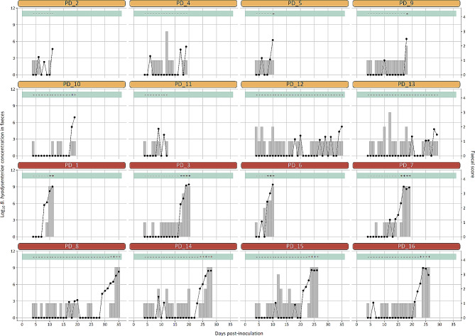
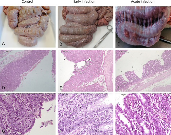
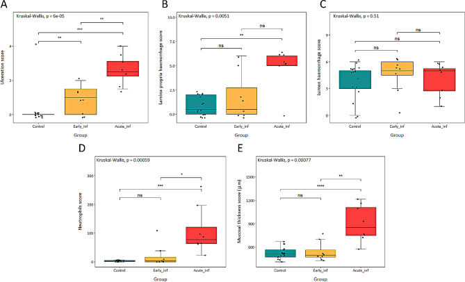
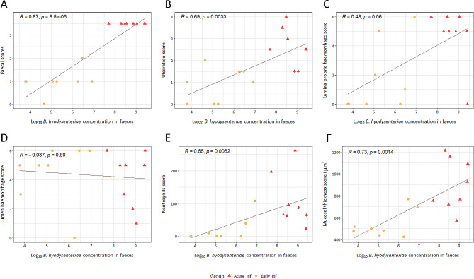
Results: First B. hyodysenteriae detection by q-PCR and first loose stools with blood and mucus occurred both at 8 days post-inoculation. The lapse between a positive q-PCR and observation of mucohaemorrhagic diarrhoea ranged from 0 to 3 days, except in a single pig in which this period lasted 5 days. Macroscopic lesions were observed in the large intestine from both infected groups although more frequent and severe in acute infection group. Microscopic observation of the apex mucosa revealed that in early infection only higher ulceration values were observed compared to healthy controls. In contrast, the acute infection group exhibited higher ulceration, neutrophils infiltration and increased mucosal thickness compared to the other two groups. Among the serum biomarkers tested, only haptoglobin, C-reactive protein, and creatine kinase showed a significant increase in pigs in the acute infection period compared to controls, whereas haptoglobin was the only factor with a significant increase at the early infection compared to non-infected animals.
Conclusions: This study provides new insights about SD and remarks the complex and limited options to perform an early detection of infected animals beyond PCR diagnosis.
Keywords: Acute phase proteins; Mucohaemorrhagic diarrhoea; Pig; Spirochaetes; Swine dysentery.
© 2024. The Author(s).
Conflict of interest statement
The authors declare no competing interests.
Figures

Similar articles
-
Comparison of sesion severity, distribution, and colonic mucin expression in pigs with acute swine dysentery following oral inoculation with "Brachyspira hampsonii" or Brachyspira hyodysenteriae.Vet Pathol. 2014 Nov;51(6):1096-108. doi: 10.1177/0300985813516646. Epub 2014 Feb 27. Vet Pathol. 2014. PMID: 24577722
-
Reproduction of mucohaemorrhagic diarrhea and colitis indistinguishable from swine dysentery following experimental inoculation with "Brachyspira hampsonii" strain 30446.PLoS One. 2013;8(2):e57146. doi: 10.1371/journal.pone.0057146. Epub 2013 Feb 27. PLoS One. 2013. PMID: 23460829 Free PMC article.
-
Experimental Infection of Pigs with a ST 245 Brachyspira hyodysenteriae Isolated from an Asymptomatic Pig in a Herd with No History of Swine Dysentery.Vet Sci. 2022 Jun 10;9(6):286. doi: 10.3390/vetsci9060286. Vet Sci. 2022. PMID: 35737338 Free PMC article.
-
A review of methods used for studying the molecular epidemiology of Brachyspira hyodysenteriae.Vet Microbiol. 2017 Aug;207:181-194. doi: 10.1016/j.vetmic.2017.06.011. Epub 2017 Jun 19. Vet Microbiol. 2017. PMID: 28757022 Review.
-
Swine Dysentery.Vet Pathol. 2017 Jan;54(1):22-31. doi: 10.1177/0300985816653795. Epub 2016 Jul 11. Vet Pathol. 2017. PMID: 27288432 Review.
Cited by
-
Development and application of a quadruplex TaqMan real-time fluorescence quantitative PCR assay for four porcine digestive pathogens.Front Cell Infect Microbiol. 2024 Nov 26;14:1468783. doi: 10.3389/fcimb.2024.1468783. eCollection 2024. Front Cell Infect Microbiol. 2024. PMID: 39660284 Free PMC article.
References
-
- Sato JPH, Daniel AGS, Leal CAG, Barcellos DESN, Guedes RMC. Diversity and potential genetic relationships amongst Brazilian Brachyspira hyodysenteriae isolates from cases of swine dysentery. Vet Microbiol. 2022;266. - PubMed
Grants and funding
LinkOut - more resources
Full Text Sources
Research Materials
Miscellaneous
