. 2024 Sep;11(33):e2402450.
doi: 10.1002/advs.202402450. Epub 2024 Jul 1.

DHPS-Mediated Hypusination Regulates METTL3 Self-m6A-Methylation Modification to Promote Melanoma Proliferation and the Development of Novel Inhibitors

Affiliations

DHPS-Mediated Hypusination Regulates METTL3 Self-m6A-Methylation Modification to Promote Melanoma Proliferation and the Development of Novel Inhibitors

Jing-Si Guo et al. Adv Sci (Weinh). 2024 Sep.

Abstract

Discovering new treatments for melanoma will benefit human health. The mechanism by which deoxyhypusine synthase (DHPS) promotes melanoma development remains elucidated. Multi-omics studies have revealed that DHPS regulates m6A modification and maintains mRNA stability in melanoma cells. Mechanistically, DHPS activates the hypusination of eukaryotic translation initiation factor 5A (eIF5A) to assist METTL3 localizing on its mRNA for m6A modification, then promoting METTL3 expression. Structure-based design, synthesis, and activity screening yielded the hit compound GL-1 as a DHPS inhibitor. Notably, GL-1 directly inhibits DHPS binding to eIF5A, whereas GC-7 cannot. Based on the clarification of the mode of action of GL-1 on DHPS, it is found that GL-1 can promote the accumulation of intracellular Cu2+ to induce apoptosis, and antibody microarray analysis shows that GL-1 inhibits the expression of several cytokines. GL-1 shows promising antitumor activity with good bioavailability in a xenograft tumor model. These findings clarify the molecular mechanisms by which DHPS regulates melanoma proliferation and demonstrate the potential of GL-1 for clinical melanoma therapy.

Keywords: DHPS; eIF5A‐Hypusine; m6A; melanoma.


Conflict of interest statement

The authors declare no conflict of interest.

Figures
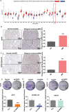
Figure 1
Figure 1
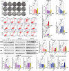
Identification of DHPS as a potential differential target in skin melanoma. A) Bioassay analysis of DHPS shows that DHPS is specifically highly expressed in a variety of cancer tissues, particularly skin cancer. B) Human melanoma microarray results show that DHPS is highly expressed in melanoma and low in healthy tissues. (Scale bar = 100 µm/50 µm, n = 129, *** p < 0.001) C) Human melanoma microarray results show that hypusination is high in melanoma and low in healthy tissues. (Scale bar = 100 µm/50 µm, n = 129 *** p < 0.001) D–F) Knockdown of DHPS effectively inhibits melanoma proliferation but does not affect normal skin cells (Scale bar = 1 cm, n = 3, *** p < 0.001).
Figure 2
Figure 2
DHPS maintains mRNA stability by m6A methylation modification in melanoma cells. A) The heat map analysis, hierarchical clustering, and volcano plot represent a differential analysis B) of the A375 cell (WT) and DHPS knockdown A375 cell (shDHPS) genes. Color scale: Red is higher expressed; blue is lower expressed. C) The mRNA metabolism and stability‐related pathways were positively associated with low DHPS expression by GSEA (P < 0.05, FDR < 0.25). D) Volcano plot of m6A peaks detected by MeRIP‐seq in the A375‐WT and the A375‐shDHPS. Red dots mean m6A peaks up, while green dots mean m6A peaks down. Note that multiple peaks may map to the same gene. E) Peak distribution of m6A modification in MeRIP‐seq results. F) The sequence motif identified from the sequencing profile. G) KEGG and GO analysis H) of genes with high m6A levels in the A375‐WT and A375‐shDHPS cells. I,J) Protein expression levels of YTHDF2, YTHDC1, and METTL3 in A375 cells after DHPS knockdown. (Treatment group vs control group, * p < 0.05, ** p < 0.01, *** p < 0.001).
Figure 3
Figure 3
DHPS regulates mRNA stability and YTHDC1, and YTHDF2 protein expression in melanoma cells through eIF5A‐Hyp‐mediated METTL3 self‐m6A‐methylation modification. A) Nine‐quadrant plot of A375‐WT compared to A375‐shDHPS. The horizontal axis is the multiplicity of difference in peak abundance values of m6A (taken as log2), the vertical axis is the multiplicity of difference in gene expression of the transcriptome (taken as log2), and the dashed lines on the horizontal and vertical axes are the default thresholds for screening for differential genes/peak in the two cohorts |log2FC| > 1. Grey dots indicate nondifferentiated genes, red and yellow dots indicate genes and m6A peak abundance values trend consistently up/down, and blue and green dots indicate the opposite trend of base gene and m6A peak abundance values. B) The m6A abundance of METTL3 transcripts in A375‐shDHPS cells compared to A375‐WT cells. C,D) Protein expression levels of YTHDF2, YTHDC1, and METTL3 in A375 or SK‐MEL‐28 cells after METTL3 overexpression. (Treatment group versus control group, ** p < 0.01, *** p < 0.001). The interaction between DHPS and METTL3 E) or eIF5A‐Hyp and METTL3 F) was studied in the Co‐IP assay. G) Co‐IP assay for inhibition of eIF5A‐Hyp interaction with METTL3 after GC‐7 (2 µm) treatment in melanoma cells A375 and SK‐MEL‐28 cells. H) The m6A peak region of METTL3 uncovered the presence of two similar “RRACH” sequences in the CDS region near the 5′UTR. I) RIP assays of METTL3 mRNA binding to eIF5A‐Hyp or METTL3. Interference with METTL3 and GC‐7 (2.0 µm) inhibited the binding of METTL3 mRNA to eIF5A‐Hyp or METTL3. J) (t1/2) of Ki67 and METTL3 in A375 and SK‐MEL‐28 cells. K) Schematic representation of the mechanism by which DHPS regulates mRNA stability in melanoma cells.
Figure 4
Figure 4
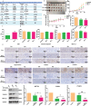
Synthesis and targeting studies of GL‐1 as a novel DHPS inhibitor. A) Synthesis Scheme: Reagents and conditions: i) NBS, H2SO4, CHCl3, 50 °C, 8 h; ii) guaiacol, K2CO3, acetonitrile, 60 °C, overnight; iii‐a) Na, CH3OH, CH2Cl2, rt, 48 h; NH4Cl, rt, overnight; or iii‐b) ethanol, acetyl chloride, CH2Cl2, rt, 24 h; Na, NH4Cl, CH3OH, rt, overnight; iv) Na, CH3OH, rt, 24 h; v) POCl3, 100 °C, overnight. B) The inhibitory effects of GL‐1 and GC‐7 on DHPS enzyme activity were compared at the same concentrations (1, 5, 10, 50, 100, 200, 400, 800, 1600, and 2000 nm). C) SPR analysis of the kinetic interactions between the GL‐1 and DHPS. D) The inhibitory effects on A375 and SK‐MEL‐28 cell viability were measured after treatment with GL‐1 or GC‐7 at different concentrations (0.001, 0.01, 0.02, 0.035, 0.0625, 0.125, 0.25, 0.50, 1.00, 2.00, 4.00, and 8.00 µm) for 24 h. E) Effect of different concentrations (0, 0.5, 1.0, 2.0 µm) of GL‐1 on hypusiantion protein expression. F,G) Western blot analysis of DARTS and CETSA samples demonstrated that GL‐1 and DHPS were able to bind stably. H) Docking pose of GL‐1 within the allosteric site of DHPS (PDB code: 6PGR). I) Cell viability rates after transfection of empty vector plasmids, DHPS‐WT plasmid, DHPS‐K329A plasmid, DHPS‐V129A plasmid, DHPS‐L281A plasmid, and DHPS‐D238A plasmid into A375 and SK‐MEL‐28 cells for 48 h and addition of different concentrations of GL‐1 for 24 h of continuous action. J) Co‐IP results showed that GL‐1 could block the binding of DHPS to the eIF5A‐Hyp protein. (Treatment group vs control group, * p < 0.05, ** p < 0.01, *** p < 0.001).
Figure 5
Figure 5
GL‐1 promotes melanoma apoptosis in a concentration‐dependent manner. A,B) GL‐1 could inhibit colony formation in a concentration‐dependent manner (Scale bar = 1 cm). C,D) FlowJ assay the induction of apoptosis in melanoma cells by GL‐1 and GC‐7. E–H) Western blot analysis of GL‐1 and GC‐7 on TYMS, Caspase9, and cleaved‐caspase3, caspase3 proteins from tumor tissue. (Treatment group vs control group, * p < 0.05, ** p < 0.01, *** p < 0.001).
Figure 6
Figure 6
Other mechanisms of anticancer effects of GL‐1 on melanoma cells. A) Changes in Cu2+ content after 24 h of intracellular GL‐1 action. B,C) Effects of GL‐1 and GC‐7 on METTL3, YTHDF2, and YTHDC1 protein expression. The effect of GL‐1 (2.0 µm) or GC‐7 (2.0 µm) on human multiple cytokines D) and angiogenesis‐related proteins E) in A375 cells was analyzed, and the average spot pixel density on the array membrane was quantified using ImageJ software to generate histogram profiles of selected analyses. (Treatment group vs control group, * p < 0.05, ** p < 0.01, *** p < 0.001).
Figure 7
Figure 7
Studies of the in vivo pharmacokinetics and anti‐melanoma activity of GL‐1. A) Evaluation of the pharmacokinetics of GL‐1. B–E) A375 cell xenograft mice treated with SPSS, GC‐7(40 mg kg−1), and GL‐1(40 mg kg−1) respectively shown in image B), tumor volume C), weight change D), and tumor weight E) analysis. n = 6 mice per group. F) TGI of GL‐1 versus GC‐7. G) Blood biochemistry of GL‐1or GC‐7 for ALT, AST, CRE, and blood glucose level. H) IHC analysis of GL‐1 and GC‐7 on Ki67, Hypusination, Cleaved‐caspase3, METTL3, YTHDF2, and YTHDC1 protein expression in tumor tissue. I) Western blot analysis of GL‐1 and GC‐7 on METTL3, YTHDF2, and YTHDC1 protein expression in tumor tissue. (Scale bar = 50 µm/20 µm, * p < 0.05, ** p < 0.01, *** p < 0.001).
Figure 8
Figure 8
Regulatory mechanisms of DHPS in melanoma cells and the action of the targeted inhibitor GL‐1. Mechanistically, DHPS catalyzes the hypusination of eIF5A. This process has two effects: it mediates the self‐m6A‐methylation modification of METTL3, then promotes METTL3 protein expression and the mRNA translation process of YTHDF2 and YTHDC1. Consequently, DHPS plays a role in maintaining intracellular mRNA homeostasis and promoting cytokine expression, thereby sustaining the proliferative state of melanoma. When DHPS is inhibited, intracellular mRNA degradation is blocked, inhibiting various cytokine expressions and subsequently inhibiting melanoma cell proliferation.

References

    1. Karimkhani C., Green A. C., Nijsten T., Weinstock M. A., Dellavalle R. P., Naghavi M., Fitzmaurice C., Brit. J. Dermatol. 2015, 177, 134. - PMC - PubMed
    1. Linos E., Swetter S. M., Cockburn M. G., Colditz G. A., Clarke C. A., J. Investigat. Dermatol. 2009, 129, 1666. - PMC - PubMed
    1. Center for Disease Control and Prevention (CDC) , Guy G. P. Jr, Thomas C. C., Thompson T., Watson M., Massetti G. M., Richardson L. C., MMWR. Morb. Mortal. Wkly Rep. 2015, 64, 591. - PMC - PubMed
    1. Whiteman D. C., Green A. C., Olsen C. M., J. Invest. Dermatol. 2016, 136, 1161. - PubMed
    1. Siegel R. L., Miller K. D., Wagle N. S., Jemal A., CA: Cancer J. Clin. 2023, 73, 17. - PubMed

Substances

LinkOut - more resources