Review
. 2024;99(4):230-247.
doi: 10.1159/000537746. Epub 2024 Jul 16.

Does a Vertebrate Morphotype of Pallial Subdivisions Really Exist?

Affiliations
Review

Does a Vertebrate Morphotype of Pallial Subdivisions Really Exist?

Kei Yamamoto et al. Brain Behav Evol. 2024.

Abstract

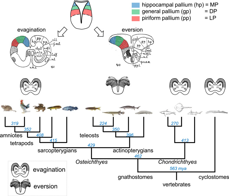
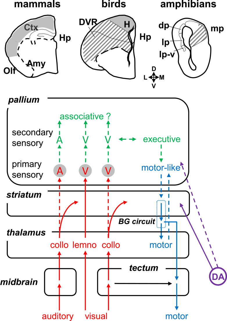
Background: Comparative neuroanatomists have long sought to determine which part of the pallium in nonmammals is homologous to the mammalian neocortex. A number of similar connectivity patterns across species have led to the idea that the basic organization of the vertebrate brain is relatively conserved; thus, efforts of the last decades have been focused on determining a vertebrate "morphotype" - a model comprising the characteristics believed to have been present in the last common ancestor of all vertebrates.

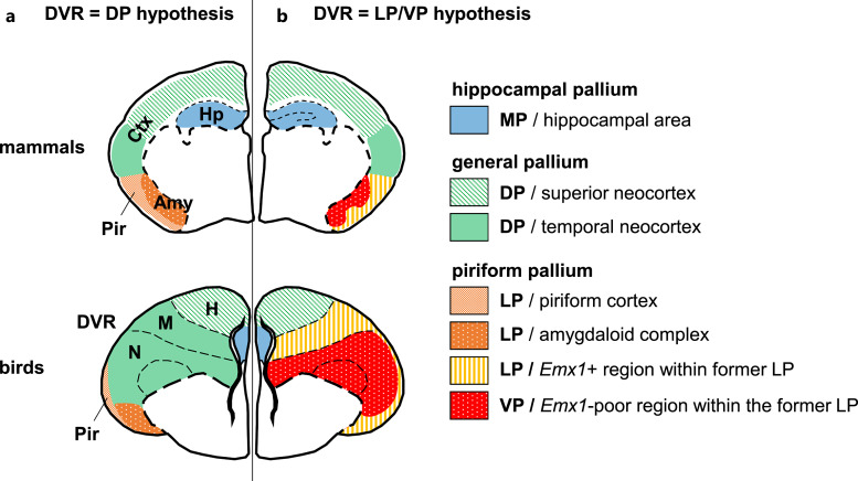
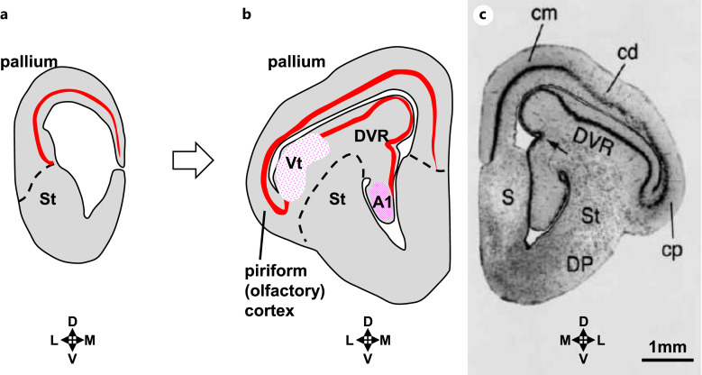
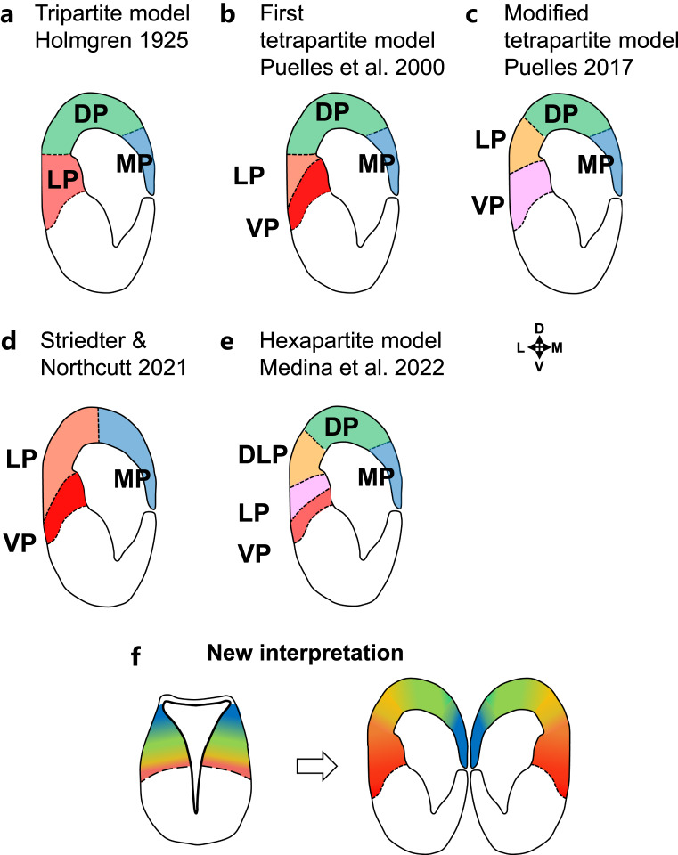
Summary: The endeavor to determine the vertebrate morphotype has been riddled with controversies due to the extensive morphological diversity of the pallium among vertebrate taxa. Nonetheless, most proposed scenarios of pallial homology are variants of a common theme where the vertebrate pallium is subdivided into subdivisions homologous to the hippocampus, neocortex, piriform cortex, and amygdala, in a one-to-one manner. We review the rationales of major propositions of pallial homology and identify the source of the discrepancies behind different hypotheses. We consider that a source of discrepancies is the prevailing assumption that there is a single "morphotype of the pallial subdivisions" throughout vertebrates. Instead, pallial subdivisions present in different taxa probably evolved independently in each lineage.

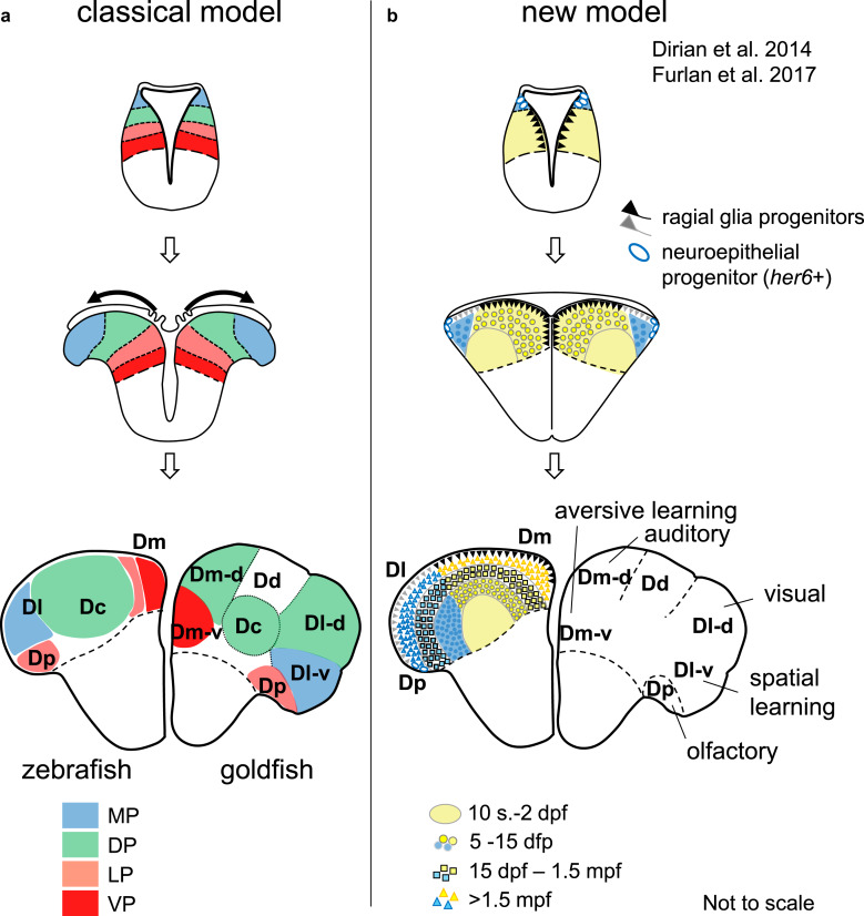
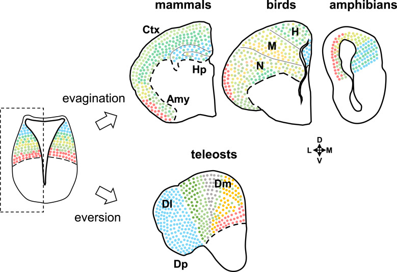
Key messages: We encounter discrepancies when we search for a single morphotype of subdivisions across vertebrates. These discrepancies can be resolved by considering that several subdivisions within the pallium were established after the divergence of the different lineages. The differences of pallial organization are especially remarkable between actinopterygians (including teleost fishes) and other vertebrates. Thus, the prevailing notion of a simple one-to-one homology between the mammalian and teleost pallia needs to be reconsidered.

Keywords: Comparative neuroanatomy; Convergent evolution; Homology; Pallium.


Conflict of interest statement

The authors have no conflict of interests to declare.

Figures

Fig. 1.
Fig. 1.
Classical view of evagination and eversion. The top schematic drawings show the comparison of pallial development in eversion (actinopterygians) and evagination (all other vertebrates) proposed by Holmgren (1925). The bottom shows a simplified phylogenetic tree of vertebrates. The italic blue numbers indicate divergence dates (million years ago [mya]) of each clade calculated using http://www.timetree.org/ [7]. Based on cladistics analysis, it is concluded that evagination is the ancestral form, and that the everted pallium specifically evolved in actinopterygians including teleost fishes.
Fig. 2.
Fig. 2.
Dorsal pallium (DP)-like connectivity found in mammals and birds. Simplified diagram of neural inputs and outputs related to the pallium, whose patterns are found to be similar between mammals and birds. Modality-specific sensory inputs are indicated in red arrows. Here visual and auditory pathways are shown as an example of modality-specific sensory afferents to the pallium. In the visual system, two parallel afferents are found: the tectofugal- or collo-pathway (“collo” stands for the colliculus, which is relayed via the midbrain roof), and thalamofugal- or lemno-pathway (“lemno” stands for lemniscal inputs, but the term is generally used to refer to non-collicular inputs). Motor outputs are indicated in blue (the detailed basal ganglia [BG] circuits are omitted for the sake of simplicity). Dopaminergic (DA) projections from the mesencephalon (A9/A10) to the telencephalon are indicated in purple. DA inputs to the striatum are considered to be critical for BG function throughout tetrapods, while those to the pallium (PFC in mammals and NCL in birds) are critical for executive functions. The solid arrows indicate the connectivity found in mammals, birds, and amphibians, while the dotted arrows indicate those found only in mammals and birds. Note that many of the pallial connections are absent in amphibians. The top brain schemas (in frontal section) show the pallial area receiving thalamic projections (circled in gray in the diagram: “A” for auditory and “V” for visual areas), similarly to the primary sensory areas of the mammalian neocortex. The brain areas with filled gray indicate the agreed homology of DP due to its “dorsal” topology. The brain areas with slanted gray lines are areas receiving thalamic sensory inputs for which homology to the mammalian DP remains debated: this area is located at the ventral end of the pallium in birds, while it is at the medial end of the pallium in amphibians. A, auditory area; Amy, amygdala; BG, basal ganglia; Ctx, neocortex; DA, dopaminergic neuron; DP, dorsal pallium (amphibian structure); DVR, dorsal ventricular ridge (avian structure); H, hyperpallium (avian structure); Hp, hippocampus; LP, lateral pallium (amphibian structure); lp-v, ventral part of the lateral pallium (amphibian structure); MP, medial pallium (amphibian structure); Olf, olfactory (piriform) cortex; V, visual area. Brain orientation; D, dorsal; V, ventral; L, lateral; M, medial.
Fig. 3.
Fig. 3.
Pallial homologies between mammals (top) and birds (bottom) debated during 1990s–early 2000s. The left side of the frontal section (a) represents “DVR = DP hypothesis,” whereas the right side of the section (b) represents “DVR = LP/VP hypothesis” based on the first tetrapartite model. The two different hypotheses propose different mammalian homolog of the avian DVR. Amy, amygdaloid complex; Ctx, cerebral cortex; DP, dorsal pallium (as morphotype); DVR, dorsal ventricular ridge; h, hyperpallium; Hp, hippocampus; LP, lateral pallium (as morphotype); M, mesopallium; MP, medial pallium (as morphotype); N, nidopallium; Pir, piriform cortex; VP, ventral pallium (as morphotype).
Fig. 4.
Fig. 4.
Topological organization of the DVR of tuatara, Sphenodon punctatus. Schematic representations of the developing (a) and mature (b) telencephalic vesicle (left side of frontal sections). c A cresyl violet-stained section (from Reiner and Northcutt 2000) showing the mirror image of b. The tectofugal visual area (Vt) and the primary auditory area (A1) are situated within the DVR. The pallium of Sphenodon possesses a three-layered cytoarchitecture that resembles the cortical cell plate (shown as a red line in the schema). This cell plate-like structure is continuous from the medial/DP, through the piriform cortex up to the ventral edge of the DVR. This clearly demonstrates that the DVR is topologically ventral to the piriform cortex, adjacent to the subpallium (striatum). A1, primary auditory area; cd, dorsal cortex; cm, medial cortex; cp, piriform cortex; DP, dorsal pallidum; DVR, dorsal ventricular ridge; S, septum; St, striatum; Vt, tectofugal visual area. Brain orientation: D, dorsal; V, ventral; L, lateral; M, medial.
Fig. 5.
Fig. 5.
Modifications of the classification of pallial subdivisions in sarcopterygians. a–e Frontal sections through the pallium (left side) summarizing different hypotheses of pallial subdivisions. See the text for details. The boundaries and number of pallial subdivisions keep being modified over time, depending on the criteria and species examined. Importantly, the first tetrapartite model (b) that was widely accepted after 2000’s has been abandoned by the authors themselves (c), and there is no consensus proposition since then. f Our new interpretation. The neural tube organization is gradient and boundaries of the pallial subdivisions are not as clear as it has classically been assumed. Amy, amygdala; DLP, dorsolateral pallium; DP, dorsal pallium; LP, lateral pallium; MP, medial pallium; VP, ventral pallium. Brain orientation: D, dorsal; V, ventral; L, lateral; M, medial.
Fig. 6.
Fig. 6.
New eversion theory of the teleost pallium. a Classical model of the pallial development of actinopterygians, color-coded with one of the prevailing pallial homology (based on the first tripartite model; Fig. 5b). The left side of the mature brain shows the zebrafish pallium, while the right side shows the goldfish pallium. Historically, pallial regions in actinopterygians have been considered as a simply reversed version of the sarcopterygian pallium (compare also with Fig. 1). b New model based on lineage tracing data, adapted from Dirian et al. [84] and Furlan et al. [85]. The construction of the actinopterygian pallium is not simply a reversed version of the sarcopterygian pallium. The teleost pallium does not develop by extension of the preexisting three or four embryonic subdivisions. Instead, newly born neurons are progressively “stacking-up” on top of the old ones. All the lateral parts of the pallium containing Dl and Dp are derived from the her6+ progenitors located at the dorsal tip of the neural tube until 2 dpf. Dc, central zone of the pallium; Dd, dorsal zone of the pallium; Dl, lateral zone of the pallium; Dl-d, dorsal part of Dl; Dl-v, ventral part of Dl; Dm, medial zone of the pallium; Dm-d, dorsal part of Dm; Dm-v, ventral part of Dm; Dp, posterior zone of the pallium; dpf, days post-fertilization; DP, dorsal pallium (as morphotype); LP, lateral pallium (as morphotype); MP, medial pallium (as morphotype); mpf, months post-fertilization; s, somites; VP, ventral pallium (as morphotype).
Fig. 7.
Fig. 7.
Schematic representation of the new view of pallial organization in vertebrates taking into account the everted pallium. A representative embryonic neural tube (both hemispheres) is shown on the left, and adult pallia (left side of the telencephalic hemisphere) of mammals, birds, and amphibians (evaginated pallium) and of teleosts (everted pallium) are shown on the right. The gradient organization along the neural tube (Fig. 5f) is shared within the tetrapod pallium (presumably all sarcopterygians), while the dorsoventral, medial-lateral organization is very different in the teleost pallium (see Fig. 6b). None of the current models can provide a comprehensive morphotype across vertebrates, and homology needs to be discussed at the level of smaller cell clusters or cell types, instead of the level of cytoarchitectonically distinct subdivisions in the mature brain (e.g., neocortex, amygdala, nidopallium, Dm, Dl). Amy, amygdala (mammalian structure); Ctx, neocortex (mammalian structure); Dl, lateral part of the pallium (teleost structure); Dm, medial part of the pallium (teleost structure); Dp, posterior part of the pallium (teleost structure); H, hyperpallium (avian structure); M, mesopallium (avian structure); N, nidopallium (avian structure). Brain orientation: D, dorsal; V, ventral; L, lateral; M, medial.

Similar articles

Cited by

References

    1. Edinger L, Rand HW. The relations of comparative anatomy to comparative psychology. Comp Neurol Psychol. 1908;18(5):437–57.
    1. Ariëns Kappers CU. The phylogenesis of the paleo-cortex and archi-cortex compared with the evolution of the visual neo-cortex. Arch Neurol Psychiatry. 1909;4:161–73.
    1. Jarvis ED, Gunturkun O, Bruce L, Csillag A, Karten H, Kuenzel W, et al. . Avian brains and a new understanding of vertebrate brain evolution. Nat Rev Neurosci. 2005;6(2):151–9. - PMC - PubMed
    1. Northcutt RG. The forebrain of gnathostomes: in search of a morphotype. Brain Behav Evol. 1995;46(4–5):275–318. - PubMed
    1. Holmgren N. Points of view concerning forebrain morphology in lower vertebrates. J Comp Neurol. 1922;34(5):391–459.

LinkOut - more resources