. 2024 Sep;58(5):249-254.
doi: 10.4132/jptm.2024.05.15. Epub 2024 Jul 3.

Intravascular schwannoma as an extremely unusual cause of vein obstruction: a case report

Affiliations

Intravascular schwannoma as an extremely unusual cause of vein obstruction: a case report

Luis Miguel Chinchilla-Tábora et al. J Pathol Transl Med. 2024 Sep.

Abstract

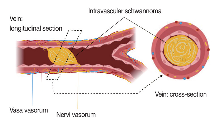
The blood vessel lumen is an extremely rare location for a benign peripheral nerve sheath tumor like schwannoma. Less than 10 cases have been previously reported. In this report, we present a case of a 68-year-old woman who had a soft tissue nodule at the posterior calf of her left leg during a physical examination. Pathological examination was performed after complete surgical excision. The patient underwent follow-up for 12 months after surgery without evidence of recurrence or any other complication. This is the first case of intravascular schwannoma reported as a cause of vein obstruction. Microscopically, the tumor was composed of Schwann spindle cells that were immunoreactive for S100 protein and SOX10. This tumor was surrounded by a well-defined vascular smooth muscle wall. Prospective series are required to improve the knowledge on the underlying mechanisms of intravascular schwannoma development.

Keywords: Nerve sheath neoplasms; Neurilemmoma; Peripheral nervous system; Peripheral nervous system neoplasms; Schwann cells.


Conflict of interest statement

Conflicts of Interest

The authors declare that they have no potential conflicts of interest.

Figures

Fig. 1.
Fig. 1.
Ultrasonographic imaging and morphological features. (A) Ultrasonographic characteristics of the tumor showing a 2.3 cm hypoechoic ovoid nodule. (B) Low blood flow around the tumor in the Doppler ultrasound view. (C) Hypercellular areas that consist of cohesive spindle cells (Antoni A zones). (D) The tumor shows an intravascular growth pattern, occupying the entire lumen of the blood vessel. (E) Hypocellular areas that consist of haphazardly arranged spindle cells in a loose myxoid stroma (Antoni B zones).
Fig. 2.
Fig. 2.
Positivity for S100 protein in intravascular tumor cells (A–D) and nervi vasorum nerves (B, D). The smooth muscle cells of the vein wall show smooth muscle actin positivity (E, F).
Fig. 3.
Fig. 3.
Immunohistochemical characteristics. (A, B) The intravascular tumor cell positivity for SOX10. (C) Some fibroblastic cells of the tumor stroma, the endothelial cells of capillaries inside the tumor, the endothelium of the vein intima, and the vasa vasorum are CD34-positive. (D) The nuclear immunoreactivity for H3K27me3 is intact in tumor cells.
Fig. 4.
Fig. 4.
Anatomical illustration image to explain the hypothetical nervi vasorum origin of intravascular schwannoma and to demonstrate the different biological structures involved. A longitudinal section view of a vessel with a schwannoma of intravascular growth and its hypothesized relation with the peripheral nerve sheath elements of the nervi vasorum network (left). Cross-section view of a blood vessel with a complete lumen obstruction by an intravascular schwannoma (right). Created in BioRender and Clip Studio Paint.

References

    1. Schwannoma Perry A. In: WHO classification of tumours: soft tissue and bone tumours. 5th ed. WHO Classification of Tumours Editorial Board, editor. Lyon: IARC Press; 2020. pp. 226–31.
    1. Evans DG. Neurofibromatosis type 2 (NF2): a clinical and molecular review. Orphanet J Rare Dis. 2009;4:16. - PMC - PubMed
    1. Casadei GP, Komori T, Scheithauer BW, Miller GM, Parisi JE, Kelly PJ. Intracranial parenchymal schwannoma: a clinicopathological and neuroimaging study of nine cases. J Neurosurg. 1993;79:217–22. - PubMed
    1. Voltaggio L, Murray R, Lasota J, Miettinen M. Gastric schwannoma: a clinicopathologic study of 51 cases and critical review of the literature. Hum Pathol. 2012;43:650–9. - PMC - PubMed
    1. Belakhoua SM, Rodriguez FJ. Diagnostic pathology of tumors of peripheral nerve. Neurosurgery. 2021;88:443–56. - PMC - PubMed

LinkOut - more resources