. 2024 May 31;27(6):110157.
doi: 10.1016/j.isci.2024.110157. eCollection 2024 Jun 21.

Fusobacterium nucleatum subsp. nucleatum RadD binds Siglec-7 and inhibits NK cell-mediated cancer cell killing

Affiliations

Fusobacterium nucleatum subsp. nucleatum RadD binds Siglec-7 and inhibits NK cell-mediated cancer cell killing

Johanna Galaski et al. iScience. .

Abstract

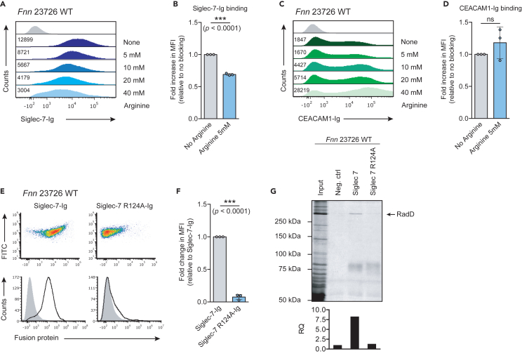
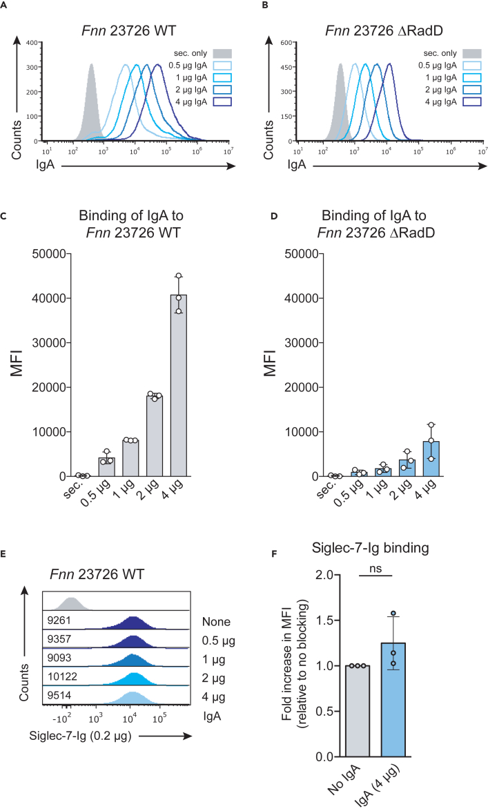
Fusobacterium nucleatum is an oral commensal bacterium that can colonize extraoral tumor entities, such as colorectal cancer and breast cancer. Recent studies revealed its ability to modulate the immune response in the tumor microenvironment (TME), promoting cancer progression and metastasis. Importantly, F. nucleatum subsp. animalis was shown to bind to Siglec-7 via lipopolysaccharides, leading to a pro-inflammatory profile in human monocyte-derived dendritic cells. In this study, we show that F. nucleatum subsp. nucleatum RadD binds to Siglec-7 on NK cells, thereby inhibiting NK cell-mediated cancer cell killing. We demonstrate that this binding is dependent on arginine residue R124 in Siglec-7. Finally, we determine that this binding is independent of the known interaction of RadD with IgA. Taken together, our findings elucidate the targeting of Siglec-7 by F. nucleatum subsp. nucleatum RadD as a means to modulate the NK cell response and potentially promoting immune evasion and tumor progression.

Keywords: cancer; immunology; microbiology; molecular biology.


Conflict of interest statement

The authors declare no competing interests.

Figures

None
Graphical abstract
Figure 1
Figure 1
Siglec-7 binding to F. nucleatum strains (A) FITC-labeled F. nucleatum subsp. nucleatum ATCC 23726 (Fnn 23726) was incubated with 2 μg of Siglec-7-Ig, NTB-A-Ig, 2B4-Ig, or CD16-Ig and binding was revealed with fluorescently labeled secondary antibodies. Filled gray histograms represent staining with secondary antibody only. One representative experiment out of three is shown. (B) Two strains of F. nucleatum subsp. nucleatum (Fnn 23726 and Fnn 25586) and two strains of F. nucleatum subsp. polymorphum (Fnp 10953 and Fnp 12230) as well as (C) a clinical strain of F. nucleatum (Fnn CTI-7) were incubated with Siglec-7-Ig. Filled gray histograms represent staining with secondary antibody only. One representative experiment out of two is shown.
Figure 2
Figure 2
F. nucleatum subsp. nucleatum confers Siglec-7-dependent protection against NK cell-mediated cancer cell killing (A) Schematic representation of target cell killing by YTS EV or YTS Siglec-7 cells. YTS empty vector (EV) or YTS Siglec-7 cells are activated almost exclusively by CD48-expressing target cells via their activating receptor 2B4. In the presence of bacteria, killing of target cells is inhibited via Siglec-7. (B) Staining of YTS EV or YTS Siglec-7 cells with Siglec-7 (upper histograms) and 2B4 (lower histograms) antibodies. Filled gray histograms represent staining with isotype control and secondary antibody only, respectively. (C) Staining of 721.221 or (D) BCBL-1 cells for Siglec-7 ligands with Siglec-7-Ig or of CD48 expression using anti-CD48 antibodies. (E) YTS EV or YTS Siglec-7 cells were precincubated or not with F. nucleatum subsp. nucleatum ATCC 23726 (Fnn 23726) at a ratio of 1:20 for 30 min at 37°C. The cells were then incubated with Calcein AM-labeled 721.221 or (F) BCBL-1 cells at an E:T ratio of 10:1 for 4 h at 37°C and the Calcein release into the supernatant was quantified. Each graph shows data from three independent experiments and each independent experiment is colored differently (white, blue, and black). Each experiment was performed in triplicates. Significance was tested using mixed-effects analysis with the Geisser-Greenhouse correction and Sidak’s multiple comparisons test (∗ p ≤ 0.05; ∗∗ p ≤ 0.01; ∗∗∗ p ≤ 0.001).
Figure 3
Figure 3
RadD of F. nucleatum subsp. nucleatum is the bacterial ligand for Siglec-7 Lysates prepared from (A) F. nucleatum subsp. nucleatum ATCC 23726 WT (Fnn 23726 WT) or (B) F. nucleatum subsp. nucleatum ATCC 23726 ΔRadD (Fnn 23726 ΔRadD) were immunoprecipitated using Siglec-7-Ig, CEACAM1-Ig, or CEACAM1 ΔN-Ig. Immunoprecipitates were visualized by Coomassie blue and immunoprecipitated bands were analyzed by mass spectrometry. RadD levels were quantified relative to the negative control. RQ, relative quantification. (C) FITC-labeled Fnn 23726 WT or its ΔRadD mutant was stained with Siglec-7-Ig. Filled gray histograms represent staining with secondary antibody only. One representative experiment out of eight is shown. (D) Quantification of Siglec-7-Ig binding to Fnn 23726 WT and the ΔRadD mutant shown as fold change in median fluorescent intensity (MFI) relative to Fnn 23726 WT. Bars represent means of eight independent experiments. Statistical significance was assessed using a two-tailed unpaired t test (∗ p ≤ 0.05; ∗∗ p ≤ 0.01; ∗∗∗ p ≤ 0.001).
Figure 4
Figure 4
RadD binding to Siglec-7 is inhibited by arginine and dependent on Siglec-7 arginine residue R124 (A) Staining of Fnn 23726 WT with Siglec-7-Ig or (C) CEACAM1-Ig in the presence of increasing concentrations of arginine. Filled gray histograms represent staining with secondary antibody only. MFI values are indicated left of each histogram. (B) Quantification of three independent experiments showing binding of Siglec-7-Ig or (D) CEACAM1-Ig as fold change in MFI in the presence of 5 mM arginine relative to no blocking. Statistical significance was assessed using a two-tailed unpaired Student’s t test (∗ p ≤ 0.05; ∗∗ p ≤ 0.01; ∗∗∗ p ≤ 0.001). (E) FITC-labeled Fnn 23726 WT were stained with Siglec-7-Ig or Siglec-7 R124A-Ig. Filled gray histograms represent staining with secondary antibody only. One representative staining out of three is shown. (F) Siglec-7-Ig and Siglec-7 R124A-Ig staining of Fnn 23726 WT quantified and shown as fold change in MFI relative to staining with Siglec-7 Ig. Bars represent means of three independent experiments. Statistical significance was assessed using a two-tailed unpaired Student’s t test (∗ p ≤ 0.05; ∗∗ p ≤ 0.01; ∗∗∗ p ≤ 0.001). (G) Lysates prepared from Fnn 23726 WT were immunoprecipitated using Siglec-7-Ig or Siglec-7 R124A-Ig. Immunoprecipitates were visualized by Coomassie. RadD levels were quantified relative to the negative control. RQ, relative quantification.
Figure 5
Figure 5
IgA does not block binding of Siglec-7-Ig to F. nucleatum (A) FITC-labeled Fnn 23726 WT or (B) Fnn 23726 ΔRadD were stained with increasing amounts of human serum IgA (0.5 μg, 1 μg, 2 μg, and 4 μg). Filled gray histograms represent staining with secondary antibody only. One representative experiment out of three is shown. Quantification of the MFI of three independent stainings of (C) Fnn 23726 WT and (D) Fnn 23726 ΔRadD with human serum IgA is shown. (E) Staining of F. nucleatum 23726 WT with Siglec-7-Ig after preincubation of bacteria with increasing amounts of human serum IgA. Filled gray histograms represent staining with secondary antibody only. MFI values are indicated left of each histogram. (F) Quantification of three independent experiments showing the fold increase in MFI in the presence of 4 μg IgA relative to no blocking. Significance was tested using Student’s t test (two-tailed and unpaired).

References

    1. Nejman D., Livyatan I., Fuks G., Gavert N., Zwang Y., Geller L.T., Rotter-Maskowitz A., Weiser R., Mallel G., Gigi E., et al. The human tumor microbiome is composed of tumor type-specific intracellular bacteria. Science. 2020;368:973–980. doi: 10.1126/science.aay9189. - DOI - PMC - PubMed
    1. Galeano Niño J.L., Wu H., LaCourse K.D., Kempchinsky A.G., Baryiames A., Barber B., Futran N., Houlton J., Sather C., Sicinska E., et al. Effect of the intratumoral microbiota on spatial and cellular heterogeneity in cancer. Nature. 2022;611:810–817. doi: 10.1038/s41586-022-05435-0. - DOI - PMC - PubMed
    1. Castellarin M., Warren R.L., Freeman J.D., Dreolini L., Krzywinski M., Strauss J., Barnes R., Watson P., Allen-Vercoe E., Moore R.A., Holt R.A. Fusobacterium nucleatum infection is prevalent in human colorectal carcinoma. Genome Res. 2012;22:299–306. doi: 10.1101/gr.126516.111. - DOI - PMC - PubMed
    1. Yamamura K., Baba Y., Nakagawa S., Mima K., Miyake K., Nakamura K., Sawayama H., Kinoshita K., Ishimoto T., Iwatsuki M., et al. Human Microbiome Fusobacterium Nucleatum in Esophageal Cancer Tissue Is Associated with Prognosis. Clin. Cancer Res. 2016;22:5574–5581. doi: 10.1158/1078-0432.ccr-16-1786. - DOI - PubMed
    1. Mitsuhashi K., Nosho K., Sukawa Y., Matsunaga Y., Ito M., Kurihara H., Kanno S., Igarashi H., Naito T., Adachi Y., et al. Association of Fusobacterium species in pancreatic cancer tissues with molecular features and prognosis. Oncotarget. 2015;6:7209–7220. doi: 10.18632/oncotarget.3109. - DOI - PMC - PubMed