Evaluation of viability, developmental competence, and apoptosis-related transcripts during in vivo post-ovulatory oocyte aging in zebrafish Danio rerio (Hamilton, 1822)
- PMID: 38952806
- PMCID: PMC11216024
- DOI: 10.3389/fvets.2024.1389070
Evaluation of viability, developmental competence, and apoptosis-related transcripts during in vivo post-ovulatory oocyte aging in zebrafish Danio rerio (Hamilton, 1822)
Abstract
Introduction: Post-ovulatory aging is a time-dependent deterioration of ovulated oocytes and a major limiting factor reducing the fitness of offspring. This process may lead to the activation of cell death pathways like apoptosis in oocytes.
Methodology: We evaluated oocyte membrane integrity, egg developmental competency, and mRNA abundance of apoptosis-related genes by RT-qPCR. Oocytes from zebrafish Danio rerio were retained in vivo at 28.5°C for 24 h post-ovulation (HPO). Viability was assessed using trypan blue (TB) staining. The consequences of in vivo oocyte aging on the developmental competence of progeny were determined by the embryo survival at 24 h post fertilization, hatching, and larval malformation rates.
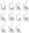
Results: The fertilization, oocyte viability, and hatching rates were 91, 97, and 65% at 0 HPO and dropped to 62, 90, and 22% at 4 HPO, respectively. The fertilizing ability was reduced to 2% at 8 HPO, while 72% of oocytes had still intact plasma membranes. Among the apoptotic genes bcl-2 (b-cell lymphoma 2), bada (bcl2-associated agonist of cell death a), cathepsin D, cathepsin Z, caspase 6a, caspase 7, caspase 8, caspase 9, apaf1, tp53 (tumor protein p53), cdk1 (cyclin-dependent kinase 1) studied, mRNA abundance of anti-apoptotic bcl-2 decreased and pro-apoptotic cathepsin D increased at 24 HPO. Furthermore, tp53 and cdk1 mRNA transcripts decreased at 24 HPO compared to 0 HPO.
Discussion: Thus, TB staining did not detect the loss of oocyte competency if caused by aging. TB staining, however, could be used as a simple and rapid method to evaluate the quality of zebrafish oocytes before fertilization. Taken together, our results indicate the activation of cell death pathways in the advanced stages of oocyte aging in zebrafish.
Keywords: apoptosis; cell death; fertilization; membrane integrity; trypan blue; zebrafish.
Copyright © 2024 Konar, Mai, Brachs, Waghmare, Samarin, Policar and Samarin.
Conflict of interest statement
The authors declare that the research was conducted in the absence of any commercial or financial relationships that could be construed as a potential conflict of interest.
Figures


Similar articles
-
Aging oocytes: exploring apoptosis and its impact on embryonic development in common carp (Cyprinus carpio).J Anim Sci. 2025 Jan 4;103:skaf002. doi: 10.1093/jas/skaf002. J Anim Sci. 2025. PMID: 39761344 Free PMC article.
-
Oocyte Ageing in Zebrafish Danio rerio (Hamilton, 1822) and Its Consequence on the Viability and Ploidy Anomalies in the Progeny.Animals (Basel). 2021 Mar 22;11(3):912. doi: 10.3390/ani11030912. Animals (Basel). 2021. PMID: 33810200 Free PMC article.
-
Alteration of mRNA abundance, oxidation products and antioxidant enzyme activities during oocyte ageing in common carp Cyprinus carpio.PLoS One. 2019 Feb 22;14(2):e0212694. doi: 10.1371/journal.pone.0212694. eCollection 2019. PLoS One. 2019. PMID: 30794661 Free PMC article.
-
Oxidative stress and ageing of the post-ovulatory oocyte.Reproduction. 2013 Oct 21;146(6):R217-27. doi: 10.1530/REP-13-0111. Print 2013 Dec. Reproduction. 2013. PMID: 23950493 Review.
-
Effects of maternal age on oocyte developmental competence.Theriogenology. 2001 Apr 1;55(6):1303-22. doi: 10.1016/s0093-691x(01)00484-8. Theriogenology. 2001. PMID: 11327686 Review.
Cited by
-
Aging oocytes: exploring apoptosis and its impact on embryonic development in common carp (Cyprinus carpio).J Anim Sci. 2025 Jan 4;103:skaf002. doi: 10.1093/jas/skaf002. J Anim Sci. 2025. PMID: 39761344 Free PMC article.
References
LinkOut - more resources
Full Text Sources
Research Materials
Miscellaneous

