. 2024 Jun 28:12:e17539.
doi: 10.7717/peerj.17539. eCollection 2024.

Effect of glucocorticoid blockade on inflammatory responses to acute sleep fragmentation in male mice

Affiliations

Effect of glucocorticoid blockade on inflammatory responses to acute sleep fragmentation in male mice

Zim Warda Hasan et al. PeerJ. .

Abstract

The association between sleep and the immune-endocrine system is well recognized, but the nature of that relationship is not well understood. Sleep fragmentation induces a pro-inflammatory response in peripheral tissues and brain, but it also activates the hypothalamic-pituitary-adrenal (HPA) axis, releasing glucocorticoids (GCs) (cortisol in humans and corticosterone in mice). It is unclear whether this rapid release of glucocorticoids acts to potentiate or dampen the inflammatory response in the short term. The purpose of this study was to determine whether blocking or suppressing glucocorticoid activity will affect the inflammatory response from acute sleep fragmentation (ASF). Male C57BL/6J mice were injected i.p. with either 0.9% NaCl (vehicle 1), metyrapone (a glucocorticoid synthesis inhibitor, dissolved in vehicle 1), 2% ethanol in polyethylene glycol (vehicle 2), or mifepristone (a glucocorticoid receptor antagonist, dissolved in vehicle 2) 10 min before the start of ASF or no sleep fragmentation (NSF). After 24 h, samples were collected from brain (prefrontal cortex, hypothalamus, hippocampus) and periphery (liver, spleen, heart, and epididymal white adipose tissue (EWAT)). Proinflammatory gene expression (TNF-α and IL-1β) was measured, followed by gene expression analysis. Metyrapone treatment affected pro-inflammatory cytokine gene expression during ASF in some peripheral tissues, but not in the brain. More specifically, metyrapone treatment suppressed IL-1β expression in EWAT during ASF, which implies a pro-inflammatory effect of GCs. However, in cardiac tissue, metyrapone treatment increased TNF-α expression in ASF mice, suggesting an anti-inflammatory effect of GCs. Mifepristone treatment yielded more significant results than metyrapone, reducing TNF-α expression in liver (only NSF mice) and cardiac tissue during ASF, indicating a pro-inflammatory role. Conversely, in the spleen of ASF-mice, mifepristone increased pro-inflammatory cytokines (TNF-α and IL-1β), demonstrating an anti-inflammatory role. Furthermore, irrespective of sleep fragmentation, mifepristone increased pro-inflammatory cytokine gene expression in heart (IL-1β), pre-frontal cortex (IL-1β), and hypothalamus (IL-1β). The results provide mixed evidence for pro- and anti-inflammatory functions of corticosterone to regulate inflammatory responses to acute sleep loss.

Keywords: Acute Sleep Fragmentation; Glucocorticoids; Metyrapone; Mifepristone; Pro-inflammatory cytokines.


Conflict of interest statement

The authors declare there are no competing interests.

Figures

Figure 1
Figure 1. Effects of acute sleep fragmentation (ASF), metyrapone, and their interaction upon cytokine gene expression in peripheral tissues.
Gene expression levels of TNF-α and IL-1β were measured in liver (A, B), spleen (C, D), EWAT (E, F) and heart (G, H) of mice injected with vehicle (0.9% NaCl) or treated with metyrapone (glucocorticoid synthesis inhibitor) after being subjected to control or ASF. Data are presented as mean ± standard error (SE) for each group. The sample size of each group was n = 10 and data were analyzed either parametrically using a two-way ANOVA, followed by Tukey’s HSD post hoc tests or non-parametrically using Kruskal-Wallis, followed by Mann Whitney U-tests. Significant differences were denoted by different letters among groups. Shared letters indicate no significant difference between groups. The level of statistical significance was set at alpha (α) = 0.05.
Figure 2
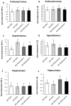
Figure 2. Effects of acute sleep fragmentation (ASF), metyrapone, and their interaction upon cytokine gene expression in select regions of brain.
Gene expression levels of TNF-α and IL-1β were measured in PFC (A, B), hypothalamus (C, D), hippocampus (E, F) of mice injected with vehicle (0.9% NaCl) or treated with metyrapone (glucocorticoid synthesis inhibitor) after being subjected to control or ASF. Data are presented as mean ± standard error (SE) for each group. The sample size of each group was n = 10 and data were analyzed either parametrically using a two-way ANOVA, followed by Tukey’s HSD post hoc tests or non-parametrically using Kruskal-Wallis, followed by Mann Whitney U-tests. Significant differences among groups denoted as by different letters. Shared letters indicate no significant difference between groups. The level of statistical significance was set at alpha (α) = 0.05.
Figure 3
Figure 3. Effects of acute sleep fragmentation (ASF), mifepristone, and their interaction upon cytokine gene expression in peripheral tissues.
Gene expression levels of TNF-α and IL-1β were measured in liver (A, B), spleen (C, D), EWAT (E, F) and heart (G, H) of mice injected with vehicle (2% ethanol in polyethylene glycol) or treated with mifepristone (glucocorticoid receptor antagonist) after being subjected to control or ASF. Data are presented as mean ± standard error (SE) for each group. The sample size of each group was n = 10 and data either parametrically using a two-way ANOVA, followed by Tukey’s HSD post hoc tests or non-parametrically using Kruskal-Wallis, followed by Mann Whitney U-tests. Significant differences were denoted by different letters among groups. Shared letters indicate no significant difference between groups. The level of statistical significance was set at alpha (α) = 0.05.
Figure 4
Figure 4. Effects of acute sleep fragmentation (ASF), mifepristone, and their interaction upon cytokine gene expression in select regions of brain.
Gene expression levels of TNF-α and IL-1β were measured in PFC (A, B), hypothalamus (C, D), hippocampus (E, F) of mice injected with vehicle (2% ethanol in polyethylene glycol) or treated with mifepristone (glucocorticoid receptor antagonist) after being subjected to control or ASF. Data are presented as mean ± standard error (SE) for each group. The sample size of each group was n = 10 and data were analyzed using a two-way ANOVA, followed by Tukey’s HSD post hoc tests or non-parametrically using Kruskal-Wallis, followed by Mann Whitney U-tests. Significant differences were observed between certain groups, as denoted by different letters. Shared letters indicate no significant difference between groups. The level of statistical significance was set at alpha (α) = 0.05.

Similar articles

References

    1. Agarwai MK. The antiglucocorticoid action of mifepristone. Pharmacology and Therapeutics. 1996;70(3):183–213. doi: 10.1016/0163-7258(96)00016-2. - DOI - PubMed
    1. Aggarwal S, Loomba RS, Arora RR, Molnar J. Associations between sleep duration and prevalence of cardiovascular events. Clinical Cardiology. 2013;36(11):671–676. doi: 10.1002/clc.22160. - DOI - PMC - PubMed
    1. Ahima RS, Harlan RE. Charting of type II glucocorticoid receptor-like immunoreactivity in the rat central nervous system. Neuroscience. 1990;39(3):579–604. doi: 10.1016/0306-4522(90)90244-X. - DOI - PubMed
    1. Ajibare AJ, Ayodele OD, Olayaki LA. Mifepristone ameliorates sleep deprivation - induced oxidative stress in the testis of rats. African Journal of Biomedical Research. 2020;23(2):239–245.
    1. Ashley NT, Sams DW, Brown AC, Dumaine JE. Novel environment influences the effect of paradoxical sleep deprivation upon brain and peripheral cytokine gene expression. Neuroscience Letters. 2016;615:55–59. doi: 10.1016/j.neulet.2016.01.013. - DOI - PMC - PubMed

MeSH terms