. 2024 Jun 17:15:1425938.
doi: 10.3389/fimmu.2024.1425938. eCollection 2024.

Different localization of P2X4 and P2X7 receptors in native mouse lung - lack of evidence for a direct P2X4-P2X7 receptor interaction

Affiliations

Different localization of P2X4 and P2X7 receptors in native mouse lung - lack of evidence for a direct P2X4-P2X7 receptor interaction

Juan Sierra-Marquez et al. Front Immunol. .

Abstract

Introduction: P2X receptors are a family of homo- and heterotrimeric cation channels gated by extracellular ATP. The P2X4 and P2X7 subunits show overlapping expression patterns and have been involved in similar physiological processes, such as pain and inflammation as well as various immune cell functions. While formation of P2X2/P2X3 heterotrimers produces a distinct pharmacological phenotype and has been well established, functional identification of a P2X4/P2X7 heteromer has been difficult and evidence for and against a physical association has been found. Most of this evidence stems, however, from in vitro model systems.

Methods: Here, we used a P2X7-EGFP BAC transgenic mouse model as well as P2X4 and P2X7 knock-out mice to re-investigate a P2X4-P2X7 interaction in mouse lung by biochemical and immunohistochemical experiments as well as quantitative expression analysis.

Results: No detectable amounts of P2X4 could be co-purified from mouse lung via P2X7-EGFP. In agreement with these findings, immuno-histochemical analysis using a P2X7-specific nanobody revealed only limited overlap in the cellular and subcellular localizations of P2X4 and P2X7 in both the native lung tissue and primary cells. Comparison of P2X4 and P2X7 transcript and protein levels in the respective gene-deficient and wild type mice showed no mutual interrelation between their expression levels in whole lungs. However, a significantly reduced P2rx7 expression was found in alveolar macrophages of P2rx4 -/- mice.

Discussion: In summary, our detailed analysis of the cellular and subcellular P2X4 and P2X7 localization and expression does not support a physiologically relevant direct association of P2X4 and P2X7 subunits or receptors in vivo.

Keywords: BAC transgenic P2X7-EGFP mouse; P2X4 receptor; P2X7 receptor; functional interaction; heteromerization; lung epithelial cells; macrophage; nanobody.


Conflict of interest statement

The authors declare that the research was conducted in the absence of any commercial or financial relationships that could be construed as a potential conflict of interest. The author(s) declared that they were an editorial board member of Frontiers, at the time of submission. This had no impact on the peer review process and the final decision.

Figures
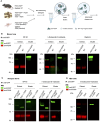
Figure 1
Figure 1
Lack of evidence for a physical interaction of P2X4 and P2X7 receptors in transgenic P2X7-EGFP mice. (A) Schematic representation of the purification of P2X7-containing complexes from mouse lung, cRNA-injected Xenopus laevis oocytes, and transfected HEK cells using anti-EGFP nanobodies (GFP-Trap®). (B) Immunoprecipitation of P2X7-EGFP complexes from lung tissue of BAC-transgenic P2X7-EGFP mice and wt controls using the indicated non-denaturing detergents for solubilization. Primary antibodies against GFP (from rat) and P2X4 (from rabbit) and secondary antibodies for infrared imaging were used for blot development and detection. Note that P2X4 has a tendency to form higher aggregates (dimers band in the middle image) but does not associate with P2X7 (absence of a band stained with both antibodies). (C) Co-purification of P2X4 with P2X7-EGFP from Xenopus laevis oocytes. cRNA encoding P2X4 subunits was injected together with cRNA encoding P2X7-EGFP or non-tagged P2X7 subunits (negative control). After 2 days, P2X7-EGFP complexes were immunoprecipitated as in B, using the indicated detergents. Blots were developed with antibodies against P2X4 and P2X7 (both derived from rabbit). (D) Immunoprecipitation of P2X7-EGFP complexes from HEK293 cells. P2X4-encoding DNA was transfected transiently into HEK293 cells stably expressing P2X7-EGFP and wt control cells. Proteins were extracted in n-dodecyl-β-D-maltoside and purified and detected as in (B). Representative results of at least two experiments are shown. Note that the harsh conditions required to elute the protein from the nanobody-coupled beads leads to a stronger protein denaturation than addition of SDS and results in a size shift of the P2X7-EGFP band as well as loss of EGFP fluorescence. Also note, that the eluate was about three times more concentrated than the extract. Panel (A) was created with BioRender.com.
Figure 2
Figure 2
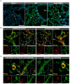
Cell type-specific P2X7 protein expression in the mouse lung. (A) 10–20 μm lung cryosections from wt mice, P2X7-EGFP mice, and P2rx7 -/- mice as negative control were prepared in parallel and immunostained with a P2X7-specifc nanobody (7E2-rbFc). Arrowheads show brighter cells, most likely macrophages. A = Alveolus, Br = Bronchiole, BV = Blood vessel. Scale bar 100 µm. (B) Lung cryosections from wt mice were co-stained with the P2X7 nanobody (7E2 nb-hFc) and antibodies against the epithelial cell marker aquaporin 5 (AQP5) and the endothelial cell marker platelet endothelial cell adhesion molecule PECAM-1 (CD31). Insets show representative areas of differential staining for the two marker proteins. The arrowhead indicates a CD31 positive cell adjacent to a red blood cell (indicated by asterisk). Scale bar 100 µm, inset scale bar 25 µm. (C) Co-staining of lung cryosections from wt mice using P2X7 nanobodies (7E2-hFc) and antibodies against ionized calcium-binding adapter molecule 1 (Iba-1) and F4/80 as macrophage markers. Insets show a representative staining of a macrophage positive for both marker proteins. Insets show Iba1 and P2X7-positive cells. Scale bar 25 µm, inset scale bar 10 µm. Nuclear staining with DAPI (in A, C) or TO-PRO-3 (in B) is shown in blue.
Figure 3
Figure 3
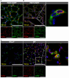
Cellular distribution of P2X4 and P2X7 protein in lung tissue. Cryosections (10–20 µm) of lung tissue from the indicated genotypes were immunostained with anti-GFP (green) and anti-P2X4 (red) antibodies and imaged by confocal microscopy (A) and Airyscan (B). Note that the enlarged inset in B was imaged separately (C) Specificity of the monoclonal rat anti-mP2X4 antibody in primary cells and co-staining with pro-surfactant protein C as a marker for AT2 epithelial cells. Nuclear staining with DAPI (in A, B) or TO-PRO-3 (in C) is shown in blue. Scale bar 100 µm. Insets show the representative subcellular distribution of the respective proteins.
Figure 4
Figure 4
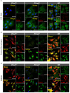
Subcellular localization of P2X4 and P2X7 in primary AT2 cells and macrophages. (A) Primary AT2 epithelial cells from wt mice and P2rx7 -/-, P2rx4 -/- controls, were stained 24 hours after plating with the P2X7-specific nanobody (7E2-hFc), rat anti-P2X4 antibody, and anti-prosurfactant protein C (SPC) as an AT2-type cell marker. (B) Alveolar macrophages and (C) bone marrow-derived macrophages (BMDM) isolated from wt and the respective P2X knock-out control mice were stained with the P2X7-specific nanobody (7E2-hFc), anti-P2X4 antibody, and an antibody against the lysosomal/endosomal marker protein macrosialin (CD68). Nuclear staining with TO-PRO-3 is shown in blue. Scale bar 25 µm. Insets show intracellular puncta and/or membrane structures. Insets in upper panels show areas of intracellular P2X4 staining and comparable areas from control cells. Insets in lower panels show plasma membrane localization. Arrowheads indicate vesicle-like structures, where P2X4 is localized.
Figure 5
Figure 5
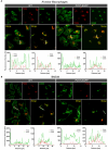
Different subcellular localization of P2X4 and P2X7 in alveolar macrophages (A) and bone marrow derived macrophages (B) from wt and P2X7-EGFP transgenic mice. Cells were stained with the P2X7-specific nanobody 7E2-hFc, the rat anti-P2X4 antibody, and the monocyte endosome marker CD68. Images were obtained using a Zeiss LSM 880 with Airyscan, and intensity profiles obtained with the built-in image processing software (ZEN black) along the indicated lines. Scale bar 10 μm.
Figure 6
Figure 6
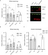
Mutual interrelation of P2X4 and P2X7 expression. (A) 50 µg total protein of membrane extracts from lung tissue was separated by SDS-PAGE and immunoblotted with a P2X4- or P2X7-specific antibody (red). Protein levels were quantified via fluorescence intensity of the secondary antibodies and vinculin was used as a loading control for normalization (green). Protein expression of each P2X subtype was not significantly altered by P2X7-EGFP overexpression or genetic ablation of the respective other P2X receptor. Data are presented as mean +/– SD from six animals analyzed in two independent experiments. Significance was analyzed using Student’s t-test. (B) Total RNA was isolated from 20–30 mg lung tissue and after reverse transcription quantified in a LightCycler 480 system. RPLP0 and PPIA were used as reference genes to calculate relative P2X4 and P2X7 mRNA levels. (C) Total RNA was isolated from alveolar macrophages and PPIA were used as reference genes to calculate relative P2X4 and P2X7 mRNA levels. Significance was analyzed using Student’s t-test. ns, not significant; ****, p<0.0001.

References

    1. Illes P, Muller CE, Jacobson KA, Grutter T, Nicke A, Fountain SJ, et al. . Update of P2X receptor properties and their pharmacology: IUPHAR Review 30. Br J Pharmacol. (2021) 178:489–514. doi: 10.1111/bph.15299 - DOI - PMC - PubMed
    1. Lewis C, Neidhart S, Holy C, North RA, Buell G, Surprenant A. Coexpression of P2X2 and P2X3 receptor subunits can account for ATP-gated currents in sensory neurons. Nature. (1995) 377:432–5. doi: 10.1038/377432a0 - DOI - PubMed
    1. Lalo U, Pankratov Y, Wichert SP, Rossner MJ, North RA, Kirchhoff F, et al. . P2X1 and P2X5 subunits form the functional P2X receptor in mouse cortical astrocytes. J Neurosci. (2008) 28:5473–80. doi: 10.1523/JNEUROSCI.1149-08.2008 - DOI - PMC - PubMed
    1. Compan V, Ulmann L, Stelmashenko O, Chemin J, Chaumont S, Rassendren F. P2X2 and P2X5 subunits define a new heteromeric receptor with P2X7-like properties. J Neurosci. (2012) 32:4284–96. doi: 10.1523/JNEUROSCI.6332-11.2012 - DOI - PMC - PubMed
    1. Torres GE, Egan TM, Voigt MM. Hetero-oligomeric assembly of P2X receptor subunits. Specificities exist with regard to possible partners. J Biol Chem. (1999) 274:6653–9. doi: 10.1074/jbc.274.10.6653 - DOI - PubMed

LinkOut - more resources