. 2024 Aug;17(4):e004614.
doi: 10.1161/CIRCGEN.124.004614. Epub 2024 Jul 2.

Rare Variant in MRC2 Associated With Familial Supraventricular Tachycardia and Wolff-Parkinson-White Syndrome

Affiliations

Rare Variant in MRC2 Associated With Familial Supraventricular Tachycardia and Wolff-Parkinson-White Syndrome

Adam S Potter et al. Circ Genom Precis Med. 2024 Aug.

Abstract

Background: Accessory pathways are a common cause of supraventricular tachycardia (SVT) and can lead to sudden cardiac death in otherwise healthy children and adults when associated with Wolff-Parkinson-White syndrome. The goal of this study was to identify genetic variants within a large family with structurally normal hearts affected by SVT and Wolff-Parkinson-White syndrome and determine causality of the gene deficit in a corresponding mouse model.

Methods: Whole exome sequencing performed on 2 distant members of a 3-generation family in which multiple members were affected by SVT or Wolff-Parkinson-White pattern (preexcitation) on ECG identified MRC2 as a candidate gene. Serial electrocardiograms, intracardiac electrophysiology studies, echocardiography, optical mapping studies, and histology were performed on both Mrc2 mutant and WT (wild-type) mice.

Results: A rare HET (heterozygous) missense variant c.2969A>G;p.Glu990Gly (E990G) in MRC2 was identified as the leading candidate gene variant segregating with the cardiac phenotype following an autosomal-dominant Mendelian trait segregation pattern with variable expressivity. In vivo electrophysiology studies revealed reentrant SVT in E990G mice. Optical mapping studies in E990G mice demonstrated abnormal retrograde conduction, suggesting the presence of an accessory pathway. Histological analysis of E990G mouse hearts showed a disordered ECM (extracellular matrix) in the annulus fibrosus. Finally, Mrc2 knockdown in human cardiac fibroblasts enhanced accelerated cell migration.

Conclusions: This study identified a rare nonsynonymous variant in the MRC2 gene in individuals with familial reentrant SVT, Wolff-Parkinson-White ECG pattern, and structurally normal hearts. Furthermore, Mrc2 knock-in mice revealed an increased incidence of reentrant SVT and bypass tract formation in the setting of preserved cardiac structure and function.

Keywords: Wolff-Parkinson-White syndrome; cell movement; fibroblasts; fibrosis; tachycardia, supraventricular.


Conflict of interest statement

Dr Lupski has stock ownership in 23andMe (Sunnyvale, CA), is a paid consultant for the Regeneron Genetics Center (Tarrytown, NY), and is a coinventor on multiple US and European patents related to molecular diagnostics for inherited neuropathies, eye diseases, and bacterial genomic fingerprinting. The Department of Molecular and Human Genetics at Baylor College of Medicine receives revenue from clinical genetic testing conducted at Baylor Genetics (BG) Laboratories (Houston, TX). Dr Lupski serves on the Scientific Advisory Board of BG. Dr Wehrens is a cofounder and board member of Elex Biotech LLC, a start-up company developing RyR2 (ryanodine receptor type-2) modifying drugs for heart disease. Dr Wehrens is also a consultant at Pfizer and Rocket Pharmaceuticals. The other authors report no conflicts.

Figures
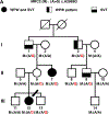
Figure 1.
Figure 1.. Pedigree of family with inherited Wolff-Parkinson-White syndrome.
A. Three generation pedigree of a family segregating a WPW and SVT phenotype. Individuals affected are marked as full black symbols if found to have both WPW and SVT, half-full left side if ECG demonstrated a WPW pattern, and half-full lower if found to have SVT. A rare missense variant c.2969A>G (p.Glu990Gly) in the MRC2 gene was found to co-segregate with affectation status in this family. Variant carriers are labeled as M:(A/G) denoting the heterozygous state of the variant, whereas homozygous reference individuals are labeled as M:(A/A). The pedigree ID is shown as well as the genotype and phenotype of each individual.
Figure 2.
Figure 2.. Electrocardiograms of family members.
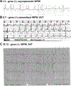
A. WPW pattern is shown in 3 lead ECG in subject I-3 (see pedigree). B. Representative sinus rhythm with intermittent WPW pattern during Holter monitoring prior to ablation in subject II:7. C. 12 lead ECG with WPW pattern at age 2 years in patient III:12, who also experienced SVT (not shown).
Figure 3.
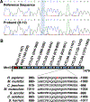
Figure 3.. Identification of MRC2 variant in patients with WPW and SVT.
A. Sanger sequencing chromatograms showing the reference wild-type sequence in an unaffected family member and the MRC2 variant confirmation in the Proband (III-12). Sanger sequencing confirmation and segregation was performed for all 14 individuals in the family. B. Schematic representation of the MRC2 protein showing its ricin domain (red), fibronectin type 2 (FN2) domain (green), and eight CTLD8 domains (blue). The identified variant in the pedigree falls within the sixth CTLD6 domain changing a glutamic acid (Glu) residue at position 990 of the protein to a glycine (Gly).
Figure 4.
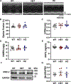
Figure 4.. Characterization of Mrc2 E990G knock-in mouse model.
A. Representative M-mode echocardiogram images showing left ventricular size and function. B. Quantification of ejection fraction and (C) left ventricular posterior wall thickness in diastole from WT (3M, 8F), HET (7F) and HZ (3M, 5F) E990G mice. D. Quantification of heart weight-to-body weight ratio and (E) heart weight-to-tibial length ratio in WT (6M, 7F), HET (2M, 7F) and HZ (2M, 5F) E990G mice. F. Representative western blot images and (G) quantifications of total Mrc2 and GAPDH loading control from murine atrial tissue harvested from WT (3M, 3F), HET (2M, 4F) and HZ (3M, 3F) E990G mice. WT, wild-type; HET, heterozygous; HZ, homozygous E990G mice; Mrc2, Mannose Receptor C-type 2 protein. No significant differences between groups or sexes were noted. Data expressed as means ± SEM.
Figure 5.
Figure 5.. E990G knock-in mice exhibit increased susceptibility to pacing-induced SVT.
A. Representative baseline surface ECG for WT, HET and HZ E990G mice. B. Quantification of baseline RR interval, (C) baseline PR interval, (D) baseline QTc interval, and (E) AVNERP in WT (4M, 2F), HET (3M, 4F) and HZ (3M, 5F) E990G mice. F. Representative intracardiac electrogram traces showing SVT in both the HET and HZ E990G mice. Peaks marked with the letter ‘S’ represent pacing stimuli. G. Bar graph showing increased incidence in SVT inducibility in HET and HZ E990G mice versus WT mice analyzed using the Chi-square two-sided test. WT, wild-type; HET, heterozygous; HZ, homozygous E990G mice; AVNERP, atrioventricular effective refractory period; SVT, supraventricular tachycardia. No significant difference was noted between sexes. Data expressed as means ± SEM.
Figure 6.
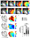
Figure 6.. E990G knock-in mice exhibit SVT and abnormal conduction pathways.
A. Representative activation maps obtained with optical mapping using potentiometric dye, di-4-ANEPPS, showing normal conduction in HET E990G isolated mouse hearts during sinus rhythm (top row of maps), and during SVT (bottom row of maps) after 1 mM isoproterenol. B. Examples of the most common types of conduction pathways with a cartoon on the left and optimal mapping example on the right, and C. Quantification of these 3 types of pathways isolated WT (6M, 1F), HET (1M, 4F) and HZ (2M, 4F) E990G mouse hearts in the presence of 1mM isoproterenol; statistics with Chi-square two-sided test. WT, wild-type; HET, heterozygous; HZ, homozygous E990G mice; RA is right atrium; LA, left atrium; V, ventricle.
Figure 7.
Figure 7.. Reduced collagen deposition and reduced extracellular matrix integrity in the annulus fibrosus in E990G murine hearts.
A. Bright field images of histologic sections of the AV groove showing annulus fibrosus stained with Picrosirius red and B. quantification showing reduced fibrosis (red staining) in HZ mice (n=4 mice per genotype). C. Polarized light images of the same sections showing birefringence visualized as red and green fluorescent signals. D. Quantification of ratio between red and green fluorescence. The reduced red-to-green ratio is indicative of a more loosely packed extracellular matrix. WT, wild-type; HET, heterozygous; HZ, homozygous E990G mice; RA, right atria; RV, right ventricle. Data expressed as means ± SEM; statistics using one-way ANOVA. Scale bar, 50 mM.
Figure 8.
Figure 8.. MRC2-knockdown activates human atrial fibroblasts (hAFs).
A. Representative immunoblots of MRC2 protein levels relative to GAPDH in hAF, human cardiomyocytes (hCM), left ventricular (LV), and left atrial appendage (LAA) tissue-samples. B. Confirmation of MRC2-knockdown by qPCR and (C) immunoblots; (D) shows MRC2 protein quantifications. E. Hydroxyproline (HPA) accumulation in hAF secretomes following silencing MRC2 with siRNA. Representative immunoblots (F) of collagen-1a protein accumulation in cell secretomes of the MRC2-knockdown hAFs quantified in (G), despite unchanged collagen-1a gene expression in MRC2-knockdown cells (H). (I-J) Accelerated cell migration (wound-healing/scratch assay) following silencing of MRC2 in hAFs (scale bar = 0.3mm). KD, knock down; NC, non-coding siRNA. Data expressed as means ± SEM; statistics using t-test (panels B, E, G, H) or Mann-Whitney U test (panel D).

References

    1. Coban-Akdemir ZH, Charng WL, Azamian M, Paine IS, Punetha J, Grochowski CM, Gambin T, Valdes SO, Cannon B, Zapata G, et al. Wolff-Parkinson-White syndrome: De novo variants and evidence for mutational burden in genes associated with atrial fibrillation. Am J Med Genet A. 2020;182:1387–1399. - PMC - PubMed
    1. Etheridge SP, Escudero CA, Blaufox AD, Law IH, Dechert-Crooks BE, Stephenson EA, Dubin AM, Ceresnak SR, Motonaga KS, Skinner JR, et al. Life-Threatening Event Risk in Children With Wolff-Parkinson-White Syndrome: A Multicenter International Study. JACC Clin Electrophysiol. 2018;4:433–444. - PubMed
    1. Wolff L, Parkinson J, White PD. Bundle-branch block with short P-R interval in healthy young people prone to paroxysmal tachycardia. Am Heart J. 1930;5:685–704. - PMC - PubMed
    1. Gaussin V, Morley GE, Cox L, Zwijsen A, Vance KM, Emile L, Tian Y, Liu J, Hong C, Myers D, et al. Alk3/Bmpr1a receptor is required for development of the atrioventricular canal into valves and annulus fibrosus. Circ Res. 2005;97:219–226. - PMC - PubMed
    1. Kolditz DP, Wijffels MC, Blom NA, van der Laarse A, Markwald RR, Schalij MJ, Gittenberger-de Groot AC. Persistence of functional atrioventricular accessory pathways in postseptated embryonic avian hearts: implications for morphogenesis and functional maturation of the cardiac conduction system. Circulation. 2007;115:17–26. - PubMed

MeSH terms

LinkOut - more resources