. 2024 Aug 14;15(8):e0154924.
doi: 10.1128/mbio.01549-24. Epub 2024 Jul 2.

Host cells reprogram lipid droplet synthesis through YY1 to resist PRRSV infection

Affiliations

Host cells reprogram lipid droplet synthesis through YY1 to resist PRRSV infection

Zifang Zheng et al. mBio. .

Abstract

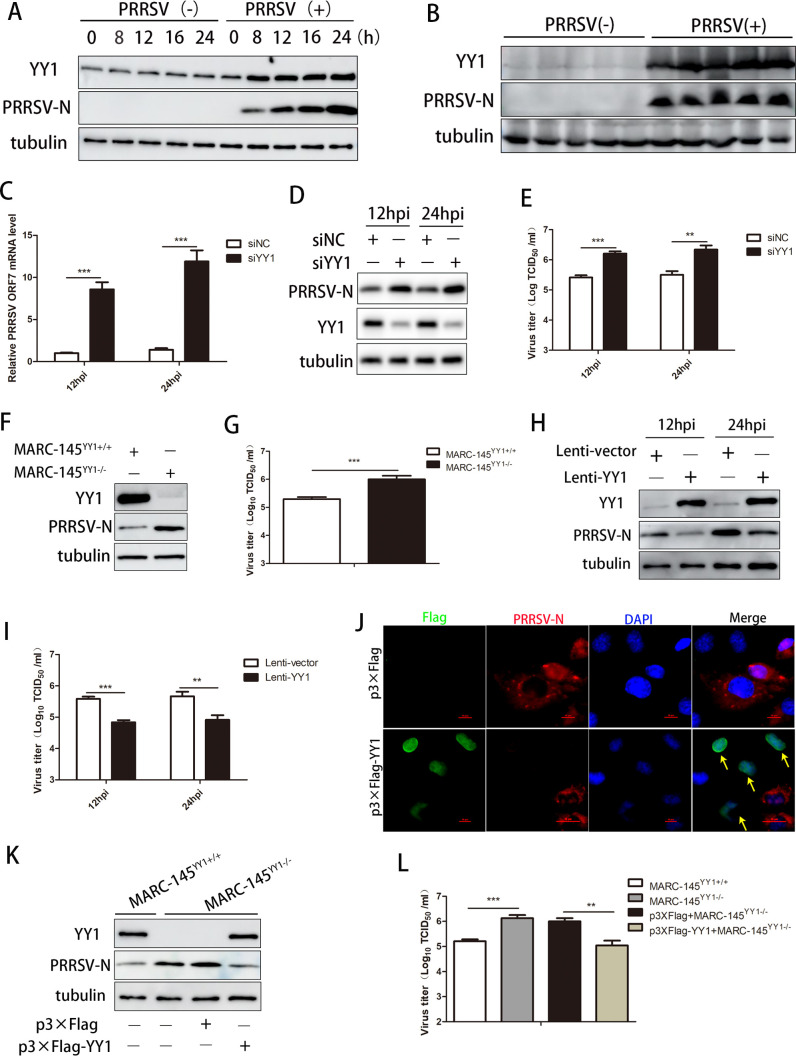
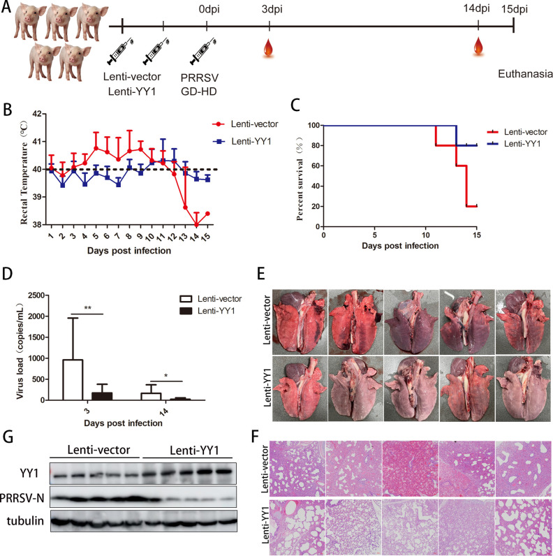
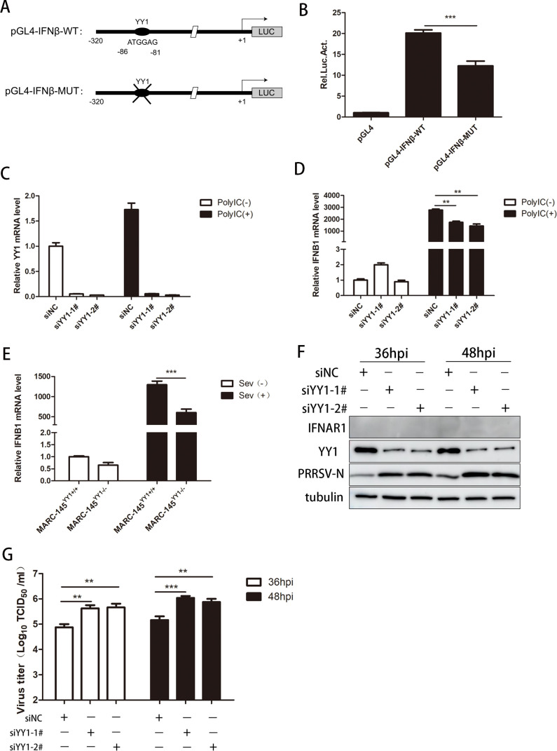
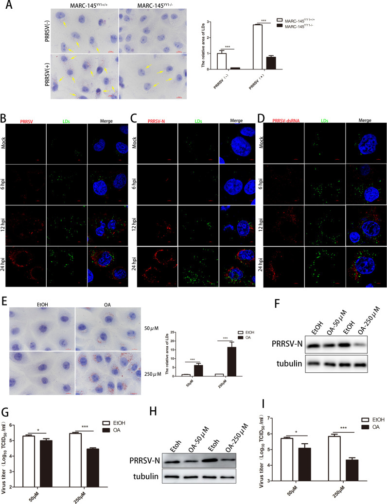
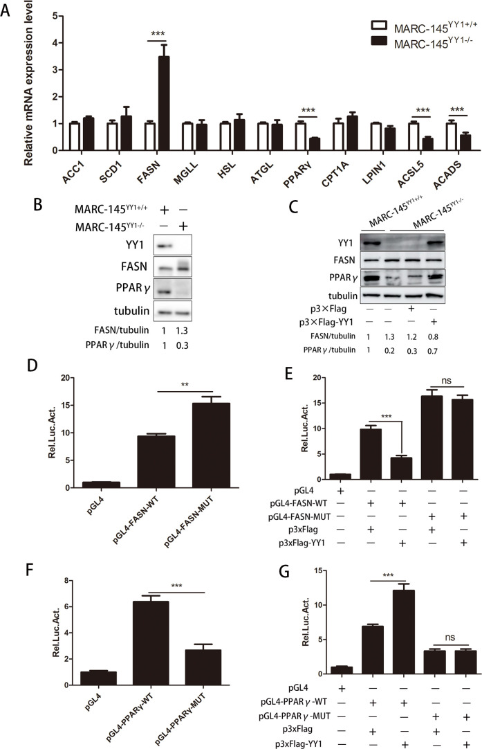
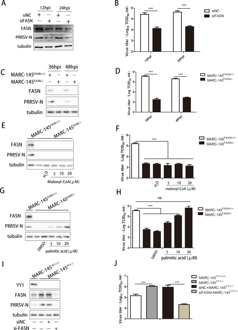
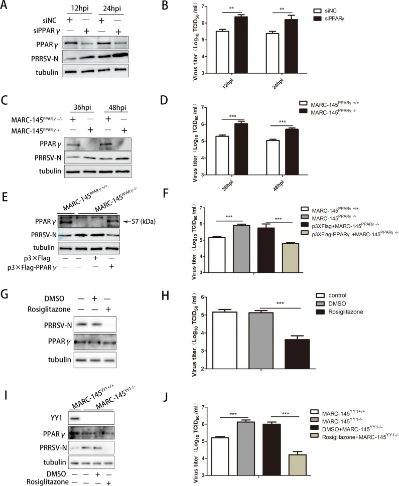
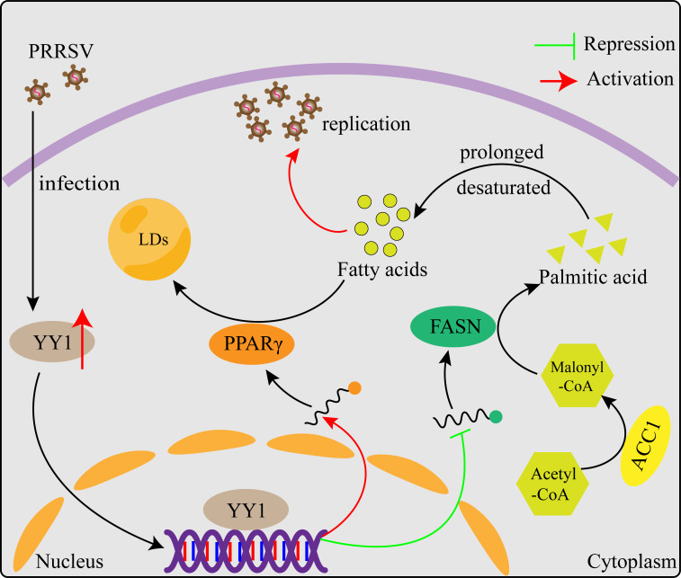
Metabolism in host cells can be modulated after viral infection, favoring viral survival or clearance. Here, we report that lipid droplet (LD) synthesis in host cells can be modulated by yin yang 1 (YY1) after porcine reproductive and respiratory syndrome virus (PRRSV) infection, resulting in active antiviral activity. As a ubiquitously distributed transcription factor, there was increased expression of YY1 upon PRRSV infection both in vitro and in vivo. YY1 silencing promoted the replication of PRRSV, whereas YY1 overexpression inhibited PRRSV replication. PRRSV infection led to a marked increase in LDs, while YY1 knockout inhibited LD synthesis, and YY1 overexpression enhanced LD accumulation, indicating that YY1 reprograms PRRSV infection-induced intracellular LD synthesis. We also showed that the viral components do not colocalize with LDs during PRRSV infection, and the effect of exogenously induced LD synthesis on PRRSV replication is nearly lethal. Moreover, we demonstrated that YY1 affects the synthesis of LDs by regulating the expression of lipid metabolism genes. YY1 negatively regulates the expression of fatty acid synthase (FASN) to weaken the fatty acid synthesis pathway and positively regulates the expression of peroxisome proliferator-activated receptor gamma (PPARγ) to promote the synthesis of LDs, thus inhibiting PRRSV replication. These novel findings indicate that YY1 plays a crucial role in regulating PRRSV replication by reprogramming LD synthesis. Therefore, our study provides a novel mechanism of host resistance to PRRSV and suggests potential new antiviral strategies against PRRSV infection.IMPORTANCEPorcine reproductive and respiratory virus (PRRSV) has caused incalculable economic damage to the global pig industry since it was first discovered in the 1980s. However, conventional vaccines do not provide satisfactory protection. It is well known that viruses are parasitic pathogens, and the completion of their replication life cycle is highly dependent on host cells. A better understanding of host resistance to PRRSV infection is essential for developing safe and effective strategies to control PRRSV. Here, we report a crucial host antiviral molecule, yin yang 1 (YY1), which is induced to be expressed upon PRRSV infection and subsequently inhibits virus replication by reprogramming lipid droplet (LD) synthesis through transcriptional regulation. Our work provides a novel antiviral mechanism against PRRSV infection and suggests that targeting YY1 could be a new strategy for controlling PRRSV.

Keywords: antiviral; lipid droplet; porcine reproductive and respiratory syndrome virus; reprogram; yin yang 1.


Conflict of interest statement

The authors declare no conflict of interest.

Figures

Fig 1
Fig 1
YY1 inhibits PRRSV replication in vitro. (A) PAMs were infected or not infected with PRRSV (MOI = 1) for the indicated periods, and the cells were harvested to detect the expression of YY1 by Western blotting. Tubulin served as an internal control, and the PRRSV N protein was used as an infection indicator. (B) The lung tissues of five piglets infected with PRRSV and five piglets not infected with PRRSV were collected and crushed, and the expression of YY1 was detected by Western blotting. Tubulin served as an internal control, and the PRRSV N protein was used as an infection indicator. (C–E) PAMs were transfected with si-NC or si-YY1 at a concentration of 50 nM for 24 h, followed by infection with PRRSV (MOI = 1) for the indicated periods. Cells and supernatants were harvested to determine (C) PRRSV ORF7 mRNA expression, (D) YY1 and PRRSV N protein expression, tubulin served as an internal control, and (E) supernatant virus titer. (F and G) YY1+/+ and YY1−/− MARC-145 cells were infected with PRRSV (MOI = 1) for 36 h, the cells and supernatants were harvested to determine (F) YY1 and PRRSV N protein expression, tubulin served as an internal control, (G) supernatant virus titer. (H and I) PAMs were infected with recombinant lentivirus expressing YY1 or control lentivirus, followed by infection with PRRSV (MOI = 1) for the indicated periods. Cells and supernatants were harvested to determine (H) YY1 and PRRSV N protein expression, tubulin served as an internal control, and (I) the supernatant virus titer. (J) MARC-145 cells were transfected with p3×Flag (vector) or p3×Flag-YY1 for 24 h, followed by infection with PRRSV (MOI = 1) for 36 h. The cells were harvested to determine the location of the PRRSV-N protein and YY1 by immunofluorescence analysis. (K and L) YY1−/− MARC-145 cells were transfected with p3×Flag (vector) or p3×Flag-YY1 for 24 h, followed by infection with PRRSV (MOI = 1) for 36 h. The cells and supernatants were harvested to determine (K) YY1 and PRRSV N protein expression, and tubulin served as an internal control, and (L) supernatant virus titer. P values were calculated using Student’s t-test. **P < 0.01; ***P < 0.001.
Fig 2
Fig 2
YY1 inhibits PRRSV replication in vivo. (A) Flow chart of the animal experiments. Ten piglets were randomly divided into two groups, with five piglets in each group. The piglets were infected with recombinant lentivirus expressing YY1 (experimental group) or control lentivirus (NC group) and reinfected 2 days later at the same dose. Two days after lentivirus reinfection, each piglet was infected with PRRSV GD-HD. Serum samples were collected from each group on 3 and 14 days after PRRSV infection, and the remaining piglets were euthanized on 15 days. (B) The rectal temperature of the piglets was recorded daily. (C) The survival rate of the piglets in each group. (D) The viral load in the serum was detected by real-time qPCR. A recombinant plasmid containing the PRRSV ORF7 gene was used to construct a standard curve, and the viral load in each serum sample was calculated according to the standard curve. (E) Pathological changes in the lung surface were observed. The extracted piglet lungs were washed with PBS and placed on clean ground for filming. (F) Lung tissue sections from piglets in the two groups were stained with hematoxylin and eosin (HE). (G) The lungs of five piglets in the experimental group and five piglets in the control group were collected and crushed, and the protein expression of YY1 and PRRSV N was detected by Western blotting. Tubulin served as an internal control. P values were calculated using student’s t-test. *P < 0.05; **P < 0.01.
Fig 3
Fig 3
The regulation of IFN-β by YY1 is not the main mechanism by which it inhibits PRRSV replication. (A) Schematic representation of the IFN-β promoter. The binding site of YY1 was annotated and inserted into the dual-luciferase reporter plasmid pGL4 with the YY1 consensus site (pGL4-IFNβ-WT) or without the YY1 consensus site (pGL4-IFNβ-MUT). (B) MARC-145 cells were transfected with the indicated plasmids and phRL-TK. The cells were collected to detect the promoter activity of IFNβ-WT and IFNβ-MUT. (C and D) MARC-145 cells were transfected with si-NC or si-YY1 at a concentration of 50 nM for 24 h, followed by transfection with poly(I:C) for 12 h, and the cells were harvested to detect (C) YY1 mRNA expression and (D) IFN-β mRNA expression. (E) YY1+/+ and YY1−/− MARC-145 cells were infected with Sev for 12 h, and the cells were harvested to detect IFN-β mRNA expression. (F and G) IFNAR1−/− MARC-145 cells were transfected with si-NC or si-YY1 at a concentration of 50 nM for 24 h, followed by infection with PRRSV (MOI = 1) for 36 and 48 h. The cells and supernatants were harvested to determine (F) IFNAR1, YY1, and PRRSV N protein expression, and tubulin served as an internal control, and (G) supernatant virus titer. P values were calculated using Student’s t-test. **P < 0.01; ***P < 0.001.
Fig 4
Fig 4
YY1 reprograms the synthesis of intracellular lipid droplets to exert antiviral effects. (A) YY1+/+ and YY1−/− MARC-145 cells were fixed, the LDs were stained with Oil Red O, and the nuclei were counterstained with hematoxylin. (B–D) MARC-145 cells were infected with PRRSV (MOI = 0.1) for 0, 6, 12, and 24 h, and the LDs were stained with a BODIPY probe (green). The viruses were labeled with PRRSV-positive serum (red), PRRSV nucleocapsid protein-specific antibody (red), and dsRNA antibody (red). Confocal microscopy was used to photograph and analyze (B) the colocalization between virus particles and LDs, (C) the colocalization between PRRSV nucleocapsid protein and LDs, and (D) the colocalization between PRRSV dsRNA and LDs. (E) MARC-145 cells were incubated with 50 or 250 µM oleic acid (dissolved in absolute ethanol) for 24 h, and the cells were fixed and stained with Oil Red O. The nuclei were counterstained with hematoxylin, and the number of lipid droplets in the cells was analyzed using a 100× oil lens. (F and G) MARC-145 cells were infected with PRRSV (MOI = 1) for 1 h, and the virus was removed and incubated with 50 or 250 µM oleic acid for 24 h. The cells and supernatants were harvested to determine (F) PRRSV N protein expression, tubulin served as an internal control, and (G) the supernatant virus titer. (H and I) PAMs were infected with PRRSV (MOI = 1) for 1 h, and the virus was removed and incubated with 50 or 250 µM oleic acid for 24 h. Cells and supernatants were harvested to determine (H) PRRSV N protein expression, tubulin served as an internal control, and (I) supernatant virus titer. P values were calculated using Student’s t-test. *P < 0.05; ***P < 0.001.
Fig 5
Fig 5
YY1 regulates the expression of the lipid metabolism genes FASN and PPARγ. (A) mRNA expression levels of various lipid metabolism-associated factors in YY1+/+ and YY1−/− MARC-145 cells, as determined using reverse transcriptase PCR (RT-qPCR). (B) Western blot analysis of FASN and PPARγ protein expression in YY1+/+ and YY1−/− MARC-145 cells, tubulin served as an internal control. (C) YY1−/− MARC-145 cells were transfected with p3×Flag (vector) or p3×Flag-YY1 for 24 h, and the protein expression levels of YY1, FASN, and PPARγ in YY1+/+ and YY1−/− MARC-145 cells were measured, tubulin served as an internal control. (D and E) MARC-145 cells were transfected with the indicated plasmids and phRL-TK. The cells were collected to detect the promoter activity of FASN-WT and FSAN-MUT. (F and G) MARC-145 cells were transfected with the indicated plasmids and phRL-TK. The cells were collected to detect the promoter activity of PPARγ-WT and PPARγ-MUT. P values were calculated using Student’s t-test. ns, not significant; **P < 0.01; ***P < 0.001.
Fig 6
Fig 6
YY1 affects PRRSV replication by regulating FASN-mediated fatty acid synthesis. (A and B) PAMs were transfected with si-NC or si-FASN at a concentration of 50 nM for 24 h, followed by infection with PRRSV (MOI = 1) for the indicated periods. Cells and supernatants were harvested to determine (A) FASN and PRRSV N protein expression, tubulin served as an internal control, and (B) supernatant virus titer. (C and D) FASN+/+ and FASN−/− MARC-145 cells were infected with PRRSV (MOI = 1) for the indicated periods, and the cells and supernatants were harvested to determine (C) FASN and PRRSV N protein expression, tubulin served as an internal control, and (D) supernatant virus titers. (E and F) FASN+/+ and FASN−/− MARC-145 cells were infected with PRRSV (MOI = 1) for 1 h, the virus was removed and incubated with different concentrations of malonyl-CoA (dissolved in distilled water) for 36 h. Cells and supernatants were harvested to determine (E) FASN and PRRSV N protein expression, tubulin served as an internal control, and (F) supernatant virus titer. (G and H) FASN+/+ and FASN−/− MARC-145 cells were infected with PRRSV (MOI = 1) for 1 h, the virus was removed and incubated with different concentrations of palmitic acid (dissolved in DMSO) for 36 h. Cells and supernatants were harvested to determine (G) FASN and PRRSV N protein expression, and tubulin served as an internal control, and (H) supernatant virus titers. (I and J) YY1+/+ and YY1−/− MARC-145 cells were transfected with si-NC or si-FASN at a concentration of 50 nM for 24 h, followed by infection with PRRSV (MOI = 1) for 36 h. The cells and supernatants were harvested to determine (I) YY1, FASN, and PRRSV N protein expression, tubulin serving as an internal control, and (J) supernatant virus titers. P values were calculated using Student’s t-test. ns, not significant; ***P < 0.001.
Fig 7
Fig 7
YY1 affects PRRSV replication by regulating PPARγ-mediated LD synthesis. (A and B) PAMs were transfected with si-NC or si-PPARγ at a concentration of 50 nM for 24 h, followed by infection with PRRSV (MOI = 1) for the indicated periods. Cells and supernatants were harvested to determine (A) PPARγ and PRRSV N protein expression, tubulin served as an internal control, and (B) supernatant virus titer. (C and D) PPARγ+/+ and PPARγ−/− MARC-145 cells were infected with PRRSV (MOI = 1) for the indicated periods, and the cells and supernatants were harvested to determine (C) PPARγ and PRRSV N protein expression, tubulin served as an internal control, and (D) the supernatant virus titer. (E and F) PPARγ−/− MARC-145 cells were transfected with p3×Flag (vector) or p3×Flag-PPARγ for 24 h, followed by infection with PRRSV (MOI = 1) for 36 h. The cells and supernatants were harvested to determine (E) PPARγ and PRRSV N protein expression, tubulin served as an internal control, and (F) the supernatant virus titer. (G and H) MARC-145 cells were infected with PRRSV (MOI = 1) for 1 h, and the virus was removed and incubated with 10 µM rosiglitazone for 36 h. The cells and supernatants were harvested to determine (G) PRRSV N protein expression, tubulin served as an internal control, and (H) supernatant virus titers. (I and J) YY1+/+ and YY1−/− MARC-145 cells were infected with PRRSV (MOI = 1) for 1 h, the virus was removed, and the cells were incubated with 10 µM rosiglitazone for 36 h. The cells and supernatants were harvested to determine (I) YY1 and PRRSV N protein expression, tubulin served as an internal control, and (J) supernatant virus titers. P values were calculated using Student’s t-test. **P < 0.01; ***P < 0.001.
Fig 8
Fig 8
Schematic model for the mechanisms by which YY1 inhibits PRRSV replication. Intracellular FFAs are required for PRRSV replication. On PRRSV infection, the expression of the host transcription factor YY1 is increased. YY1 promotes the synthesis of intracellular LDs and reduces the synthesis of intracellular FFAs by positively regulating the expression of PPARγ and negatively regulating the expression of FASN, thereby inhibiting PRRSV replication.

Similar articles

Cited by

References

    1. Xu K, Zhou Y, Mu Y, Liu Z, Hou S, Xiong Y, Fang L, Ge C, Wei Y, Zhang X, Xu C, Che J, Fan Z, Xiang G, Guo J, Shang H, Li H, Xiao S, Li J, Li K. 2020. CD163 and pAPN double-knockout pigs are resistant to PRRSV and TGEV and exhibit decreased susceptibility to PDCoV while maintaining normal production performance. eLife 9. doi:10.7554/eLife.57132 - DOI - PMC - PubMed
    1. Du T, Nan Y, Xiao S, Zhao Q, Zhou EM. 2017. Antiviral strategies against PRRSV infection. Trend Microbiol 25:968–979. doi:10.1016/j.tim.2017.06.001 - DOI - PubMed
    1. Petersen GEL, Buntjer JB, Hely FS, Byrne TJ, Doeschl-Wilson A. 2022. Modeling suggests gene editing combined with vaccination could eliminate a persistent disease in livestock. Proc Natl Acad Sci U S A 119:e2107224119. doi:10.1073/pnas.2107224119 - DOI - PMC - PubMed
    1. Ke W, Zhou Y, Lai Y, Long S, Fang L, Xiao S. 2022. Porcine reproductive and respiratory syndrome virus nsp4 positively regulates cellular cholesterol to inhibit type I interferon production. Redox Biol 49:102207. doi:10.1016/j.redox.2021.102207 - DOI - PMC - PubMed
    1. Guo R, Yan X, Li Y, Cui J, Misra S, Firth AE, Snijder EJ, Fang Y. 2021. A swine arterivirus deubiquitinase stabilizes two major envelope proteins and promotes production of viral progeny. PLoS Pathog 17:e1009403. doi:10.1371/journal.ppat.1009403 - DOI - PMC - PubMed

MeSH terms

Substances

LinkOut - more resources