. 2024 Aug 14;15(8):e0104924.
doi: 10.1128/mbio.01049-24. Epub 2024 Jul 2.

Brincidofovir inhibits polyomavirus infection in vivo

Affiliations

Brincidofovir inhibits polyomavirus infection in vivo

Arrienne B Butic et al. mBio. .

Abstract

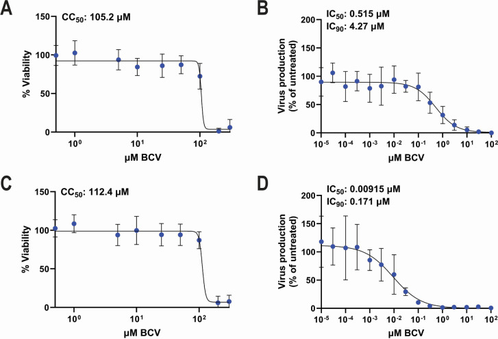
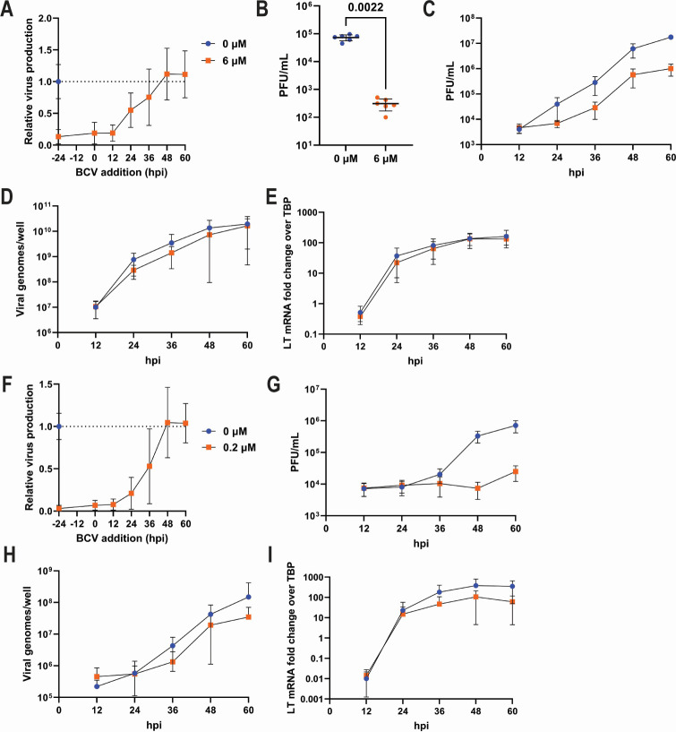
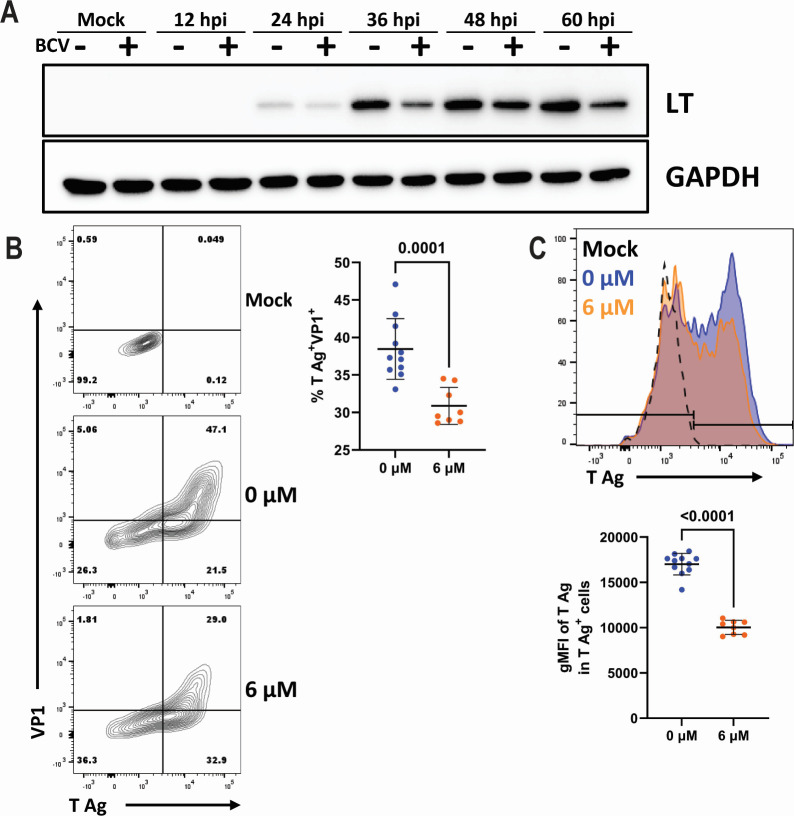
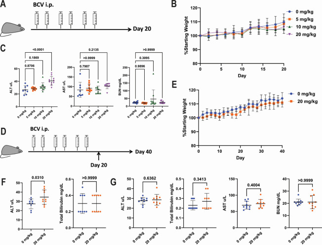
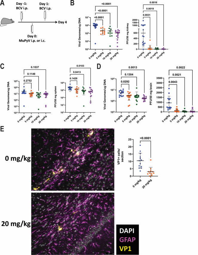
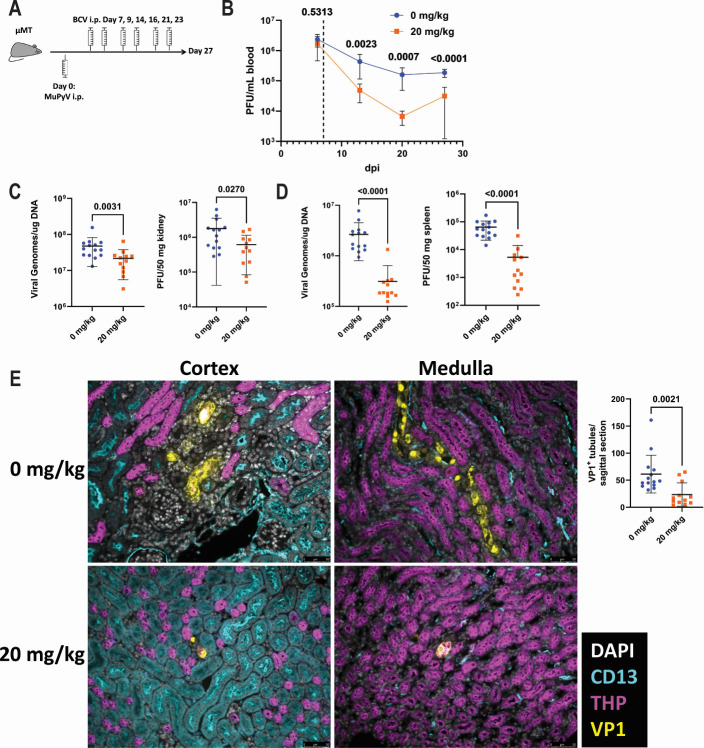
Polyomaviruses are species-specific DNA viruses that can cause disease in immunocompromised individuals. Despite their role as the causative agents for several diseases, there are no currently approved antivirals for treating polyomavirus infection. Brincidofovir (BCV) is an antiviral approved for the treatment of poxvirus infections and has shown activity against other double-stranded DNA viruses. In this study, we tested the efficacy of BCV against polyomavirus infection in vitro and in vivo using mouse polyomavirus (MuPyV). BCV inhibited virus production in primary mouse kidney cells and brain cortical cells. BCV treatment of cells transfected with MuPyV genomic DNA resulted in a reduction in virus levels, indicating that viral inhibition occurs post-entry. Although in vitro BCV treatment had a limited effect on viral DNA and RNA levels, drug treatment was associated with a reduction in viral protein, raising the possibility that BCV acts post-transcriptionally to inhibit MuPyV infection. In mice, BCV treatment was well tolerated, and prophylactic treatment resulted in a reduction in viral DNA levels and a potent suppression of infectious virus production in the kidney and brain. In mice with chronic polyomavirus infection, therapeutic administration of BCV decreased viremia and reduced infection in the kidney. These data demonstrate that BCV exerts antiviral activity against polyomavirus infection in vivo, supporting further investigation into the use of BCV to treat clinical polyomavirus infections.

Importance: Widespread in the human population and able to persist asymptomatically for the life of an individual, polyomavirus infections cause a significant disease burden in the immunocompromised. Individuals undergoing immune suppression, such as kidney transplant patients or those treated for autoimmune diseases, are particularly at high risk for polyomavirus-associated diseases. Because no antiviral agent exists for treating polyomavirus infections, management of polyomavirus-associated diseases typically involves reducing or discontinuing immunomodulatory therapy. This can be perilous due to the risk of transplant rejection and the potential development of adverse immune reactions. Thus, there is a pressing need for the development of antivirals targeting polyomaviruses. Here, we investigate the effects of brincidofovir, an FDA-approved antiviral, on polyomavirus infection in vivo using mouse polyomavirus. We show that the drug is well-tolerated in mice, reduces infectious viral titers, and limits viral pathology, indicating the potential of brincidofovir as an anti-polyomavirus therapeutic.

Keywords: antiviral agents; brain; brincidofovir; kidney; polyomavirus.


Conflict of interest statement

K.F. and M.H. are employees of Symbio, which owns the license to BCV.

Figures

Fig 1
Fig 1
BCV inhibits MuPyV in primary renal and cortical cells. (A) Cell viability in AMK cells after 84 hours of BCV treatment. Data are from three independent experiments (n = 18). (B) Dose-response curve of viral inhibition in AMK cells. Cells were pretreated with BCV for 24 hours and then infected at a multiplicity of infection (MOI) of 0.1. Viral titers were measured at 60 hpi by plaque assay and normalized to viral titers in untreated cells. Data are from three independent experiments (n = 7). (C) Cell viability in primary cortical cells after 84 hours of BCV treatment. Data are from two independent experiments (n = 12). (D) Dose-response curve of viral inhibition in primary cortical cells. Cells were pretreated with BCV for 24 hours and then infected at an MOI of 0.1. Viral titers were measured at 60 hpi and normalized to viral titers in untreated cells. Data are from three independent experiments (n = 6).
Fig 2
Fig 2
BCV inhibits MuPyV post-entry but does not inhibit viral DNA or RNA production. (A) Effect of BCV addition at different time points on virus production. AMK cells were infected at an MOI of 0.1, and BCV was added to the media at the indicated time points. Virus production was assessed at 60 hpi by plaque assay and normalized to untreated cells. Data are from five independent repeats (n = 15). (B) Effect of BCV on virus production after viral genome transfection. AMK cells were pretreated with BCV for 24 hours, transfected with viral DNA, and returned to BCV-containing media. Viral lysates were collected at 60 hours post-transfection and titered by plaque assay. Data are from two independent repeats (n = 6). (C) Effect of BCV on virus production throughout infection. AMK cells were pretreated with BCV, infected at an MOI of 0.1, and returned to BCV-containing media. Cell lysates were collected at the indicated time points, and virus production was assessed. Data are from two independent experiments (n = 6). (D and E) Effect of BCV on viral DNA and RNA production. AMK cells were treated as in panel C, and viral DNA (D) and RNA (E) were quantified at the indicated time points. Data are from two independent experiments (n = 6). (F–I) Effect of BCV on cortical cells. (F) Effect of BCV addition at different time points on virus production. Cortical cells were infected with an MOI of 0.1, and BCV was added to the media at the indicated time points. Virus production was assessed at 60 hpi by plaque assay and normalized to untreated cells. Data are from two independent repeats (n = 5). (G) Effect of BCV on virus production throughout infection. Cortical cells were pretreated with BCV, infected at an MOI of 0.1, and returned to BCV-containing media. Cell lysates were collected at the indicated time points and virus production was assessed. Data are from two independent experiments (n = 9). (H and I) Effect of BCV on viral DNA and RNA production. Cortical cells were treated as in panel G, and viral DNA (H) and RNA (I) were quantified at the indicated time points. Data are from two independent experiments (n = 7–9). Data were analyzed by the Mann-Whitney test (B).
Fig 3
Fig 3
BCV reduces MuPyV large T antigen levels. (A) Western blot for LT antigen. AMK cells were pretreated with BCV, infected at an MOI of 0.1, and returned to BCV-containing media. Cell lysates were collected at the indicated time points, and protein levels were assessed. Data are representative of two independent experiments. (B and C) Analysis of T Ag and VP1 protein levels. AMK cells were pretreated with BCV, infected at an MOI of 0.1, and returned to BCV-containing media. Cells were trypsinized at 24 hpi and stained for T Ag and VP1. Protein expression was quantified by flow cytometry. (B, left) Representative plots of T Ag and VP1 expression in uninfected cells or infected cells treated with 0 or 6 µM of BCV. (B, right) Frequency of T ag+VP1+ cells. Data are from two independent experiments (n = 8–11). (C, top) Representative histogram of T Ag expression. (C, bottom) Levels of T Ag expression in the T ag+ cells. Data are from two independent experiments (n = 8–11). Data were analyzed by the Mann-Whitney test (B and C).
Fig 4
Fig 4
In vivo tolerability of BCV. (A) Schematic of BCV tolerability analysis. Mice received two doses of BCV a week for 3 weeks and were weighed every 2 days. After 20 days, mice were euthanized, and serum was collected for the analysis of ALT, AST, and BUN levels. (B) Weights of mice treated biweekly with the indicated doses of BCV for 3 weeks. (C) Serum ALT (left), AST (center), and BUN (right) levels in the mice after 3 weeks of BCV treatment. (D) Schematic of extended BCV tolerability analysis. Mice received BCV treatments as in panel A and were followed until 40 days after BCV initiation. Serum was collected at days 20 and 40. (E) Weights of mice treated biweekly with the indicated doses of BCV for 3 weeks. (F) Serum ALT (left) and total bilirubin (right) at day 20. (G) Serum ALT, total bilirubin, AST, and BUN at day 40. Data were analyzed by one-way ANOVA (C), unpaired t test: (F) ALT and (G) ALT/AST/BUN, or Mann-Whitney test: (F) bilirubin and (G) bilirubin. Data are from two independent experiments (n = 8–10).
Fig 5
Fig 5
BCV treatment limits MuPyV kidney and brain infection. (A) Schematic of acute infection experiments with BCV treatment. Mice received BCV on days −1 and 1 and were infected with MuPyV either i.p. or i.c. on day 0. Mice were euthanized on day 4, and virus levels were assessed. Virus levels were quantified by qPCR for viral genomes or by plaque assay. (B) Kidney virus levels in i.p.-infected mice. Data are from four independent experiments (n = 10–15). (C) Spleen virus levels in i.p.-infected mice. Data are from four independent experiments (n = 11–15). (D) Brain virus levels in i.c.-infected mice. Data are from three independent experiments (n = 13–14). (E) GFAP and VP1 immunofluorescence staining of brain sections from vehicle- or 20 mg/kg BCV-treated mice. Left: representative images of brain regions with infected VP1+ cells. Right: quantification of the frequency of VP1+ cells. Data shown are the average number of VP1+ cells in two sections from different brain regions per mouse. Data are from two independent experiments (n = 11–13). Data were analyzed by Brown-Forsythe and Welch ANOVA tests (B–D) or Mann-Whitney test (E).
Fig 6
Fig 6
BCV reduces viral burden and limits kidney pathology during chronic infection. (A) µMT mice were infected with MuPyV, and viral load in the blood was measured weekly by plaque assay. Starting at 7 dpi, mice received biweekly injections of 0 or 20 mg BCV/kg of body weight. At 27 dpi, mice were euthanized, and virus levels in the spleen and kidney were quantified. (B) Blood was collected weekly, and virus titers were quantified by plaque assay. The dashed line indicates the start of BCV administration. Data are from three independent experiments (n = 12–14). (C) Viral load in the kidney quantified by qPCR for viral DNA (left) or plaque assay (right). Data are from three independent experiments (n = 12–14). (D) Viral load in the spleen quantified by qPCR (left) or plaque assay (right). Data are from three independent experiments (n = 12–14). (E) Sites of virus infection in the kidneys of vehicle- or BCV-treated µMT mice. Left: sites of virus infection in the kidney identified by VP1 immunofluorescence. Right: quantification of the number of VP1+ tubules per kidney section. Data are from three independent experiments (n = 12–14). Data were analyzed by two-way ANOVA (B) or Mann-Whitney test (C–E).

Similar articles

Cited by

References

    1. Buck CB, Van Doorslaer K, Peretti A, Geoghegan EM, Tisza MJ, An P, Katz JP, Pipas JM, McBride AA, Camus AC, McDermott AJ, Dill JA, Delwart E, Ng TFF, Farkas K, Austin C, Kraberger S, Davison W, Pastrana DV, Varsani A. 2016. The ancient evolutionary history of polyomaviruses. PLoS Pathog 12:e1005574. doi: 10.1371/journal.ppat.1005574 - DOI - PMC - PubMed
    1. Gardner SD, Field AM, Coleman DV, Hulme B. 1971. New human papovavirus (B.K.) isolated from urine after renal transplantation. Lancet 1:1253–1257. doi: 10.1016/s0140-6736(71)91776-4 - DOI - PubMed
    1. Kant S, Dasgupta A, Bagnasco S, Brennan DC. 2022. BK virus nephropathy in kidney transplantation: a state-of-the-art review. Viruses 14:1616. doi: 10.3390/v14081616 - DOI - PMC - PubMed
    1. Padgett BL, Walker DL, ZuRhein GM, Eckroade RJ, Dessel BH. 1971. Cultivation of papova-like virus from human brain with progressive multifocal leucoencephalopathy. Lancet 1:1257–1260. doi: 10.1016/s0140-6736(71)91777-6 - DOI - PubMed
    1. Kamminga S, van der Meijden E, Feltkamp MCW, Zaaijer HL. 2018. Seroprevalence of fourteen human polyomaviruses determined in blood donors. PLoS ONE 13:e0206273. doi: 10.1371/journal.pone.0206273 - DOI - PMC - PubMed

MeSH terms

LinkOut - more resources