. 2024 Jul 23;98(7):e0049824.
doi: 10.1128/jvi.00498-24. Epub 2024 Jul 2.

Identification of the proteolytic signature in CVB3-infected cells

Affiliations

Identification of the proteolytic signature in CVB3-infected cells

Marli Vlok et al. J Virol. .

Abstract

Coxsackievirus B3 (CVB3) encodes proteinases that are essential for processing of the translated viral polyprotein. Viral proteinases also target host proteins to manipulate cellular processes and evade innate antiviral responses to promote replication and infection. While some host protein substrates of the CVB3 3C and 2A cysteine proteinases have been identified, the full repertoire of targets is not known. Here, we utilize an unbiased quantitative proteomics-based approach termed terminal amine isotopic labeling of substrates (TAILS) to conduct a global analysis of CVB3 protease-generated N-terminal peptides in both human HeLa and mouse cardiomyocyte (HL-1) cell lines infected with CVB3. We identified >800 proteins that are cleaved in CVB3-infected HeLa and HL-1 cells including the viral polyprotein, known substrates of viral 3C proteinase such as PABP, DDX58, and HNRNPs M, K, and D and novel cellular proteins. Network and GO-term analysis showed an enrichment in biological processes including immune response and activation, RNA processing, and lipid metabolism. We validated a subset of candidate substrates that are cleaved under CVB3 infection and some are direct targets of 3C proteinase in vitro. Moreover, depletion of a subset of TAILS-identified target proteins decreased viral yield. Characterization of two target proteins showed that expression of 3Cpro-targeted cleaved fragments of emerin and aminoacyl-tRNA synthetase complex-interacting multifunctional protein 2 modulated autophagy and the nuclear factor kappa-light-chain-enhancer of activated B cells (NF-κB) pathway, respectively. The comprehensive identification of host proteins targeted during virus infection provides insights into the cellular pathways manipulated to facilitate infection.

Importance: RNA viruses encode proteases that are responsible for processing viral proteins into their mature form. Viral proteases also target and cleave host cellular proteins; however, the full catalog of these target proteins is incomplete. We use a technique called terminal amine isotopic labeling of substrates (TAILS), an N-terminomics to identify host proteins that are cleaved under virus infection. We identify hundreds of cellular proteins that are cleaved under infection, some of which are targeted directly by viral protease. Revealing these target proteins provides insights into the host cellular pathways and antiviral signaling factors that are modulated to promote virus infection and potentially leading to virus-induced pathogenesis.

Keywords: AIMP2; EMD; N-terminomics; degradomics; picornavirus; protease.


Conflict of interest statement

The authors declare no conflict of interest.

Figures

Fig 1
Fig 1
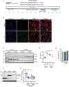
Strategy for identifying proteolytic substrates in CVB3-infected cells using TAILS. (A) Flowchart of TAILS approach for CVB3-infected HeLa and HL-1 cells. Immunofluorescence of mock- and CVB3-infected (B) HeLa cells (MOI 10, 5 h.p.i., scale bar 20 µm) or (C) HL-1 cells (MOI 50, 20 h.p.i., scale bar 10 µm) stained with 4′,6-diamidino-2-phenylindole (DAPI) (blue) and VP1 (green).
Fig 2
Fig 2
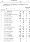
N-terminal peptides of the CVB3 polyprotein detected by TAILS. (A) Graphical presentation of cleavage sites detected in the polyprotein inferred by TAILS analysis, with known cleavage sites indicated above. TAILS analysis also detected other potential cleavage site indicated below. Colors depict data sets in which TAILS peptides were detected: both HeLa and HL-1 (purple), HL-1 (red), and HeLa (blue). (B) Table showing TAILS-identified peptides within the CVB3 polyprotein including the peptide locations and the high-to-low (H/L) ratios in both biological replicates. Number of biological replicates and technical replicates in which the peptides were detected are also included. Peptide modifications are indicated by asterisk (*, dimethylated), underlined (acetylation), and italics (carbamidomethylation). Predicted upstream peptides are included (P10–P1) with those displaying an arginine in P1 in gray. Known CVB3 cleavage peptides are indicated in bold.
Fig 3
Fig 3
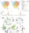
Identification of host cellular proteome in CVB3-infected cells. Venn diagrams of high-confidence proteins (A) and peptides (B) identified in TAILS analysis of CVB3-infected HeLa and HL-1 cells with comparisons with in vitro TAILS analysis of CVB3 3Cpro in HL-1 cell lysates (32). High-confidence substrates that had a positive H/L ratio outside the linear range were analyzed. (C) Ice logo of amino acid adjacent to the cleavage sites of candidate 2Apro and 3Cpro substrates from TAILS HeLa and HL-1 data sets. (D) Bar graph of cellular protease cleavage sites using TopFinder inferred by TAILS high candidate substrates in CVB3-infected cells. Both positive and negative H/L ratio substrates were graphed. Calpain-2 (CAN2), caspase 1 (CASP1), caspase 3 (CASP3), caspase 6 (CASP6), caspase 7 (CASP7), caspase 8 (CASP8), caspase 9 (CASP9), cathepsin B (CATB), cathepsin (CATD), cathepsin E (CATE), procathepsin L (CATL1), cathepsin S (CATS), granzyme A (GRAA), granzyme B (GRAB), granzyme C (GRAC), granzyme M (GRAM), serine protease HTRA2, mitochondrial (HTRA2), meprin A subunit B (MEP1B), collagenase type IV (MMP2), matrix metalloproteinase-9 (MMP9), and stromelysin-3 (MMP11). (E) Network analysis of TAILS high-confidence proteins in CVB3-infected HeLa and HL-1 cells. Databases used for pathway and GO term analyses are depicted by node shapes with GO term biological processes (ellipse), GO term molecular function (rectangle), and Kyoto Genes and Genomes (KEGG) pathways (triangle).
Fig 4
Fig 4
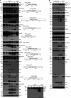
Validation of cleavage of candidate TAILS protein targets in CVB3-infected cells. Immunoblotting of indicated proteins in (A) CVB3-infected HeLa cells (MOI 10) and (B) poliovirus-infected HeLa cells (MOI 10). Shown in (A) (right) are the schematic of proteins, the predicted mass of cleavage fragments, and the predicted cleavage site with P4-P1 and P1′-P4’ amino acids based on the TAILS detected peptide. cp, cleavage product; *, non-specific, unclassified product. Representative immunoblots from at least two independent experiments.
Fig 5
Fig 5
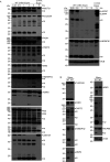
Validation of candidate of TAILS protein targets using In vitro cleavage assays. Immunoblots of indicated proteins in HeLa cell lysates incubated with purified recombinant (A) wild-type or catalytically inactive (C174A) CVB3 3C protease or (B) wild-type or catalytically inactive CVB3 2A protease (C57A). cp, cleavage product. Representative immunoblots from at least two independent experiments.
Fig 6
Fig 6
Depletion of TAILS candidate proteins in CVB3-infected cells. Immunoblots of HeLa cells transfected with control siRNA (siSCR) or siRNAs directed against (A) AIMP2, (B) EMD, (C) CNOT2, or (D) RAI. siRNA-treated cells (48 h) were infected with CVB3 with an MOI 1.0 (A, B, C) or 0.1 (D) for 16 h. Extracellular and intracellular viral yields were detected by plaque assay. Shown are box plots of log10 PFU with means and 95% confidence intervals from three independent experiments with at least two technical replicates. (E) Extracellular and intracellular viral titers collected from CVB3-infected (MOI 0.1) HeLa cells were pre-treated with cerulenin (45 M) and ACA (20 M). P-values from nested one-way t-test are indicated.
Fig 7
Fig 7
EMD promotes autophagy and CVB3 infection. (A) Schematic of EMD with annotated domains and the TAILS peptides identified in CVB3-infected HeLa and HL-1 cells. (B) Immunofluorescence of EMD in mock- or CVB3-infected HeLa cells (MOI 10). Scale bar 20 µm. (C) Immunoblots of indicated proteins in control siRNA or EMD siRNA-treated mock- or CVB3-infected cells. (D) Quantitation of (C) LC3-II immunoblots. (E) Immunoblots of GFP-N-EMD and GFP-C-EMD from HeLa cells transfected with expression plasmids containing N- and C-terminal EMD cleavage fragments. (F) Viability of cells transfected with GFP-tagged N-terminal or C-terminal plasmids (24 h post transfection) by trypan blue dye exclusion. (G) Extracellular and intracellular virus titers from CVB3-infected (MOI 1.0) HeLa cells transfected with GFP-N-EMD and GFP-C-EMD, collected at 20 h post infection. Shown are box plots of plaque assay (PFU) from at least three independent experiments of CVB3-infected cells transfected with the indicated plasmids normalized to that of CVB3-infected cells transfected with GFP-expressing plasmid.
Fig 8
Fig 8
Cleavage fragments of AIMP2 promotes CVB3 infection. (A) Schematic of AIMP2 with annotated binding domains and the TAILS peptides identified in CVB3-infected HeLa and HL-1 cells. (B) Immunofluorescence of AIMP2 in mock- or CVB3-infected HeLa cells (MOI 10). Scale bar 20 µm. (C) Immunoblots of HeLa cells transfected with expression plasmids containing GFP-tagged N-terminal and C-terminal AIMP2 cleavage fragments. (D) Viability of cells transfected with GFP-tagged N-terminal or C-terminal AIMP2 plasmids by trypan blue dye exclusion assay. Viral yield measured by plaque assay (PFU) of CVB3-infected cells transfected with GFP control plasmid or GFP-tagged (E) N-terminal or (F) C-terminal AIMP2 cleavage fragment plasmids. Shown are three independent experiments, displaying the averages of at least three technical measurements. (G) Quantitation of (E) and (F) by box plots and normalizing PFU of HeLa cells transfected N-terminal and C-terminal AIMP2 cleavage fragment plasmids normalized to that of cells transfected with GFP.
Fig 9
Fig 9
Cleavage fragments of AIMP2 modulate NFκB signaling. (A) Immunofluorescence of p65 in mock- and CVB3-infected HeLa cells. Scale bar 20 µm. (B) Induction of NFκB-responsive firefly luciferase reporter in TNFα-treated HeLa cells transfected with plasmids expressing GFP-tagged wild-type AIMP2, N-terminal or C-terminal cleavage fragments of AIMP2. Cells were co-transfected with a Renilla luciferase reporter for normalization. Shown are the ratio of firefly luciferase activities normalized to GFP-expressing cells. (C) Viability of cells treated with indicated drug concentrations. (D) Normalized NFκB-responsive firefly luciferase reporter in TNFα-treated HeLa cells with the indicated concentrations of BAY11-7082 for 20 min. (E) Relative viral titers of HeLa cells transfected with either N-terminal or C-terminal AIMP2 and treated with BAY11-7082 (10 µm) 30 min prior to infection (MOI 0.1) for 16 h. The values are normalized with GFP-transfected infected cells and presented as averages ± SD from at least three independent experiments.

References

    1. Olejniczak M, Schwartz M, Webber E, Shaffer A, Perry TE. 2020. Viral myocarditis-incidence diagnosis and management. J Cardiothorac Vasc Anesth 34:1591–1601. doi:10.1053/j.jvca.2019.12.052 - DOI - PubMed
    1. Collaborators GBoDS . 2015. Global, regional, and national incidence, prevalence, and years lived with disability for 301 acute and chronic diseases and injuries in 188 countries, 1990-2013: a systematic analysis for the global burden of disease study 2013. Lancet 386:743–800. doi:10.1016/S0140-6736(15)60692-4 - DOI - PMC - PubMed
    1. Buggey J, ElAmm CA. 2018. Myocarditis and cardiomyopathy. Curr Opin Cardiol 33:341–346. doi:10.1097/HCO.0000000000000514 - DOI - PubMed
    1. Esfandiarei M, McManus BM. 2008. Molecular biology and pathogenesis of viral myocarditis. Annu Rev Pathol 3:127–155. doi:10.1146/annurev.pathmechdis.3.121806.151534 - DOI - PubMed
    1. Fung G, Luo H, Qiu Y, Yang D, McManus B. 2016. Myocarditis. Circ Res 118:496–514. doi:10.1161/CIRCRESAHA.115.306573 - DOI - PubMed

LinkOut - more resources